Maharashtra Board Class 11 Biology Important Questions Chapter 15 Excretion and Osmoregulation

Balbharti Maharashtra State Board 11th Biology Important Questions Chapter 15 Excretion and Osmoregulation Important Questions and Answers.

Maharashtra State Board 11th Biology Important Questions Chapter 15 Excretion and Osmoregulation

Question 1.
What are metabolic waste products?
Answer:
Metabolism produces a variety of by-products, some of which need to be eliminated. Such by-products are called metabolic waste products.

Question 2.
Define excretion.
Answer:
The process of eliminating waste products from the body is called excretion.

Maharashtra Board Class 11 Biology Important Questions Chapter 15 Excretion and Osmoregulation

Question 3.
Where are metabolic wastes produced?
Answer:
Metabolic wastes are produced inside body cells.

Question 4.
Enlist the various excretory products produced in the human body.
Answer:
The various excretory products produced by the human body are as follows:

  1. Fluids such as water; gaseous wastes like CO2; nitrogenous wastes like ammonia, urea and uric acid, creatinine; minerals; salts of sodium, potassium, calcium, etc. if present in body in excess are excreted through urine, faeces and sweat.
  2. Pigments formed due to breakdown of haemoglobin like bilirubin (excreted through faeces) and urochrome (eliminated through urine).
  3. The pigments present in consumed foodstuffs like beet root or excess of vitamins, hormones and drugs.
  4. Volatile substances present in spices (eliminated through lungs).

Question 5.
Write a note on deamination.
Answer:

  1. Deamination is the process of breakdown of excess amino acids.
  2. It is an essential process, since the body of an organism is unable to store excess amino acids.
  3. In this process, amino group is separated from the amino acid and ammonia is formed.
  4. Toxic ammonia is either excreted or converted to less toxic forms like urea or uric acid before excretion.

Maharashtra Board Class 11 Biology Important Questions Chapter 15 Excretion and Osmoregulation

Question 6.
Availability of water plays a key role in deciding the mode of excretion of an organism. Justify.
Answer:

  1. Ammonia is the basic product of deamination process.
  2. Ammonia is highly toxic and needs to be diluted immediately.
  3. If there is no or limited access to water, need for conversion of ammonia becomes necessary.
    Hence, the availability of water plays a key role in deciding the mode of excretion of an organism.

Question 7.
What are the three main modes of excretion in animals?
Answer:
The three main modes of excretion in animals are as follows:
i. Ammonotelism
ii. Ureotelism
iii. Uricotelism

  1. Ammonotelism:
    • Elimination of nitrogenous wastes in the form of ammonia is called as ammonotelism.
    • Ammonia is basic in nature and hence it can disturb the pH of the body, if not eliminated immediately.
    • Any change in pH would disturb all enzyme catalyzed reactions in the body and would also make the plasma membrane unstable.
    • Ammonia is readily soluble in water and needs large quantity of water to dilute and reduce its toxicity.
    • This is however an energy saving mechanism of excretion and hence all animals that have plenty of water available for dilution of ammonia, excrete nitrogenous wastes in the form of ammonia.
    • Animals that follow this mode of excretion are known as ammonotelic animals.
    • 1 gm ammonia needs about 300 – 500 ml of water for elimination.
    • Ammonotelic animals excrete ammonia through general body surface (skin), gills and kidneys.
      e.g. Ammonotelism is found in aquatic invertebrates, bony fishes, and aquatic / larval amphibians. Animals without excretory system (Protozoa) are also ammonotelic.
  2. Ureotelism:
    • Elimination of nitrogenous wastes in the form of urea is called as ureotelism.
    • Urea is comparatively less toxic and less water-soluble than ammonia. Hence, it can be concentrated to some extent in body.
    • The body requires less water for elimination.
    • Since it is less toxic and less water soluble, ureotelism is suitable for animals that need to conserve water to some extent. Hence, ureotelism is common in terrestrial animals, as they have to conserve water.
    • It takes about 50 ml H2O for removal of 1 gm NH2 in form of urea.
    • Ureotelic animals generally convert ammonia to urea in the liver by operating ornithine / urea cycle in which 3 ATP molecules are used to produce one molecule of urea.
      e.g. Mammals, cartilaginous fishes (sharks and rays), many aquatic reptiles, most of the adult amphibians, etc. are ureotelic.
  3. Uricotelism:
    • Elimination of nitrogenous wastes in the form of uric acid is called as uricotelism.
    • Uric acid is least toxic and hence, it can be retained in the body for some time in concentrated form.
    • It is least soluble in water. Hence there is minimum (about 5 – 10 ml for 1 gm) or no need of water for its elimination.
    • Those animals which need to conserve more water follow uricotelism. However, these animals need to spend more energy.
    • Ammonia is converted into uric acid by ‘inosinic acid pathway’ in the liver of birds, e.g. Birds, some insects, many reptiles, land snails, are uricotelic.

Question 8.
Fill in the blanks:
i. ________ is the basic product of deamination process.
ii. Aquatic amphibians excrete nitrogenous waste in the form of _______.
iii. Uricotelic organisms, convert ammonia to urea in the _______ by operating _____ cycle.
iv. Ammonia is converted into uric acid by ______ pathway in birds.
Answer:
i. Ammonia
ii. Ammonia
iii. liver, omithine/urea
iv. inosinic acid

Question 9.
Explain the following sentences,
i. Humans are ureotelic.
Answer:

  • Urea is comparatively less toxic and less water – soluble than ammonia. Hence, it can be concentrated to some extent in the body.
  • The body requires less water for elimination of urea.
    c. Due to these properties, ureotelism is suitable for animals which need to conserve water to some extent.
    Thus, humans are ureotelic.

ii. Sharks retain more urea in their blood.
Answer:

  • Sharks retain more urea in their body fluid (blood) to make their blood isotonic to surrounding marine water (in order to maintain osmotic balance).
  • This helps them to prevent possible loss of water by exosmosis.

Question 10.
Distinguish between Ureotelism and Uricotelism.
Answer:

No. Ureotelism Uricotelism
i. It is the elimination of nitrogenous waste in the form of urea. It is the elimination of nitrogenous waste in the form of uric acid.
ii. Excretion of urea requires less (moderate ) amount of water. Excretion of uric acid requires negligible amount of water.
iii. Removal of 1 gm of urea requires 50 ml of water. Removal of 1 gm of uric acid requires 5 – 10 ml of water, j
iv. Urea is less toxic. Uric acid is least toxic.
e.g. It is generally seen in terrestrial animals. Mammals, cartilaginous fishes (sharks and rays), many aquatic reptiles, most adult amphibians, etc. It is seen in birds, some insects, many reptiles, land snails, etc.

Maharashtra Board Class 11 Biology Important Questions Chapter 15 Excretion and Osmoregulation

Question 11.
Distinguish between Ammonotelism and Uricotelism.
Answer:

No.

Ammonotelism

Uricotelism

i. It is the elimination of nitrogenous waste in the form of ammonia. It is the elimination of nitrogenous waste in the form of uric acid.
ii. Excretion of ammonia requires plenty of water. Excretion of uric acid requires negligible amount of water.
iii. Removal of 1 gm of ammonia requires 300 – 500 ml of water. Removal of 1 gm of uric acid requires 10ml of water.
iv. Ammonia is very toxic. Uric acid is less toxic.
e.g. It is found in aquatic invertebrates, bony fishes and aquatic/ larval amphibians, etc. It is seen in birds, some insects, many reptiles, land snails, etc.

Question 12.
Terrestrial animals are generally either ureotelic or uricotelic, not ammonotelic. Why?
Answer:

  1. Ammonia is highly toxic to animals.
  2. An animal requires large amount of water to dissolve and eliminate ammonia.
  3. Terrestrial animals cannot lose such a large amount of water.
  4. Ureotelic and uricotelic animals require less amount of water for removal of nitrogenous waste. Hence, to conserve water, ureotelism and uricotelism is adapted by terrestrial animals.

Question 13.
What is plasma creatinine? Why is it used as an index of kidney function?
Answer:

  1. Plasma creatinine is produced from catabolism of creatinine phosphate during skeletal muscle contraction.
  2. It provides a ready source of high energy phosphate.
  3. Normally blood creatinine levels remain steady because the rate of production matches its excretion in urine.
  4. Hence, plasma creatinine is used as an index of kidney function and its level above normal is an indication of poor renal function.
    [Note: Plasma creatinine is a waste product produced by muscles from the breakdown of a compound called ‘creatine phosphate.]

Question 14.
How can excretion play a role in homeostasis?
Answer:

  1. Homeostasis is the maintenance of constant internal environment of the body.
  2. Homeostasis is however dependent on osmoregulation, which is the process of controlling solute concentrations and water balance.
  3. The composition of blood and therefore the internal environment is highly dependent on what the excretory organs retain in the body.
    Hence, excretion plays an important role in homeostasis.

Question 15.
How do different organisms carry out excretion?
Answer:
Different organisms carry out excretion in the following manner:

  1. Unicellular organisms have contractile vacuoles which collect and discharge waste products outside the cell.
  2. Excretion in sponges takes place by diffusion of waste material in water. This waste is discharged through the osculum.
  3. True organs of excretion are found in those animals that show bilateral symmetry.
  4. The most common type of excretory organ is a simple or branching tube that opens to the exterior, through pores called nephridiopores. This system is generally found in some annelids, Amphioxus, earthworms, etc.
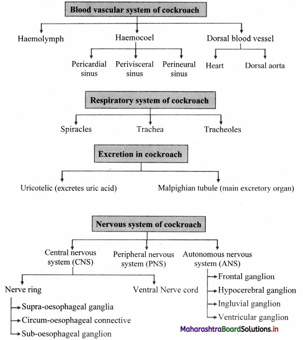
  5. In most of the insects, excretion takes place by a set of blind ended tubules called malpighian tubules.
  6. Crustaceans have green glands as excretory organs.
  7. Members of phylum Echinodermata do not have any specialised excretory organs. Waste materials directly diffuse into water or are excreted through tube feet.
  8. The mammalian kidneys are a collection of functional units called nephrons, which are well designed to excrete metabolic waste.

Maharashtra Board Class 11 Biology Important Questions Chapter 15 Excretion and Osmoregulation

Question 16.
What are nephridia? Explain the major types of nephridia in detail.
Answer:
Nephridia are simple or branching tubules used for excretion which open to the exterior through pores called nephridiopores.
Two major types of nephridia are as follows:
i. Protonephridia:
These are network of dead end tubes called flame cells. They are mostly found in animals that lack a true body cavity, e.g. Platyhelminthes, rotifers, some annelids and Amphioxus.

ii. Metanephridia:
These are unbranched coiled tubes that are connected to the body cavity through funnel like structures called nephrostomes. Body fluid enters the nephridium through nephrostome and gets discharged ‘ through nephridiopore. e.g. Earthworms.

Question 17.
Distinguish between the Ureter and Urethra.
Answer:

No. Ureter Urethra
i. Ureters are two duct-like structures arising from the hilum of the kidney. Urethra is a single tube-like structure arising from the urinary bladder.
ii. Ureter carries urine from the kidney to the urinary bladder. Urethra carries urine from the urinary bladder to the exterior of the body.
iii. Ureters are paired structures. Urethra is unpaired structure.

Question 18.
Write a short note on micturition.
Answer:

  1. The process of release of urine from the urinary bladder is called micturition.
  2. The average capacity of urinary bladder is 700 ml.
  3. When urinary bladder is almost half filled, stretch receptors in urinary bladder transmit impulses to spinal cord, initiating a conscious desire to expel urine.
  4. Micturition reflex center of spinal cord transmit impulses to the wall of urinary bladder and internal urethral sphincter.
  5. Bladder muscles contract and muscles of internal urethral sphincter relax.
  6. The external sphincter receives impulses from conscious centre of brain and relaxes.
  7. This leads to elimination of urine from the bladder.

Question 19.
Explain the L.S of kidney with a neat and labelled diagram.
Answer:

  1. Each kidney is covered by three layers of tissue, namely the outermost renal fascia, middle adipose capsule and innermost renal capsule.
    • The outermost layer, renal fascia is made up of a thin layer of fibrous connective tissue. It anchors the kidney to the abdominal wall as well as surrounding tissue.
    • The middle layer is a mass of fatty tissue called adipose capsule. It protects the kidneys by shock absorption.
    • The innermost layer, renal capsule is a smooth transparent fibrous membrane that is continuous with outer layer of ureters. It acts as a barrier against spread of infections in kidney.
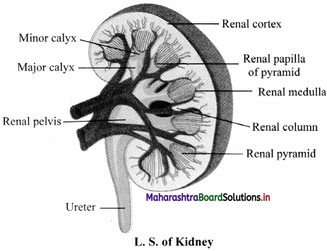
  2. The L.S. of kidney shows two distinct regions within the capsule. Histologically, kidney is divisible into two regions as renal cortex and renal medulla.
    • Renal cortex is the outer / peripheral, red coloured and granular region. It contains Malpighian bodies, convoluted tubules and blood vessels.
    • Medulla is inner region of kidney with pale red colour and striated appearance. Medulla mainly consists of Loops of Henle and collecting ducts. All these are arranged in conical manner to form renal pyramids.
    • Cortex extends in medulla as columns of Bertini / renal columns between pyramids. Narrow tip of pyramid is called as renal papilla. There are several pyramids.
    • Renal papilla open into the minor calyx. Minor calyces merge together to form major calyces and major calyces unite together to form renal pelvis.
    • Renal pelvis (renal sinus) is funnel-shaped area in the region of medulla of kidney. Renal pelvis
      Maharashtra Board Class 11 Biology Important Questions Chapter 15 Excretion and Osmoregulation 1

Question 20.
What is nephrology?
Answer:
Nephrology is branch of biology that deals with the structure, function and disorders of male and female urinary system.

Maharashtra Board Class 11 Biology Important Questions Chapter 15 Excretion and Osmoregulation

Question 21.
Write a note on nephron.
Answer:

  1. Nephrons are structural and functional units of kidney.
  2. Each nephron consists of a 4 – 6 cm long, thin-walled tube called the renal tubule and a bunch of capillaries known as the glomerulus.
  3. The wall of the renal tubule is made up of a single layer of epithelial cells.
  4. Its proximal end is wide, blind, cup-like and is called as Bowman’s capsule, whereas the distal end is open.
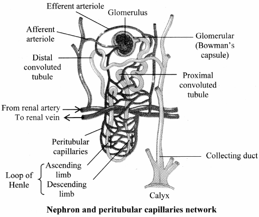
  5. The nephron is divisible into Bowman’s capsule, neck, proximal convoluted tubule (PCT), Loop of Henle (LoH), distal convoluted tubule (DCT) and collecting tubule (CT).
  6. The glomerulus is present in the cup-like cavity of Bowman’s capsule and both are collectively known as renal corpuscle or Malpighian body.

Question 22.
With the help of a well labelled diagram, describe the structure of nephron.
Answer:
Nephron is the structural and functional unit of kidney.
Structure of nephron:
A nephron (uriniferous tubule) is a thin walled, coiled duct, lined by a single layer of epithelial cells. Each nephron is divided into two main parts:
i. Malpighian body
ii. Renal tubule
Maharashtra Board Class 11 Biology Important Questions Chapter 15 Excretion and Osmoregulation 2

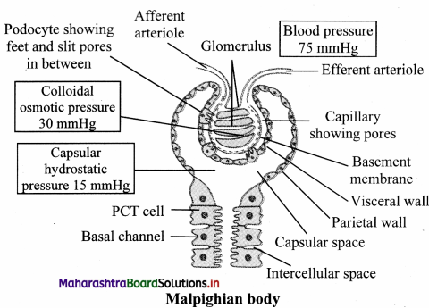
i. Malpighian body: Each Malpighian body is about 200pm in diameter and consists of a Bowman’s capsule and glomerulus.
a. Glomerulus:
Glomerulus is a bunch of fine blood capillaries located in the cavity of Bowman’s capsule.
A small terminal branch of the renal artery, called as afferent arteriole enters the cup cavity (Bowman capsule) and undergoes extensive fine branching to form network of several capillaries. This bunch is called as glomerulus.
The capillary wall is fenestrated (perforated).
All capillaries reunite and form an efferent arteriole that leaves the cup cavity.
The diameter of the afferent arteriole is greater than the efferent arteriole. This creates a high hydrostatic pressure essential for ultrafiltration, in the glomerulus.

b. Bowman’s capsule:
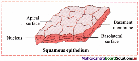
It is a cup-like structure having double walls composed of squamous epithelium.
The outer wall is called as parietal wall and the inner wall is called as visceral wall.
The parietal wall is thin consisting of simple squamous epithelium.
There is a space called as capsular space / urinary space in between two walls.
Visceral wall consists of special type of squamous cells called podocytes having a foot-like pedicel. These podocytes are in close contact with the walls of capillaries of glomerulus.
There are small slits called as filtration slits in between adjacent podocytes.

ii. Renal tubule:
a. Neck:
The Bowman’s capsule continues into the neck. The wall of neck is made up of ciliated epithelium. The lumen of the neck is called the urinary pole. The neck leads to proximal convoluted tubule.

b. Proximal Convoluted Tubule :
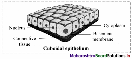
This is highly coiled part of nephron which is lined by cuboidal cells with brush border (microvilli) and surrounded by peritubular capillaries. Selective reabsorption occurs in PCT. Due to convolutions (coiling), filtrate flows slowly and remains in the PCT for longer duration, ensuring that maximum amount of useful molecules are reabsorbed.

c. Loop of Henle :
This is ‘U’ shaped tube consisting of descending and ascending limb.
The descending limb is thin walled and permeable to water and lined with simple squamous epithelium.
The ascending limb is thick walled and impermeable to water and is lined with simple cuboidal epithelium.
The LoH is surrounded by capillaries called vasa recta.
Its function is to operate counter current system – a mechanism for osmoregulation.
The ascending limb of Henle’s loop leads to DCT.

d. Distal convoluted tubule:
This is another coiled part of the nephron.
Its wall consists of simple cuboidal epithelium.
DCT performs tubular secretion / augmentation / active secretion in which, wastes are taken up from surrounding capillaries and secreted into passing urine.
DCT helps in water reabsorption and regulation of pH of body fluids.

e. Collecting tubule:
This is a short, straight part of the DCT which reabsorbs water and secretes protons.
The collecting tubule opens into the collecting duct.

Question 23.
What is the difference between Cortical nephrons and Juxtamedullary nephrons.
Answer:

Cortical nephrons

Juxtamedullary nephrons

i. They have a shorter loop of Henle. They have a longer loop of Henle.
ii. Loop of Henle of these nephrons extends very little into the medulla. Loop of Henle of these nephrons run deep into the medulla.
iii. Most nephrons are cortical nephrons. Few nephrons are juxtamedullary nephrons.
iv. Efferent arteriole forms peritubular capillary network around DCT, PCT and Henle’s loop of cortical nephrons Efferent arteriole forms loop-shaped vasa recta around  Henle’s loop of juxtamedullary nephrons.

Question 24.
Sketch and label Malpighian body.
Answer:
Maharashtra Board Class 11 Biology Important Questions Chapter 15 Excretion and Osmoregulation 3

Question 25.
What are podocytes? In which part of the nephron are they present?
Answer:
Podocytes are a special type of squamous cells that have a foot-like pedicel. They are present in the visceral wall of the Bowman’s capsule and are in close contact with the walls of capillaries of glomerulus

Maharashtra Board Class 11 Biology Important Questions Chapter 15 Excretion and Osmoregulation

Question 26.
Write a short note on Juxta Glomerular Apparatus.
Answer:

  1. Some smooth muscle cells of the wall of afferent arteriole are modified in such a way that their sarcoplasm is granular. These cells are called juxtaglomerular (JG) cells.
  2. In each nephron, initial part of DCT makes contact with the afferent arteriole of same nephron.
  3. Cells in the wall of DCT in this region are packed more densely than those in other region of DCT. This is called macula densa.
  4. Macula densa and the JG cells together form Juxta Glomerular Apparatus (JGA).
  5. The JGA plays an important role in blood pressure regulation within the kidney.

Question 27.
Explain the mechanism of urine formation in detail.
Answer:
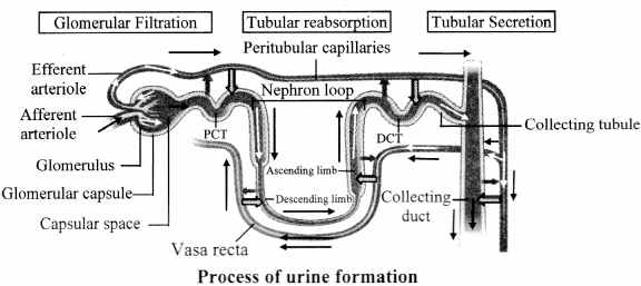
Process of urine formation is completed in three steps, namely;
i. Ultrafiltration/ Glomerular filtration,
ii. Selective reabsorption,
iii. Tubular secretion / Augmentation

i. Ultrafiltration / Glomerular filtration :
Diameter of afferent arteriole is greater than the efferent arteriole. The diameter of capillaries is still smaller than both arterioles. Due to the difference in diameter, blood flows with greater pressure through the glomerulus. This is called as glomerular hydrostatic pressure (GHP) and normally, it is about 55 mmHg. GIIP is opposed by osmotic pressure of blood (normally, about 30 mm Hg) and capsular pressure (normally, about 15 mm Hg).

Hence net / effective filtration pressure (EFP) is 10 mm Hg.
EFP = Hydrostatic pressure in glomerulus – (Osmotic pressure of blood + Filtrate Hydrostatic pressure)
= 55 – (30 +15)
= 10 mm Hg

Under the effect of high pressure, the thin walls of the capillary become permeable to major components of blood (except blood cells and macromolecules like protein).
Thus, plasma except proteins oozes out through wall of capillaries.
About 600 ml blood passes through each kidney per minute.

The blood (plasma) flowing through kidney (glomeruli) is filtered as glomerular filtrate, at a rate of 125 ml / min. (180 L/d).

Glomerular filtrate / deproteinized plasma / primary urine is alkaline, contains urea, amino acids, glucose, pigments, and inorganic ions.

Glomerular filtrate passes through filtration slits into capsular space and then reaches the proximal convoluted tubule.

ii. Selective reabsorption :
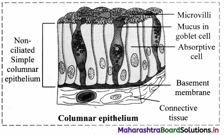
Selective reabsorption occurs in proximal convoluted tubule (PCT). It is highly coiled so that glomerular filtrate passes through it very slowly. Columnar cells of PCT are provided with microvilli due to which absorptive area increases enormously.

This makes the process of reabsorption very effective.
These cells perform active (ATP mediated) and passive (simple diffusion) reabsorption.
Substances with considerable importance (high threshold) like – glucose, amino acids, vitamin C, Ca++, K+, Na+, Cl are absorbed actively, against the concentration gradient. Low threshold substances like water, sulphates, nitrates, etc., are absorbed passively.
In this way, about 99% of glomerular filtrate is reabsorbed in PCT and DCT.

iii. Tubular secretion / Augmentation :
Finally filtrate reaches the distal convoluted tubule via loop of Henle. Peritubular capillaries surround DCT. Cells of distal convoluted tubule and collecting tubule actively absorb the wastes like creatinine and ions like K+, H+ from peritubular capillaries and secrete them into the lumen of DCT and CT, thereby augmenting the concentration of urine and changing its pH from alkaline to acidic.

Secretion of H+ ions in DCT and CT is an important homeostatic mechanism for pH regulation of blood. Tubular secretion is the only process of excretion in marine bony fishes and desert amphibians.
Maharashtra Board Class 11 Biology Important Questions Chapter 15 Excretion and Osmoregulation 4

Question 28.
Marine bony fishes and desert amphibians rely on which process of excretion?
Answer:
Tubular secretion

Question 29.
Where does selective reabsorption of glomerular filtrate take place?
Answer:
Selective reabsorption of glomerular filtrate takes place in the proximal convoluted tubule (PCT).

Question 30.
What is glomerular filtration pressure or net effective filtration pressure?
Answer:
Glomerular filtration pressure (GFP)/ Effective filtration pressure (EFP) is the difference between the hydrostatic pressure (GHP) and the sum of osmotic pressure of blood and capsular pressure (CHP).
It can be represented as:
EFP = Glomerular hydrostatic pressure – (Osmotic pressure of blood + Filtrate Flydrostatic pressure)
= 55 – (30 + 15)
= 10 mmHg
[Note: Net filtration pressure = Glomerular blood hydrostatic pressure – (Capsular hydrostatic pressure + Blood colloid osmotic pressure)
Source: Tort or a, G., Derrickson, B. Principles of Anatomy and Physiology. 11th Edition.]

Question 31.
Distinguish between Selective reabsorption and Tubular secretion.
Answer:

No.

Selective rcabsorption

Tubular secretion

i. Selective reabsorption is concerned with the selective absorption of useful substances from the glomerular filtrate. Tubular secretion is transfer of materials from peritubular capillaries to the renal tubular lumen.
ii. Substances with considerable importance (high threshold) like – glucose, amino acids, vitamin C, Ca++, K+, Na+, Cl are absorbed actively, against concentration gradient In this process, substances like urea. amino acids, glucose, pigments, and inorganic ions arc removed from the blood and discharged along with the urine.
iii. Selective reabsorption occurs in Proximal convoluted tubule, Henle’s loop, Distal convoluted tubule and collecting duct. Tubular secretion occurs in Distal convoluted tubules only.

Maharashtra Board Class 11 Biology Important Questions Chapter 15 Excretion and Osmoregulation

Question 32.
Explain the process of concentration of urine in deai1.
OR
Explain counter current mechanism in detail.
Answer:
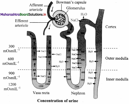
Under the conditions like low water intake or high water loss due to sweating, humans can produce concentrated urine. This urine can be concentrated around four times i.e. 1200 mOsm/L, than the blood (300 mOsm/L). hence, a mechanism called countercurrent mechanism is operated in the human kidneys. The countercurrent mechanism operating in the Limbs of Henle’s loop of juxtamedullarv nephrons and vasa recta is as follows:
i. It involves the passage of fluid from descending to ascending limb of Henle’s loop.

ii. This mechanism is called countercurrent mechanism, since the flow of tubular fluid is in opposite direction through both limbs.

iii. In case of the vasa recta, blood flows from ascending to descending parts of itself.

iv. Wall of descending limb is thin and permeable to water, hence, water diffuses from tubular fluid into tissue fluid due to which, tubular fluid becomes concentrated.
v. The ascending limb is thick and impermeable to water. Its cells can reabsorb Na+ and Cl from tubular fluid and release into tissue fluid.

vi. Due to this, tissue fluid around descending limb becomes concentrated. This makes more water to move out from descending limb into tissue fluid by osmosis.

vii. Thus, as tubular fluid passes down through descending limb, its osmolarity (concentration) increases gradually due to water loss and on the other hand, progressively decreases due to Na+ and Cl secretion as it flows up through ascending limb.

viii. Whenever retention of water is necessary, the pituitary secretes ADH. ADH makes the cells in the wall of collecting ducts permeable to water.

ix. Due to this, water moves from tubular fluid into tissue fluid, making the urine concentrated.

x. Cells in the wall of deep medullar part of collecting ducts are permeable to urea. As concentrated urine flows through it, urea diffuses from urine into tissue fluid and from tissue fluid into the tubular fluid flowing through thin ascending limb of Henle’s loop.

xi. This urea cannot pass out from tubular fluid while flowing through thick segment of ascending limb, DCT and cortical portion of collecting duct due to impermeability for it in these regions.

xii. However, while flowing through collecting duct, water reabsorption is operated under the influence of ADII. Due to this, urea concentration increases in the tubular fluid and same urea again diffuses into tissue fluid in deep medullar region.

xiii. Thus, same urea is transferred between segments of renal tubule and tissue fluid of inner medulla. This is called urea recycling; operated for more and more water reabsorption from tubular fluid and thereby excreting small volumes of concentrated urine.

xiv. Osmotic gradient is essential in the renal medulla for water reabsorption by counter current multiplier system.

xv. This osmotic gradient is maintained by vasa recta by operating counter current exchange system.

xvi. Vasa recta also have descending and ascending limbs. Blood that enters the descending limb of the vasa recta has normal osmolarity of about 300 mOsm/L.

xvii. As it flows down in the region of renal medulla where tissue fluid becomes increasingly concentrated, Na+, Cl and urea molecules diffuse from tissue fluid into blood and water diffuse from blood into tissue fluid.

xviii. Due to this, blood becomes more concentrated which now flows through ascending part of vasa recta. This part runs through such region of medulla where tissue fluid is less concentrated.

xix. Due to this, Na+, Cl and urea molecules diffuse from blood to tissue fluid and water from tissue fluid to blood. This mechanism helps to maintain the osmotic gradient.
Maharashtra Board Class 11 Biology Important Questions Chapter 15 Excretion and Osmoregulation 5

Question 33.
Camel excretes concentrated urine. Give reason.
Answer:
In order to reabsorb water to maximum capacity, loop of Henle is longer in desert mammals like camel. Hence, camel excretes concentrated urine.

Question 34.
Why is urine yellow in colour?
Answer:
Normal urine is pale yellow coloured transparent liquid, due to the pigment urochrome.

Question 35.
How is the composition of urine regulated?
Answer:
The composition of urine depends upon food and fluid consumed by an individual. There are two ways in which it the composition is regulated. They are as follows:

  1. Regulating water reabsorption through ADH
  2. Electrolyte reabsorption though RAAS
  3. Atrial Natriuretic Peptide

i) Regulating water reabsorption through ADH:
Hypothalamus in the midbrain has special receptors called osmoreceptors which can detect change in osmolarity (measure of total number of dissolved particles per liter of solution) of blood.

If osmolarity of blood increases due to water loss from the body (after eating namkeen or due to sweating), osmoreceptors trigger release of Antidiuretic hormone (ADH) from neurohypophysis (posterior pituitary). ADH stimulates reabsorption of water from last part of DCT and entire collecting duct by increasing the permeability of cells.

This leads to reduction in urine volume and decrease in osmolarity of blood.

Once the osmolarity of blood comes to normal, activity of osmoreceptor cells decreases leading to decrease in ADH secretion. This is called negative feedback.

In case of hemorrhage or severe dehydration too, osmoreceptors stimulate ADH secretion. ADH is important in regulating water balance through kidneys.

In absence of ADH, diuresis (dilution of urine) takes place and person tends to excrete large amount of dilute urine. This condition called as diabetes insipidus.
[Note: Hypothalamus is a part of forebrain]

ii) Electrolyte reabsorption through RAAS:
Another regulatory mechanism is RAAS (Renin Angiotensin Aldosterone System) by Juxta Glomerular Apparatus (JGA).

Whenever blood supply (due to change in blood pressure or blood volume) to afferent arteriole decreases (e.g. low BP/dehydration), JGA cells release Renin. Renin converts angiotensinogen secreted by hepatocytes in liver to Angiotensin I. ‘Angiotensin converting enzyme’ further modifies Angiotensin I to Angiotensin II, the active form of hormone. It stimulates adrenal cortex to release another hormone called aldosterone that stimulates DCT and collecting ducts to reabsorb more Na and water, thereby increasing blood volume and pressure.

iii) Atrial natriuretic peptide (ANP):
A large increase in blood volume and pressure stimulates atrial wall to produce atrial natriuretic peptide (ANP). ANP inhibits Na+ and Cl reabsorption from collecting ducts inhibits release of renin, reduces aldosterone and ADH release too. This leads to a condition called Natriuresis (increased excretion of Na+ in urine) and diuresis.

Maharashtra Board Class 11 Biology Important Questions Chapter 15 Excretion and Osmoregulation

Question 36.
What is renin? Give its function.
Answer:
Renin is an enzyme secreted by juxtaglomerular cells of afferent arteriole.
Function: It activates Angiotensinogen to Angiotensin-I.

Question 37.
What is the function of Angiotensin II?
Answer:
Functions of Angiotensin II:

  1. It constricts arterioles in kidney thereby reducing blood flow and increasing blood pressure.
  2. It stimulates PCT cells to enhance reabsorption of Na+, Cl and water.
  3. It stimulates adrenal cortex to release another hormone called aldosterone that stimulates DCT and collecting ducts to reabsorb more Na and water, thereby increasing blood volume and pressure.

Question 38.
Which hormones and factors are involved in regulation of kidney function?
Answer:
Hormones like ADH, Renin, Angiotensin and Atrial Natriuretic Peptide (ANP) are involved in regulation of kidney function.

Question 39.
Can improper kidney function lead to brittle bones? Justify your answer.
Answer:
Yes, improper kidney function lead to brittle bones.

  1. Kidneys participate in synthesis of calcitriol, the active form of Vitamin D which is needed for absorption of dietary calcium.
  2. Deficiency of calcitriol can lead to brittle bones.

Question 40.
Do organs other than kidney participate in excretion? Explain.
Answer:
Yes, various organs other than the kidney participate in excretion. They are as follows:
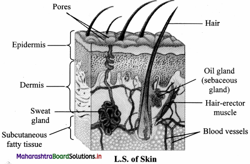
i. Skin:
Skin acts as an accessory excretory organ. The skin of many organisms is thin and permeable. It helps in diffusion of waste products like ammonia.
Human skin however is thick and impermeable. It shows presence of two types of glands namely, sweat glands and sebaceous glands.
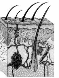
a. Sweat glands are distributed all over the skin. They are abundant in the palm and facial regions. These simple, unbranched, coiled, tubular glands open on the surface of the skin through an opening called sweat pore. Sweat is primarily produced for thermoregulation but it also excretes substances like water, NaCl, lactic acid and urea.
b. Sebaceous glands are present at the neck of hair follicles. They secrete oily substance called sebum. It forms a lubricating layer on skin making it softer. It protects skin from infection and injury.

ii. Lungs:
Lungs are the accessory excretory organs. They help in excretion of volatile substances like C02 and water vapour produced during cellular respiration. Along with CO2, lungs also remove excess of H2O in the form of vapours during expiration. They also excrete volatile substances present in spices and other food stuff.

Question 41.
Enlist the human excretory organs and their excretory products.
Answer:
Excretory OrgAnswer:

  1. Lungs: Remove CO2 and also water vapour to a considerable extent. Volatile substances present in spices and other food stuff are excreted through lungs
  2. Kidneys: Remove nitrogenous waste products like ammonia, urea and uric acid, creatinine. They also remove excessive amount of water, salts and certain minerals.
  3. Skin: Remove water, NaCl, lactic acid and urea by through of sweat.

Question 42.
What is albuminaria? What are its causes?
Answer:

  • Albuminaria is the presence of excess albumin in the urine.
  • Causes: Injury to the endothelial capsular membrane as a result of increased blood pressure, injury or irritation of kidney cells by substances such as toxins or heavy metals.

Maharashtra Board Class 11 Biology Important Questions Chapter 15 Excretion and Osmoregulation

Question 43.
A patient report indicates presence of excessive quantities of ketone bodies in the urine. What does this indicate? How is it caused?
Answer:
Presence of excessive quantities of ketone bodies in the urine indicates that the patient is suffering from diabetes mellitus, starvation or too little carbohydrates in the diet.

Question 44.
Sheela is suffering from kidney infection. Presence of which type of cells in the urine can be indicative of this?
Answer:
Presence of leucocytes in the urine can be indicative of infection of kidney or other urinary organs.

Question 45.
Enlist the various disorders and diseases related to the excretory system.
Answer:
Some disorders and diseases related to the excretory system are as follows:

  1. Kidney stones
  2. Uremia
  3. Nephritis
  4. Renal Failure
  5. Ketonuria
  6. Albuminaria

Question 46.
Write a note on kidney stones with reference to types, symptoms and diagnosis.
Answer:
Kidney stones are also called renal calculi. They may be formed in any portion of urinary tract, from kidney tubules to external opening.
Types:
Depending on their composition, kidney stones are classified into the following types.

  1. Calcium stones : These are usually calcium oxalate or calcium phosphate stones.
  2. Struvite stones : These are formed in response to bacterial infection caused by urea – splitting bacteria. They grow in size quickly and become quite large.
  3. Uric acid stones : These stones usually affect people drinking less water or consuming high protein diet.
  4. Cystine stones : It is a genetic disorder that causes the kidney to excrete too much of certain amino acid.

Symptoms:
Intermittent pain below rib cage in back and side ways. Hazy, brownish/reddish/ pinkish urine. Frequent urge to pass urine. Pain during micturition.

Diagnosis:
Uric acid content of blood, colour of urine, kidney X-ray, sonography of kidney are different diagnostic tests prescribed depending on symptoms.

Question 47.
What is uremia?
Answer:
If the level of urea in blood rises above 0.05%, the condition is known as uremia. It may lead to kidney failure.

Question 48.
What is the normal content of urea in blood?
Answer:
The normal content of urea in blood is 0.01 to 0.03 %.

Question 49.
Write a note on nephritis.
Answer:

  1. Nephritis is the inflammation of kidneys characterised by proteinuria.
  2. It is caused due to increased permeability of glomerular capsular membrane, permitting large amounts of proteins to escape from blood to urine.
  3. This leads to change in blood colloidal osmotic pressure, leading to movement of fluid from blood to interstitial spaces.
  4. It is reflected as edema.

Maharashtra Board Class 11 Biology Important Questions Chapter 15 Excretion and Osmoregulation

Question 50.
What is renal failure? Describe its types.
Answer:
Renal failure is the decrease or cessation of glomerular filtration and ¡s classified into two types.
i) Acute Renal failure (ARF):
ARF is sudden worsening of renal function that most commonly happens after severe bleeding. There is a decrease in urine output (oligouriaf scanty urine i.e., less than 400 mi/day or less than 0.5 ml/kg/li in children). Other causes of ARF may include acute obstruction of both ureters or nephrotoxic drugs. ARF can be detected biochemically by elevated serum creatinine levels.

ii) Chronic kidney disease (CKD) :
It is the progressive and generally irreversible decline in glomerular filtration rate (GFR). It may be caused due to chronic glomerulonephritis. It can be detected by reduced kidney size and possibility of anaemia.

Question 51.
When does a patient need to undergo haemodialysis? Explain the process in detail.
Answer:

  1. When renal function of a person falls below 5 – 7 %, accumulation of harmful substances in blood begins. In such a condition, the person has to go for artificial means of filtration of blood i.e. haemodialysis.
  2. In haemodialysis, a dialysis machine is used to filter blood. The blood is filtered outside the body using a dialysis unit.
  3. In this procedure, the patients’ blood is removed; generally from the radial artery and passed through a cellophane tube that acts as a semipermeable membrane.
  4. The tube is immersed in a fluid called dialysate which is isosmotic to normal blood plasma. Hence, only excess salts if present in plasma pass through the cellophane tube into the dialysate.
  5. Waste substances being absent in the dialysate, move from blood into the dialyzing fluid.
  6. Filtered blood is returned to vein.
  7. In this process it is essential that anticoagulant like heparin is added to the blood while it passing through the tube and before resending it into the circulation, adequate amount of anti-heparin is mixed.
  8. Also, the blood has to move slowly through the tube and hence the process is slow.

Question 52.
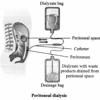
Write a note on peritoneal dialysis.
Answer:

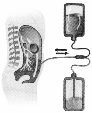
  1. In this method, the dialyzing fluid is introduced in abdominal cavity or peritoneal cavity.
  2. The peritoneal membrane acts as semipermeable dialyzing membrane.
  3. Toxic wastes and extra solutes pass into the fluid.
  4. This fluid is drained out after a prescribed period of time.
  5. Peritoneal dialysis can be repeated as per the need of the patient.
  6. It can be carried out at home at work or while travelling. But it is not as efficient as haemodialysis.

Question 53.
What are the drawbacks of haemodialysis?
Answer:

  • Kidneys are associated with secretion of erythropoietin, renin and calcitriol which is not possible using dialysis machine.
  • During dialysis, the blood has to move slowly through the tube and hence the process is slow.

Question 54.
What is kidney transplant?
Answer:

  1. It is the organ transplant of a healthy kidney into a patient with end – stage renal disease.
  2. Kidney transplantation is classified as cadaveric (deceased donor) or living donor kidney transplant.
  3. Living donor kidney transplant are further classified as genetically related (living-related) or non-related (living non-related) transplants.

Maharashtra Board Class 11 Biology Important Questions Chapter 15 Excretion and Osmoregulation

Question 55.
Complete the diagram / chart with correct labels / information. Write the conceptual details regarding it.
i)
Maharashtra Board Class 11 Biology Important Questions Chapter 15 Excretion and Osmoregulation 6
Answer:
Maharashtra Board Class 11 Biology Important Questions Chapter 15 Excretion and Osmoregulation 7
Skin:
Skin acts as an accessory excretory organ. The skin of many organisms is thin and permeable. It helps in diffusion of waste products like ammonia.
Human skin however is thick and impermeable. It shows presence of two types of glands namely, sweat glands and sebaceous glands.
a. Sweat glands are distributed all over the skin.
They are abundant in the palm and facial regions. These simple, unbranched, coiled, tubular glands open on the surface of the skin through an opening called sweat pore. Sweat is primarily produced for thermoregulation but it also excretes substances like water, NaCI, lactic acid and urea.

b. Sebaceous glands are present at the neck of hair follicles.
They secrete oily substance called sebum. It forms a Lubricating layer on skin making it softer. It protects skin from infection and injury.

ii)
Maharashtra Board Class 11 Biology Important Questions Chapter 15 Excretion and Osmoregulation 8
Answer:
Maharashtra Board Class 11 Biology Important Questions Chapter 15 Excretion and Osmoregulation 9

  1. In this method, the dialyzing fluid is introduced in abdominal cavity or peritoneal cavity.
  2. The peritoneal membrane acts as semipermeable dialyzing membrane.
  3. Toxic wastes and extra solutes pass into the fluid.
  4. This tluid is drained out after a prescribed period of time.
  5. Peritoneal dialysis can be repeated as per the need of the patient.
  6. It can be carried out at home at work or while travelling. But it is not as efficient as haemodialysis.

Question 55.
Invertebrates, bony fishes, tadpoles, etc. are ammonotelic. Whereas birds, reptiles, land snails, etc. are uricotelic. What would be the probable reason for the difference in mode of excretion?
i. Ammonotelic organisms conserve water as conversion of ammonia to uric acid requires large amount of water.
ii. Elimination of ammonia requires large quantity of water, thus ammonotelism is seen in aquatic animals.
iii. Uricotelic organisms require moderate water for eliminating urea and formation of ammonia requires expenditure of energy. Thus, to conserve water and energy, these animals have uricotelism mode of excretion.
iv. Aquatic animals can retain ammonia and store it in the body for long time.
(A) i and iii
(B) only ii
(C) only iv
(D) ii and iii
Answer:
(B)

Question 56.
A lab technician was evaluating blood reports of some patients. She observed the following values of the report:

Sr. No. Patient Comments on urine/ blood report
i. A High levels of glucose in urine
ii. B Presence of excess quantities of ketone in urine
iii. C Presence of leucocytes in urine
iv. D 0.06% urea in blood
V. E High level of proteins in blood

Read the given comments and discuss what disorder/disease the patient must be suffering from.

Answer:

Sr. No. Patient Comments on urine/ blood report Disorder/ Disease
i. A High levels of glucose in urine Glucosuria
ii. B Presence of excess quantities of ketone in urine Ketonuria (Indicative of diabetes mellitus)
iii. C Presence of leucocytes in urine Infection of kidney or urinary organs
iv. D 0.06% urea in blood Uremia
V. E High level of proteins in blood Proteinuria/ Nephritis

Multiple Choice Questions

Question 1.
Uric acid is the main nitrogenous waste in
(A) birds
(B) cartilaginous fish
(C) mammals
(D) larval amphibians
Answer:
(A) birds

Question 2.
Protonephridia is the excretory organ of
(A) platyhelminthes
(B) coelenterates
(C) arthropods
(D) aschelminthes
Answer:
(A) platyhelminthes

Maharashtra Board Class 11 Biology Important Questions Chapter 15 Excretion and Osmoregulation

Question 3.
The glomerulus receives blood through the
(A) vasa recta
(B) renal artery
(C) afferent arteriole
(D) efferent arteriole
Answer:
(C) afferent arteriole

Question 4.
More ADH results in
(A) reduced permeability of DCT
(B) dilute urine
(C) reduced blood pressure
(D) concentrated urine
Answer:
(D) concentrated urine

Competitive Corner

Question 1.
Match the items in Column-I with those in Column-II [NEET Odisha 2019]

Column-I Column-II
i. Podocytes a. Crystallised oxalates
ii. Protonephridia b. Annelids
iii. Nephridia c. Amphioxus
iv. Renal calculi d. Filtration slits

Select the correct option from the following:
(A) i-d, ii-b, iii-c, iv-a
(B) i-c, ii-d, iiii-b, iv-a
(C) i-c, ii-b, iii-d, iv-a
(D) i-d, ii-c, iii-b, iv-a
Answer:
(D) i-d, ii-c, iii-b, iv-a

Question 2.
Match the following parts of a nephron with their function: [NEET Odisha 2019]

i. Descending limb of Henle’s loop a. Reabsorption of salts only
ii. Proximal convoluted tubule b. Reabsorption of water only
iii. Ascending limb of Henle’s loop c. Conditional reabsorption of sodium ions and water
iv. Distal convoluted tubule d. Reabsorption of ions, water and organic nutrients

Select the correct option from the following:
(A) i-d, ii-a, iii-c, iv-b
(B) i-a, ii-c, iii-b, iv-d
(C) i-b, ii-d, iii-a, iv-c
(D) i-a, ii-d, iii-b, iv-c
Answer:
(C) i-b, ii-d, iii-a, iv-c

Question 3.
Which of the following factors is responsible for the formation of concentrated urine? [NEET(UG) 2019]
(A) Secretion of erythropoietin by Juxtaglomerular complex.
(B) Hydrostatic pressure during glomerular filtration.
(C) Low levels of antidiuretic hormone.
(D) Maintaining hyperosmolarity towards inner medullary interstitium in the kidneys.
Answer:
(D) Maintaining hyperosmolarity towards inner medullary interstitium in the kidneys.

Maharashtra Board Class 11 Biology Important Questions Chapter 15 Excretion and Osmoregulation

Question 4.
Portions of renal cortex, which are projected into renal medulla, among the renal pyramids are called as _______. [MHT CET 2019]
(A) Columns of Bertini
(B) Columnae Camae
(C) Ampullae
(D) Ducts of Bellini
Answer:
(A) Columns of Bertini

Question 5.
Renal failure is typically detected by biochemical analysis of blood which shows _______. [MHT CET 2019]
(A) increased level of amino acids
(B) lower level of uric acid
(C) lower level of serum creatinine
(D) higher level of serum creatinine
Answer:
(D) higher level of serum creatinine

Question 6.
Which of the following statements is CORRECT with reference to nephron? [MHT CET 2019]
(A) ADH hormone increases permeability of PCT cells to reabsorb water.
(B) Efferent arteriole forms peritubular network all around tubule.
(C) Podocytes occur in ascending limb of loop of Henle.
(D) Descending limb of Henle’s loop is impenneable to water.
Answer:
(B) Efferent arteriole forms peritubular network all around tubule.

Question 7.
Match the items given in Column I with those in Column li and select the correct option given below. [NFET (UG) 2018]

Column I (Function)

Column II (Part of excretory system)

i. Ultrafiltration a. Henle’s loop
ii. Concentration of urine b. Ureter
iii. Transport of urine c. Urinary bladder
iv. Storage of urine d. Malpighian corpuscle
v. e. Proximal convoluted tubule

(A) i-e, ii-d, iii-a, iv-b
(B) i-d, ii-a, iii-b, iv-c
(C) i-d, ii-e, iii-b, iv-c
(D) i-e, ii-d, iii-a, iv-c
Answer:
(B) i-d, ii-a, iii-b, iv-c

Question 8.
Match the items given in Column I with those in Column II and select the correct option given below. [NEET (UG) 2018]

Column I 

Column II

i. Glycosuria a. Accumulation of uric acid in joints
ii. Gout b. Mass of crystalised salts within the kidney
iii. Renal calculi c. Inflammation of glomeruli
iv. Glomerular nephritis d. Presence of glucose in urine

(A) i-b, ii-c, iii-a, iv-d
(B) i-a, ii-b, iii-c, iv-d
(C) i-c, ii-b, iii-d, iv-a
(D) i-d, ii-a, iii-b, iv-c
Answer:
(D) i-d, ii-a, iii-b, iv-c

Maharashtra Board Class 11 Biology Important Questions Chapter 15 Excretion and Osmoregulation

Question 9.
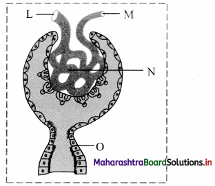
In the given diagram of Malpighian body, blood is filtered from part labelled ________
Maharashtra Board Class 11 Biology Important Questions Chapter 15 Excretion and Osmoregulation 15
(A) L
(B) M
(C) N
(D) O
Hint: L – Afferent arteriole
M – Efferent arteriole
N – Glomerulus
O – Proximal convoluted tubule
Blood filtration occurs in glomerulus. Afferent arteriole is the blood vessel leading to glomerulus. Efferent arteriole carries blood away from the glomerulus. Proximal convoluted tubule is involved in reabsorption of useful substances from the filtrate.
Answer:
(C) N

Question 10.
Majority of kidney stones consist crystals of [MHT CET 2018]
(A) calcium oxalate, sodium bicarbonate
(B) calcium oxalate, calcium phosphate
(C) calcium phosphate, sodium chloride
(D) calcium carbonate, copper sulphate
Answer:
(B) calcium oxalate, calcium phosphate

Question 11.
Which of the following group of animals is guanotelic? [MHT CEE 2018]
(A) Labeo, turtle, camel
(B) Lizard, snake, scorpion
(C) Penguin, spider, scorpion
(D) Spider, scorpion, snake
Answer:
(C) Penguin, spider, scorpion

Question 12.
Which of the following statements is CORRECT? [NEET (UG) 2017]
(A) The ascending limb of loop of Henle is impermeable to water.
(B) The descending limb of loop of Henle is impermeable to water.
(C) The ascending limb of loop of Henle is permeable to water.
(D) The descending limb of loop of Henle is permeable to electrolytes.
Hint: Descending limb of loop of Henle is permeable to water but impermeable to electrolytes. While ascending limb of loop of Henle is impermeable to water and permeable to electrolytes.
Answer:
(A) The ascending limb of loop of Henle is impermeable to water.

Question 13.
A decrease in blood pressure/volume will not cause the release of [NEET (UG) 2017]
(A) Renin
(B) Atrial Natriuretic Factor
(C) Aldosterone
(D) ADH
Hint: Decrease in blood pressure or volume stimulates the release of renin, aldosterone and ADH. ANF is released when blood pressure increases or blood volume increases. Release of ANF causes vasodilation and also inhibit renin angiotensin mechanism that decreases blood pressure and blood volume.
Answer:
(B) Atrial Natriuretic Factor

Maharashtra Board Class 11 Biology Important Questions Chapter 15 Excretion and Osmoregulation

Question 14.
Formation of urea takes place in the _________. [MHT CET 2017]
(A) Heart
(B) Kidney
(C) Liver
(D) Lung
Answer:
(C) Liver

Question 15.
The yellow colour of normal urine is due to [MHT CET 2017]
(A) Bilirubin
(B) Biliverdin
(C) Urochrome
(D) Uric acid
Answer:

Question 16.
Uremia is indicated when the blood urea level rises above _______ [MHT CET 2017]
(A) 0.05%
(B) 0.04%
(C) 0.03%
(D) 0.02 %
Answer:
(A) 0.05%
Maharashtra Board Class 11 Biology Important Questions Chapter 14 Human Nutrition

Balbharti Maharashtra State Board 11th Biology Important Questions 14 Human Nutrition Important Questions and Answers.

Maharashtra State Board 11th Biology Important Questions Chapter 14 Human Nutrition

Question 1.
Explain various steps involved in nutrition.
Answer:
The various steps involved in nutrition are as follows:

  1. Ingestion: It is the introduction of food into the mouth, i.e. intake of food (eating) inside the body.
  2. Digestion: The process during which the complex, non-diffusible and non-absorbable food substances are converted into simple, diffusible, and absorbable substances by the action of enzymes is called digestion.
  3. Absorption: The process of diffusion of digested food into blood and lymph is called absorption.
  4. Assimilation: The process by which protoplasm is synthesized into each cell of the body by utilizing simple food substances is called assimilation.
  5. Egestion: The elimination of undigested food from the body is called egestion.

Question 2.
What are the dietary needs of human being?
Answer:
Carbohydrates, proteins, fats, vitamins, minerals, water and fibres in adequate amount are the dietary needs of human being.

Maharashtra Board Class 11 Biology Important Questions Chapter 14 Human Nutrition

Question 3.
Fill in the blanks:
i. Food provides _________ for growth and tissue repair.
ii. ________ are also required in small quantities for nutrition.
Answer:
i. energy, organic material.
ii. Vitamins, minerals.

Question 4.
Define: Digestion
Answer:
Digestion is defined as the process by which the complex, non-diffusible and non-absorbable food substances are converted into simple, diffusible and assimilable substances.

Question 5.
What is dentition?
Answer:
The study of teeth with respect to their number, arrangement, development etc. is known as dentition.

Maharashtra Board Class 11 Biology Important Questions Chapter 14 Human Nutrition

Question 6.
Describe the structure and functions of the various parts of the alimentary canal.
Answer:
Human Digestive system:
Human digestive system consists of alimentary canal and associated digestive glands.
Alimentary canal:
Alimentary canal is a long tube-like structure of varying diameter starting from mouth and ending with anus. It is about 8-10m long.
Alimentary canal consists of mouth, buccal cavity, pharynx, oesophagus, stomach, small intestine, large intestine and anus.
Mouth:

  • It is also called oral or buccal cavity and is bounded by fleshy lips.
  • Its side walls are formed of cheeks, roof is formed by palate and floor by tongue.
  • It is internally lined by a mucous membrane.
  • Salivary glands open into the buccal cavity.

Function: It helps in ingestion of food.

Teeth:

  • 32 teeth are present in the buccal cavity of an adult human being.
  • Human dentition is described as thecodont, diphyodont and heterodont.
  • It is called thecodont type because each tooth is fixed in a separate socket present in jaw bones by gomphosis type of joint.
  • In our life time, we get only two sets of teeth, milk teeth and permanent teeth. This is called diphyodont dentition.
  • We have four different type of teeth hence we are heterodont.
  • Types of teeth are incisors (I) canines (C) premolars (PM) and molar (M).
  • Each half of each jaw has two incisors, one canine, two premolars and three molars.
  • Thus, dental formula of adult human can be represented as:
    i\(\frac{2}{2}\), c\(\frac{1}{1}\), pm\(\frac{2}{2}\), m\(\frac{3}{3}\) = \(\frac{8}{8}\) = 16 × 2 = 32

Tongue:
It is the muscular fleshy organ and is roughly triangular in shape. It lies along the floor of the buccal cavity.

Functions: The upper surface of the tongue bears numerous projections called papillae.
These papillae contain sensory receptors called taste buds.

ii. Pliary nx:

  • The buccal cavity leads to a short pharynx.
  • Pharynx is a common passage for food and air.
  • The pharynx opens into trachea through an opening called glottis.
  • The glottis is guarded by a cartilaginous flap called epiglottis. The epiglottis closes during the swallowing (deglutition) action and pre vents entry of food into the trachea.
  • The lower region of pharynx is called oropharynx.
  • Oropharynx opens into oesophagus through gullet.

iii. Oesophagus:

  • The oesophagus is a thin, muscular tube.
  • It lies behind the trachea.
  • It is approximately 25cm long tube passes through the neck, central aspect of rib cage, pierces the diaphragm and joins the stomach.
  • It is lined by mucus cells.
  • Mucus lubricates the passageway of food.
  • Oesophagus is made up of longitudinal and circular muscles.

Function: The rhythmic wave of contraction and relaxation of these muscles is called peristalsis that helps in passage of food through oesophagus.

iv. Stomach:
The stomach is located in the upper left portion of the abdominal cavity.
It is a muscular sac-like ‘J1 shaped organ, around 25 to 30cm in length.
It is divided into upper cardiac region and lower pyloric region.

  • Cardia or Cardiac: It is first part in which oesophagus opens. The cardia surrounds the band of circular muscles present at the junction of oesophagus and stomach called cardiac sphincter. The cardiac sphincter prevents back flow or regurgitation of food from stomach to oesophagus.
  • Fundus: It is the dome shaped region above and left of cardia.
  • Body: It forms the large central portion of stomach that stores the food.
  • Pylorus: It is a narrow posterior region of stomach.
    It opens into duodenum, the initial part of small intestine.
    This opening is guarded by a set of sphincter muscles called pyloric sphincter.
    It regulates the flow of food from stomach to small intestine.

Function: The stomach temporarily stores the food.
It chums the food and helps in mixing the food with gastric juice.

v. Small Intestine:

  • It is about 6 meters long and 2.5 cm broad tube coiled within abdominal cavity.
  • The coils are held together by mesenteries, supporting the blood vessels, lymph vessels and nerves.
  • It is divided into three parts: Duodenum, jejunum and ileum.

vi. Large Intestine:

  • It is 1.5 meters in length.
  • It is wider in diameter and shorter than small intestine.
  • It consists of caecum, colon and rectum.

vii. Anus:

  • Anus is the terminal opening of alimentary canal.
  • It is guarded by sphincter.

Function: It expels faecal matter by a process called egestion or defecation.

Question 7.
Draw a neat and labelled diagram of stomach and write a short note on it.
Answer:
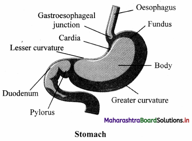
Stomach:
The stomach is located in the upper left portion of the abdominal cavity.
It is a muscular sac-like ‘J1 shaped organ, around 25 to 30cm in length.
It is divided into upper cardiac region and lower pyloric region.

  1. Cardia or Cardiac: It is first part in which oesophagus opens. The cardia surrounds the band of circular muscles present at the junction of oesophagus and stomach called cardiac sphincter. The cardiac sphincter prevents back flow or regurgitation of food from stomach to oesophagus.
  2. Fundus: It is the dome shaped region above and left of cardia.
  3. Body: It forms the large central portion of stomach that stores the food.
  4. Pylorus: It is a narrow posterior region of stomach.
    It opens into duodenum, the initial part of small intestine.
    This opening is guarded by a set of sphincter muscles called pyloric sphincter.
    It regulates the flow of food from stomach to small intestine.

Function: The stomach temporarily stores the food.
It chums the food and helps in mixing the food with gastric juice.
Maharashtra State Board Class 11 Biology Important Questions Chapter 14 Human Nutrition 1

Question 8.
Describe the structure of Small Intestine.
Answer:
It is about 6 meters long and 2.5 cm broad tube coiled within abdominal cavity.
The coils are held together by mesenteries, supporting the blood vessels, lymph vessels and nerves.
It is divided into three parts.

  1. Duodenum:
    • It is about 26 cm long ‘U’ shaped structure.
    • The duodenum turns towards left side of abdominal cavity below the stomach.
  2. Jejunum:
    • It is about 2.5 meters long, coiled middle portion of small intestine.
    • It is narrower than the duodenum.
  3. Ileum:
    • It is about 3.5 meters long.
    • It is highly coiled and little broader than jejunum.
    • The ileum opens into the caecum of large intestine at ileocaecal junction.

Maharashtra Board Class 11 Biology Important Questions Chapter 14 Human Nutrition

Question 9.
Explain anatomy of different parts of Large Intestine.
Answer:
Ileum opens into large intestine.
It is 1.5 meters in length.
It is wider in diameter and shorter than small intestine.
It consists of caecum, colon and rectum.

  1. Caecum:
    • Caecum is a small, blind sac present at the junction of ileum and colon.
    • It is 6cm in length.
    • It hosts some symbiotic microorganisms.
    • An elongated worm like vermiform appendix arises from the caecum.
    • Appendix is vestigial organ in human beings and functional in herbivorous animals for the digestion of cellulose.
  2. Colon:
    • Caecum opens into colon.
    • Colon is tube like-organ consist of three parts, ascending colon, transverse colon and descending colon.
    • The colon is internally lined by mucosal cells.
  3. Rectum:
    • It is posterior region of large intestine.
    • It temporarily stores the undigested waste material called faeces till it is egested out through anus.

Question 10.
Differentiate between Small Intestine and Large Intestine
Answer:

Small Intestine Large Intestine
i. It is about 6 meters long. It is about 1.5 meters long.
ii Small intestine is 2.5 cm broad tube. Large intestine is broader than the small intestine. !
iii. It is divided into three parts, as Duodenum, Jejunum, Ileum. It is divided into three parts as – caecum, colon I and rectum.
iv Absorbs the digested nutrients. Takes part in absorption of water and minerals.
V. Villi present. Villi absent.
vi. Digestion is completed in small intestine. No role in digestion.

Question 11.
Explain in detail the layers of gastrointestinal tract.
Answer:
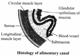
The entire gastrointestinal tract is lined by four basic layers from inside to outside namely, mucosa, submucosa, muscularis and serosa.
These layers show modification depending on the location and function of the organ concerned.
Maharashtra State Board Class 11 Biology Important Questions Chapter 14 Human Nutrition 2

  1. Serosa:
    • It is the outermost layer.
    • It is made up of a layer of squamous epithelium called mesothelium and inner layer of connective tissue.
  2. Muscularis:
    • This layer is formed of smooth muscles.
    • These muscles are usually arranged in three concentric layers.
    • Outermost layer shows longitudinal muscles, middle circular muscles and inner oblique muscles.
    • This layer is wider in stomach and comparatively thin in intestinal region.
    • The layer of oblique muscles is absent in the intestine.
  3. Submucosa:
    • It is formed of loose connective tissue containing blood vessels, lymph vessels and nerves.
    • Duodenal submucosa shows presence of glands.
  4. Mucosa:
    • The lumen of the alimentary canal is lined by mucosa.
    • Throughout the length of alimentary canal, the mucosa layer shows presence of goblet cells that secrete mucus.
    • This lubricates the lumen of alimentary canal.
    • This layer shows modification in different regions of alimentary canal. In stomach, it is thrown into irregular folds called rugae.
    • In stomach mucosa layer forms gastric glands that secrete gastric juice.

Maharashtra Board Class 11 Biology Important Questions Chapter 14 Human Nutrition

Question 12.
Write a short note on villi.
Answer:

  1. Mucosa of small intestine forms finger like folding called villi.
  2. The intestinal villi are lined by brush border or epithelial cells having microvilli at the free surface.
  3. Villi are supplied with a network of capillaries and lymph vessels called lacteals.
  4. Mucosa forms crypis in bctween the bases of vifli in intestine called crvpís of Licberkuhn which arc intestinal glands.

Question 13.
Describe the various digestive glands associated with alimentary canal.
Answer:
The digestive glands associated with the alimentary canal include the salivary glands, liver and pancreas.

  1. Salivary Glands:
    • There are three pairs of salivary glands which open in buccal cavity.
    • Parotid glands are present in front of the ear.
    • The submandibular glands are present below the lower jaw.
    • The glands present below the tongue are called sublingual.
    • Salivary glands are made up of two types of cells.
    • Serous cells secrete a fluid containing digestive enzyme called salivary amylase.
    • Mucous cells produce mucus that lubricates food and helps swallowing.
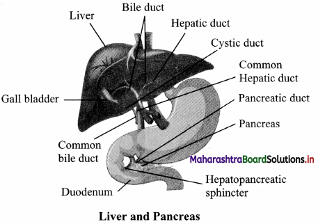
  2. Liver:
    • Liver is dark reddish-brown coloured largest gland of the body, weighing 1.2 to 1.5 kg, in adults.
    • Situated in right upper portion of the abdominal cavity, below the diaphragm.
    • Divided into 2 lobes, right and left.
    • A thin connective tissue sheath called Glisson’s capsule covers the liver and invaginates inside to divide the liver into cord like structures called hepatic lobules which are functional units of liver containing hepatic cells (hepatocytes).
    • Each hepatic lobule is polygonal in shape. At the junction of adjacent lobules, a triangular portal area is present.
    • In this portal area a branch of each of hepatic artery, hepatic portal vein and bile duct are present. Lobule consist of cords of hepatic cells which are arranged around a central vein.
    • In between the cords of hepatic cells, spaces called sinusoids are present through which the blood flows. In the sinusoids, phagocytic cells called Kupffer cells are present.
    • Hepatic cells secrete bile. Bile is carried by hepatic ducts in a thin muscular sac called gall bladder.
    • The duct of the gall bladder and hepatic duct together form common bile duct.
    • Liver synthesizes vitamins A, D, K and B12, blood proteins.
  3. Pancreas:
    • Pancreas is a leaf shaped heterocrine gland present in the gap formed by bend of duodenum under the stomach.
    • Exocrine part of pancreas is made up of acini, the acinar cells secrete alkaline pancreatic juice that contains various digestive enzymes.
    • Pancreatic juice is collected and carried to duodenum by pancreatic duct.
    • The common bile duct joins pancreatic duct to form hepato-pancreatic duct. It opens into duodenum.
    • Opening of hepato-pancreatic duct is guarded by sphincter of Oddi.
    • Endocrine part of pancreas is made up of islets of Langerhans situated between the acini.
    • It contains three types of cells a-cells which secrete glucagon, P-cells which secretes insulin and 5 cells secrete somatostatin hormone.
    • Glucagon and insulin together control the blood-sugar level.
    • Somatostatin hormone inhibits glucagon and insulin secretion.

Question 14.
Observe the diagram given below and explain the structure and functions of the gland which stores glycogen and is involved in detoxification.
Maharashtra State Board Class 11 Biology Important Questions Chapter 14 Human Nutrition 3
Answer:
The gland which stores glycogen and helps in detoxification is liver.

  1. Liver:
    • Liver is dark reddish-brown coloured largest gland of the body, weighing 1.2 to 1.5 kg, in adults.
    • Situated in right upper portion of the abdominal cavity, below the diaphragm.
    • Divided into 2 lobes, right and left.
    • A thin connective tissue sheath called Glisson’s capsule covers the liver and invaginates inside to divide the liver into cord like structures called hepatic lobules which are functional units of liver containing hepatic cells (hepatocytes).
    • Each hepatic lobule is polygonal in shape. At the junction of adjacent lobules, a triangular portal area is present.
    • In this portal area a branch of each of hepatic artery, hepatic portal vein and bile duct are present. Lobule consist of cords of hepatic cells which are arranged around a central vein.
    • In between the cords of hepatic cells, spaces called sinusoids are present through which the blood flows. In the sinusoids, phagocytic cells called Kupffer cells are present.
    • Hepatic cells secrete bile. Bile is carried by hepatic ducts in a thin muscular sac called gall bladder.
    • The duct of the gall bladder and hepatic duct together form common bile duct.
    • Liver synthesizes vitamins A, D, K and B12, blood proteins.
  2. Kupffer cells of liver destroy toxic substances, dead and worn-out blood cells and microorganisms.
  3. Bile juice secreted by liver emulsifies fats and makes food alkaline.
  4. Liver stores excess of glucose in the form of glycogen.
  5. Deamination of excess amino acids to ammonia and its further conversion to urea takes place in liver.
  6. Synthesis of vitamins A, D, K and B12 takes place in liver.
  7. It also produces blood proteins like prothrombin and fibrinogen.
  8. During early development, it acts as haemopoietic organ.

Therefore, liver is a vital organ.

Maharashtra Board Class 11 Biology Important Questions Chapter 14 Human Nutrition

Question 15.
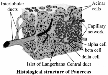
Explain heterocrine nature of pancreas with the help of histological structure.
Answer:
Pancreas:

  1. Pancreas is a leaf shaped heterocrine gland present in the gap formed by bend of duodenum under the stomach.
  2. Exocrine part of pancreas is made up of acini, the acinar cells secrete alkaline pancreatic juice that contains various digestive enzymes.
  3. Pancreatic juice is collected and carried to duodenum by pancreatic duct.
  4. The common bile duct joins pancreatic duct to form hepato-pancreatic duct. It opens into duodenum.
  5. Opening of hepato-pancreatic duct is guarded by sphincter of Oddi.
  6. Endocrine part of pancreas is made up of islets of Langerhans situated between the acini.
  7. It contains three types of cells a-cells which secrete glucagon, P-cells which secretes insulin and 5 cells secrete somatostatin hormone.
  8. Glucagon and insulin together control the blood-sugar level.
  9. Somatostatin hormone inhibits glucagon and insulin secretion.
    Maharashtra State Board Class 11 Biology Important Questions Chapter 14 Human Nutrition 4

Question 16.
Digestion is carried out by both mechanical and chemical methods. Justify.
Answer:

  1. Mechanical digestion includes various movements of alimentary canal that help chemical digestion.
  2. Mastication or chewing of food by teeth, churning in stomach and peristaltic movements of gastrointestinal tract bring about mechanical digestion in human body.
  3. Chemical digestion is a series of catabolic (breaking down) reactions that hydrolyze the food.
    Thus, Digestion is carried out by both mechanical and chemical methods.

Question 17.
Write a short note on digestion in the mouth.
Answer:
Digestion in the mouth (buccal cavity):

  1. Both mechanical and chemical digestion processes take place in mouth.
  2. Mastication or chewing of food takes place with the help of teeth and tongue.
  3. Teeth crush and grind the food while tongue manipulates the food.
  4. Crushing of food becomes easier when it gets moistened by saliva.
  5. Mucus in the saliva lubricates the food as well as it helps in binding the food particles into a mass of called bolus which is swallowed by deglutition.
  6. The tongue presses against the palate and pushes the bolus into pharynx which further passes oesophagus.
  7. The only chemical digestion that takes place in mouth is by the action of salivary amylase.
  8. It helps in conversion of starch into maltose. About 30% starch gets converted to maltose in mouth.
  9. The bolus further passes down through oesophagus by peristalsis.
  10. Food from the oesophagus enters the stomach.
    Maharashtra State Board Class 11 Biology Important Questions Chapter 14 Human Nutrition 5

Maharashtra Board Class 11 Biology Important Questions Chapter 14 Human Nutrition

Question 18.
Name all the constituents of saliva.
Answer:
Saliva contains 98% water and 2% other constituents like electrolytes (sodium, potassium, calcium, chloride, bicarbonates), digestive enzyme, salivary amylase and lysozyme.

Question 19.
Which sphincter controls the passage of food into stomach?
Answer:
The gastro-oesophageal sphincter controls the passage of food into the stomach.

Question 20.
Explain the process of digestion taking place in muscular-sac like ‘J’ shaped organ.
Answer:
The muscular-sac like ‘J’ shaped organ is stomach.

  1. Both mechanical and chemical digestion takes place in stomach.
  2. The stomach stores the food for 4-5 hours.
  3. The physical digestion take place by churning of food which done by thick muscular wall of stomach.
  4. Churning further breaks down the food particles and also helps in thorough mixing of gastric juice with food.
  5. The mucosa layer of stomach has gastric gland which shows presence of three major types of cells namely, mucus cells, peptic or chief cells and parietal or oxyntic cells.
  6. Mucus cells secrete mucus; peptic or chief cells secrete proenzyme pepsinogen and parietal cells secrete HCl and intrinsic factor which is essential for absorption of vitamin B12. Thus, gastric juice contains mucus, inactive enzyme pepsinogen, HCl and intrinsic factor.
  7. Mucus protects the inner lining of stomach from HCl present in gastric juice.
  8. HCl in gastric juice makes the food acidic and stops the action of salivary amylase, and also kills the germs
    that might be present in the food.
  9. Pepsinogen gets converted into active enzyme pepsin in the acidic medium provided by HCl.
  10. In presence of pepsin, proteins in the food get converted into simpler forms like peptones and proteoses.
  11. At the end of gastric digestion, food is converted to a semifluid acidic mass of partially digested food is
    called chyme.
  12. The chyme from stomach is pushed in the small intestine through pyloric sphincter for further digestion.
    Maharashtra State Board Class 11 Biology Important Questions Chapter 14 Human Nutrition 6

Question 21.
What is the role of rennin in infants?
Answer:

  1. Rennin found in gastric juice of infants acts on casein, a protein present in milk.
  2. It brings about curdling of milk proteins with the help of calcium.
  3. The coagulated milk protein is further digested with the help of pepsin.
  4. Rennin is absent in adults.

Question 22.
Describe the process of digestion in small intestine.
Answer:

  1. In the small intestine, intestinal juice, bile juice and pancreatic juice are mixed with food. Peristaltic movements of muscularis layer help in proper mixing of digestive juices with chyme.
  2. Bile juice and pancreatic juice are poured in duodenum through hepato-pancreatic duct.
  3. Bile salts present in the bile juice neutralize the acidic chyme and make it alkaline. II brings about emulsification of fats.
  4. Pancreatic juices are secreted by pancreas whereas the intestinal mucosa secretes digestive enzymes. The goblet cells produce mucus.
  5. The intestinal juice contains various enzymes like dipeptidases, lipases, disaccharidases, maltase, sucrase and lactase.
  6. Both pancreatic and intestinal lipases initially convert fats into fatty acid and diglycerides.
  7. Diglycerides are further converted to monoglycerides by removal of fatty acid from glycerol.
  8. The mucus and bicarbonates present in pancreatic juice protect the intestinal mucosa and provide alkaline medium for enzymatic action.
  9. Sub-mucosal Brunner’s glands help in the action of goblet cells.
  10. Most of the digestion gets over in small intestine.

Maharashtra Board Class 11 Biology Important Questions Chapter 14 Human Nutrition

Question 23.
Write a short note on bile.
Answer:

  1. Bile juice is dark green coloured fluid that contains bile pigments (bilirubin and biliverdin), bile salts (Na- glycocholate and Na-taurocholate), cholesterol and phospholipid.
  2. Bile does not contain any digestive enzyme.
  3. Bile salts neutralise the acidity of chyme and make it alkaline.
  4. It brings about emulsification of fats.
  5. It also activates lipid digesting enzymes or lipases.
  6. Bile pigments impart colour to faecal matter.

Question 24.
What are the constituents of pancreatic juice?
Answer:

  • Pancreatic juice secreted by pancreas contains pancreatic amylases, lipases and inactive enzymes trypsinogen and chymotrypsinogen.
  • Pancreatic juice also contains nucleases- the enzymes that digest nucleic acids.

Question 25.
List the constituents of intestinal juice.
Answer:
The intestinal juice contains various enzymes like dipeptidases, lipases, disaccharidases, maltase, sucrase and lactase.

Question 26.
Fill in the blanks:
i. The ________ of mucosa produce mucus.
ii. Mucus plus intestinal enzymes together constitute intestinal juice or ________.
iii. Bile juice and pancreatic juice are poured in duodenum through _________ duct.
Answer:
i. goblet cells.
ii. Succus entericus.
iii. hepato-pancreatic.

Question 27.
Give the significance of peristaltic movement of muscularis in small intestine.
Answer:
Peristaltic movements of muscularis layer help in proper mixing of digestive juices with chyme.

Question 28.
Name the juices which are mixed with food in small intestines.
Answer:
Intestinal juice, bile juice and pancreatic juice.

Question 29.
Write a note on hunger hormone.
Answer:
i. Hunger hormone is also called Ghrelin.
ii. It is hormone that is produced mainly by the stomach and small intestine, pancreas and brain.
iii. It stimulates appetite, increases food intake and promotes fat storage.

Question 30.
Explain in detail the action of pancreatic juice.
Answer:
Maharashtra State Board Class 11 Biology Important Questions Chapter 14 Human Nutrition 7

Question 31.
Explain the role of large intestine in digestion process.
Answer:

  1. Conversion of proteins into amino acids, fats to fatty acids and monoglycerides, nucleic acids to sugar and nitrogenous base and carbohydrates to monosaccharides marks the end of digestion of food.
  2. Food is now called chyle. Chyle is an alkaline slurry which contains various nutrients ready for absorption.
  3. The nutrients are absorbed and undigested remains are transported to large intestine.
  4. Mucosa of large intestine produces mucus but no enzymes.
  5. Some carbohydrates and proteins do enter the large intestine.
  6. These are digested by the action of bacteria that live in the large intestine.
  7. Carbohydrates are fermented by bacterial action and hydrogen, carbon dioxide and methane gas are produced in colon.
  8. Protein digestion in large intestine ends up into production of substances like indole, skatole and H2S.
  9. These are the reason for the odour of faeces. These bacteria synthesize several vitamins like B vitamins and vitamin K.

Maharashtra Board Class 11 Biology Important Questions Chapter 14 Human Nutrition

Question 32.
What causes pancreatitis?
Answer:

  1. Pancreatitis is inflammation of the pancreas.
  2. It may occur due to alcoholism and chronic gallstones.
  3. Other reasons include high levels of calcium, fats in blood.
  4. However, in 70% of people with pancreatitis, main reason is alcoholism.

Question 33.
Match the following:

Column I Column II
i. Cardiac Sphincter Pyloric a. Regulates the flow of food from stomach to small intestine.
ii. sphincter b. Controls the passage of food from oesophagus into the stomach.
iii. Gastro-oesophageal sphincter c. Prevents back flow of food from stomach to oesophagus.

Answer:
(i – c)
(ii – a)
(iii – b)

Question 34.
Identify ‘X’, ‘Y’ and ‘Z’ in the given diarani and explain the regulation of gastric function.
Maharashtra State Board Class 11 Biology Important Questions Chapter 14 Human Nutrition 8
Answer:

  1. ‘X’- Vagus nerve, ‘Y’- Gastrin ‘Z’- Sympathetic nerve
  2. Intestinal mucosa produces hormones like secretin, cholecystokinin (CCK) and gastric inhibiting peptide (G1P).
  3. Secretin inhibits secretion of gastric juice.
  4. It stimulates secretion of bile juice from liver, pancreatic juice and intestinal juice.
  5. CCK brings about similar action and induces satiety that is feeling of fullness or satisfaction.
  6. GIP also inhibits gastric secretion.

Question 35.
What is absorption? Mention the absorption of nutrients and other substances in alimentary canal?
Answer:
The passage of end products of digestion through the mucosal lining of alimentary canal into blood and lymph is called absorption. 90% of absorption takes place in small intestine and the rest in mouth, stomach and large intestine.

  1. Month: Absorption takes place through mucosa of mouth and lower side of tongue into the blood capillaries, e.g. Some drugs like certain painkillers.
  2. Stomach: Gastric mucosa is impermeable to most substances hence nutrients reach unabsorbed till small intestine. Little water, electrolytes, alcohol and drugs like aspirin get absorbed in stomach.
  3. Small Intestine: Glucose, fructose, galactose, amino acids, minerals and water-soluble vitamins are absorbed in blood capillaries in villi. Lipids and fat-soluble vitamins ( A, D, E, K) are absorbed in lacteals.
  4. Large Intestine: Absorption of water, electrolytes like sodium and chloride, drugs and some vitamins take place.

Question 36.
Name the various ways by which absorption takes place.
Ans
Simple diffusion, osmosis, facilitated transport and active transport.

Maharashtra Board Class 11 Biology Important Questions Chapter 14 Human Nutrition

Question 37.
Write the various mechanisms of absorption of compounds.
Answer:

  1. Absorption of part of glucose, amino acids and some electrolytes like chloride ions are absorbed by simple diffusion depending on concentration gradient.
  2. Some amino acids as well as substances like fructose are absorbed by facilitated transport.
  3. In this method, carrier ions like Na+ bring about absorption.
  4. Some ions are absorbed against concentration gradient. It requires energy. This type of absorption of mineral like sodium is called active transport.
  5. Water is absorbed along the concentration gradient.
    [Note: Glucose and galactose are transported into absorptive cells of the villi through secondary active transport that is coupled to the active transport ofNa . Amino acids are transported via active transport.]

Question 38.
Write the transportation mechanism for monoglycerides and fatty acids.
Answer:

  1. Monoglycerides and fatty acids cannot be absorbed in blood.
  2. These dissolve in the centre of spherical aggregates fonned by bile salts called micelles.
  3. Micelles enter into intestinal villi where they are reformed into chylomicrons.
  4. Chylomicrons are small protein coated fat globules.
  5. They are transported into lymph vessels called lacteals.
  6. From here, they are transported to blood stream.

Question 39.
Observe the chart given on textbook page no. 169 to find out absorption in various parts of alimentary canal.
Answer:
The passage of end products of digestion through the mucosal lining of alimentary canal into blood and lymph is called absorption. 90% of absorption takes place in small intestine and the rest in mouth, stomach and large intestine.

  1. Month: Absorption takes place through mucosa of mouth and lower side of tongue into the blood capillaries, e.g. Some drugs like certain painkillers.
  2. Stomach: Gastric mucosa is impermeable to most substances hence nutrients reach unabsorbed till small intestine. Little water, electrolytes, alcohol and drugs like aspirin get absorbed in stomach.
  3. Small Intestine: Glucose, fructose, galactose, amino acids, minerals and water-soluble vitamins are absorbed in blood capillaries in villi. Lipids and fat-soluble vitamins ( A, D, E, K) are absorbed in lacteals.
  4. Large Intestine: Absorption of water, electrolytes like sodium and chloride, drugs and some vitamins take place.

Question 40.
What is assimilation?
Answer:
The absorbed food material finally reaches the tissue and becomes a part of protoplasm. This is called as assimilation.

Question 41.
Write a short note on egestion?
Answer:

  1. Undigested waste is converted to faeces in colon and reaches rectum.
  2. Faeces contain water, inorganic salts, sloughed of mucosal cells, bacteria and undigested food.
  3. Distension of rectum stimulates pressure sensitive receptors that initiate a neural reflex for defecation or egestion.
  4. It is a voluntary process that takes place through anal opening guarded by sphincter muscles.

Question 42.
How are nutrition related disorders categorised?
Answer:

  • Little extra or less of nutrition can lead to dietary’ disorder (nutrition related disorder).
  • Nutrition related disorders can be categorized based on the food that an individual consumes and conditions that develop due to malfunctioning of the organ/s or glands associated with digestive system.

Maharashtra Board Class 11 Biology Important Questions Chapter 14 Human Nutrition

Question 43.
What is PEM?
Answer:
Protein Energy malnutrition (PEM):

  • Protein Energy Malnutrition is caused due to inadequate intake of proteins.
  • It can be associated with inadequacy of vitamins and minerals in diet.
  • PEM causes disease like Kwashiorkor and Marasmus.

Question 44.
What is Marasmus? What are the symptoms and causes?
Answer:

  1. Marasmus is a prolonged protein energy malnutrition (PEM) found in infants under one year of age.
  2. In this disease, protein deficiency is coupled with lower total food calorific value.
  3. Inadequate diet impairs physical growth and retards mental development, subcutaneous fat disappears, ribs become prominent, limbs become thin, skin becomes dry, thin and wrinkled, loss of weight, digestion and absorption of food stops due to atrophy of digestive glands. There is no oedema.

Question 45.
What are the major causes of disorders like Kwashiorkor and Marasmus?
Answer:
Major causes of disorders like Kwashiorkor and Marasmus are unavailability of nutritious food. Poverty, large family size, ill spacing of children, early termination of breast feeding and overdiluted milk arc a few causes.

Question 46.
Write a short note on:
i. Indigestion
ii. Constipation
iii. Vomiting
Answer:

  1. Indigestion:
    • Overeating, inadequate enzyme secretion, spicy food, anxiety can cause discomfort and various symptoms. It is called indigestion.
    • Improperly digested food or food poisoning also can cause indigestion.
    • It leads to loss of appetite, acidity (acid reflux), heart burn, regurgitation, dyspepsia (upper abdominal pain), stomach pain.
    • Avoiding eating large meal, lying down after meal, spicy, oily, junk food, smoking, alcohol are the.preventive measures for indigestion.
  2. Constipation:
    • When frequency of defaecation is reduced to less than once per week the condition is called constipation.
    • Difficulty in defaecation may result in abdominal pain distortion, rarely perforation.
    • The causes are, affected colonic mobility due to neurological dysfunction like spinal cord injury, low fibre diet, inadequate fluid intake and inactivity.
    • Roughage, sufficient fluids in diet, exercise can help improve the conditions.
  3. Vomiting
    • In this condition, the stomach contents are thrown out of the mouth due to reverse peristaltic movements of gastric wall.
    • It is controlled by non-vital vomiting center of medulla.
    • It is typically associated with nauseatic feeling.

Maharashtra Board Class 11 Biology Important Questions Chapter 14 Human Nutrition

Question 47.
What is diarrhoea? What are the symptoms and causes of diarrhoea?
Answer:

  • Passing loose watery stools more than three times a day is called diarrhoea. Diarrhoea can lead to dehydration.
  • The symptoms of diarrhoea are blood in stool, nausea, bloating, fever depending on cause and severity of the disorder.
  • The causes of diarrhoea are infection through food and water or disorders like ulcer, colitis, inflammation of intestine or irritable bowel syndrome.

Question 48.
Distinguish between Kwashiorkor and Marasnius.
Answer:

Kwashiorkor Marasnius
i. It is caused due to insufficient amount of proteins. It is caused due to deficiency of fats, proteins and carbohydrates.
ii. Oedema, fatty liver, lethargy are symptoms. No oedema is observed. Thinning of limb is observed.
iii. It is observed in children between 1 to 3 years of age. It is observed in infants under one year of age.

Apply Your Knowledge

Question 49.
A person visited a pediatrician with his one-year old child complaining about the child’s weight loss and diarrhoea. The doctor examines the child and finds that his limbs have become thin, the skin has become dry as well as thin and wrinkled but there is no oedema on the body.
From this information answer the following questions:
i. Which disease child is suffering from?
ii. What is the probable reason for the disease?
iii. What would be the remedies and diet suggested by the doctor?
Answer:

  1. The child is suffering from Marasmus.
  2. The probable reason for the disease is Prolonged Protein Energy Malnutrition (PEM). This may cause if mother’s milk is replaced too early with foods having low protein content and calorific value.
  3. Diet with adequate proteins and proper calorific value should be given to the infants.

Question 50.
Ramesh had dinner at his favorite Chinese restaurant. His menu included salad, large plate of paneer tikka masala, tandoori roti and red wine. For dessert, he consumed dark chocolate ice-cream and a glass of milkshake. He returned home and while lying on his couch watching TV he experienced chest pain and vomiting. Ramesh was taken to hospital and he was advised to watch his diet. What was the reason for Ramesh’s illness?
Answer:
Ramesh experienced reverse spasmodic peristalsis. The contents of the stomach backed up (refluxed) into Ramesh’s oesophagus. The HCL from the stomach irritated the walls of the oesophagus that resulted in burning sensation which is commonly known as heartburn. Ramesh’s heavy meal worsened the problem. Additionally, lying down immediately after meal intensified the problem.

Maharashtra Board Class 11 Biology Important Questions Chapter 14 Human Nutrition

Multiple Choice Questions

Question 1.
The roof of buccal cavity is called
(A) lingua
(B) tongue
(C) palate
(D) maxilla
Answer:
(C) palate

Question 2.
How many canine teeth are there in a normal human adult?
(A) 2
(B) 3
(C) 4
(D) 1 or 2
Answer:
(C) 4

Question 3.
What is the human dental formula?
(A) I 2/2, C 1/1, PM 2/2, M 3/3
(B) I 3/3, C2/2, PM 1/1, M 3/3
(C) I 1/1, C 3/3, PM 2/2, M 1/1
(D) T 2/2, C 2/2,PM 2/2, M 3/3
Answer:
(A) I 2/2, C 1/1, PM 2/2, M 3/3

Question 4.
The common passage of air and food is called
(A) pharynx
(B) larynx
(C) oesophagus
(D) trachea
Answer:
(A) pharynx

Question 5.
The long, thin and narrow tube connecting pharynx to the stomach is called
(A) Stomach
(B) Alimentary canal
(C) Oesophagus
(D) Duodenum
Answer:
(C) Oesophagus

Question 6.
The length of small intestine is________ metres.
(A) 15
(B) 6
(C) 2
(D) more than 30
Answer:
(B) 6

Maharashtra Board Class 11 Biology Important Questions Chapter 14 Human Nutrition

Question 7.
Main function of rectum is
(A) absorption of water from the undigested matter
(B) digestion and absorption of fats
(C) temporary storage of undigested matters
(D) both(A) and (C)
Answer:
(C) temporary storage of undigested matters

Question 8.
Vestigial organ of human body is
(A) caecum
(B) ileum
(C) appendix
(D) rectum
Answer:
(C) appendix

Question 9.
Find the odd one out.
(A) Parotid
(B) Sub – lingual
(C) Sub – maxillary
(D) Acinar
Answer:
(D) Acinar

Question 10.
The name of salivary glands present in front of ear is
(A) parotid
(B) sub maxillary
(C) sub lingual
(D) parietal
Answer:
(A) parotid

Question 11.
The largest gland of the human body is
(A) pancreas
(B) liver
(C) salivary glands
(D) thyroid
Answer:
(B) liver

Question 12.
Emulsification of fats is done by
(A) saliva
(B) gastric juice
(C) bile
(D) intestinal juice
Answer:
(C) bile

Question 13.
Kupffer cells are found in
(A) Liver
(B) Pancreas
(C) Buccal cavity
(D) Pharynx
Answer:
(A) Liver

Question 14.
The _____ cells present in pancreas secrete somatostatin hormone.
(A) Alpha
(B) Beta
(C) Delta
(D) Omega
Answer:
(C) Delta

Maharashtra Board Class 11 Biology Important Questions Chapter 14 Human Nutrition

Question 15.
Salivary amylase brings about the digestion of
(A) proteins
(B) fats
(C) carbohydrates
(D) vitamins
Answer:
(C) carbohydrates

Question 16.
Which component of saliva acts as an antibacterial agent?
(A) Lysozyme
(B) electrolytes
(C) salivary amylase
(D) water
Answer:
(A) Lysozyme

Question 17.
The enzyme in saliva that digests starch is
(A) pepsin
(B) amylase
(C) rennin
(D) maltase
Answer:
(B) amylase

Question 18.
Gastric juice contains
(A) H2SO4
(B) HCl
(C) ptyalin
(D) bile
Answer:
(B) HCl

Question 19.
_______ stops the activity of salivary amylase.
(A)H2SO4
(B) HCl
(C) Pepsin
(D) Protease
Answer:
(B) HCl

Question 20.
Proteins are broken down into Peptones by the action of
(A) Pepsin
(B) Proteases
(C) Trypsin
(D) Peptidase
Answer:
(A) Pepsin

Question 21.
Digestion in the small intestine occurs in
(A) acidic medium
(B) alkaline medium
(C) neutral medium
(D) isotonic solution
Answer:
(B) alkaline medium

Question 22.
Acidic medium of chyme is made alkaline by
(A) succus entericus
(B) pancreatic juice
(C) bile
(D) all of these
Answer:
(C) bile

Question 23.
Succus entericus is the name given to
(A) a junction between ileum and large intestine
(B) intestinal juice
(C) swelling in the gut
(D) appendix
Answer:
(B) intestinal juice

Question 24.
Protein deficiency in children causes
(A) kwashiorkor
(B) gigantism
(C) dwarfism
(D) jaundice
Answer:
(A) kwashiorkor

Maharashtra Board Class 11 Biology Important Questions Chapter 14 Human Nutrition

Question 25.
Protruding belly is a characteristic symptom of
(A) Marasmus
(B) Diarrhoea
(C) Jaundice
(D) Kwashiorkor
Answer:
(D) Kwashiorkor

Question 26.
PEM can cause disease like
(A) Marasmus
(B) jaundice
(C) diarrhea
(D) constipation
Answer:
(A) Marasmus

Question 27.
Among the following, which is a symptom of constipation?
(A) Loose motion
(B) Difficulty in defecation
(C) Vomiting
(D) Yellowing of eyes
Answer:
(B) Difficulty in defecation

Question 28.
Jaundice is caused due to
(A) abnormal bilirubin metabolism
(B) abnormal carbohydrate metabolism
(C) abnormal lipid metabolism
(D) abnormal protein metabolism
Answer:
(A) abnormal bilirubin metabolism

Question 29.
Vomiting is caused due to
(A) peristalsis
(B) reverse epistasis
(C) reverse spasmodic peristalsis
(D) osmosis
Answer:
(C) reverse spasmodic peristalsis

Competitive Corner

Question 1.
Match the items given in column-I with those in column-II and choose the correct option: [NEET Odisha 2019]

Column I Column II
i. Rennin a. Vitamin B12
ii. Enterokinase b. Facilitated transport
iii. Oxyntic cells c. Milk proteins
iv. Fructose d. Trypsinogen

(A) i – c, ii – d, iii – a, iv – b
(B) i – c, ii – d, iii – b, iv – a
(C) i – d, ii – c, iii – a, iv – b
(D) i – d, ii – c, iii – b, iv – a
Hint: Rennin is an enzyme which digests milk proteins. Enterokinase enzyme helps in conversion of trypsinogen into trypsin. Fructose is transported through facilitated transport. Oxyntic cells secrete Hydrochloric acid and intrinsic factors that play significant role in absorption of vitamin B12.
Answer:
(A) i – c, ii – d, iii – a, iv – b

Question 2.
Kwashiorkor disease is due to – [NEET Odisha 2019]
(A) protein deficiency not accompanied by calorie deficiency
(B) simultaneous deficiency of proteins and fats
(C) simultaneous deficiency of proteins and calories
(D) deficiency of carbohydrates
Answer:
(A) protein deficiency not accompanied by calorie deficiency

Maharashtra Board Class 11 Biology Important Questions Chapter 14 Human Nutrition

Question 3.
Match the following structures with their respective location in orgAnswer: [NEET (UG) 2019]

i. Crypts of Lieberkuhn P Pancreas
ii. Glisson’s Capsule q. Duodenum
iii. Islets of Langerhans r. Small intestine
iv. Brunner’s Glands s. Liver

Select the correct option from the following:
(A) i – r, ii – s, iii – p, iv – q
(B) i – r, ii – q, iii – p, iv – s
(C) i – r, ii – p, iii – q, iv – s
(D) i – q, ii – s, iii – p, iv – r
Answer:
(A) i – r, ii – s, iii – p, iv – q

Question 4.
Identify the cells whose secretion protects the lining of gastro – intestinal tract from various enzymes. [NEET (UG) 2019]
(A) Oxyntic cells
(B) Duodenal cells
(C) Chief cells
(D) Goblet cells
Answer:
(D) Goblet cells

Question 5.
Which of the following terms describe human dentition? [NEET (UG) 2018]
(A) Pleurodont, monophyodont, homodont
(B) Thecodont, diphyodont, heterodont
(C) Thecodont, diphyodont, homodont
(D) Pleurodont, diphyodont, heterodont
Answer:
(B) Thecodont, diphyodont, heterodont

Question 6.
Lacteals absorb _________ [MHT CET 2018]
(A) amino acids
(B) fatty acids and glycerol
(C) glucose and fructose
(D) amylose and maltose
Answer:
(B) fatty acids and glycerol

Question 7.
Following are various symptoms of marasmus except, [MHT CET 2018]
(A) oedema of lower legs and face
(B) dry, wrinkled skin
(C) extreme leanness
(D) atrophy of digestive glands
Answer:
(A) oedema of lower legs and face

Question 8.
One of the following groups of enzymes forms contents of succus entericus [MHT CET 2018]
(A) maltase, enterokinase, trypsin
(B) trypsin, pepsin, lactase
(C) nuclease, amylase, chymotrypsin
(D) sucrase, maltase, dipeptidase
Answer:
(D) sucrase, maltase, dipeptidase

Maharashtra Board Class 11 Biology Important Questions Chapter 14 Human Nutrition

Question 9.
A baby boy aged two years is admitted to play school and passes through a dental check-up. The dentist observed that the boy had twenty teeth. Which teeth were absent? [NEET (UG) 2017]
(A) Incisors
(B) Canines
(C) Pre-molars
(D) Molars
Answer:
(C) Pre-molars

Question 10.
Which of the following options best represents the enzyme composition of pancreatic juice? [NEET (UG) 2017]
(A) amylase, peptidase, trypsinogen, rennin
(B) amylase, pepsin, trypsinogen, maltase
(C) peptidase, amylase, pepsin, rennin
(D) lipase, amylase, trypsinogen, procarboxypeptidase
Answer:
(D) lipase, amylase, trypsinogen, procarboxypeptidase

Maharashtra Board Class 11 Biology Important Questions Chapter 11 Study of Animal Type

Balbharti Maharashtra State Board 11th Biology Important Questions Chapter 11 Study of Animal Type: Cockroach Important Questions and Answers.

Maharashtra State Board 11th Biology Important Questions Chapter 11 Study of Animal Type: Cockroach

Question 1.
Why are cockroaches said to be omnipresent?
Answer:
Cockroaches are said to be omnipresent as they are present everywhere, all over the world. They are usually seen in damp and moist places, crevices.

Maharashtra Board Class 11 Biology Important Questions Chapter 11 Study of Animal Type : Cockroach

Question 2.
Cockroaches are nocturnal and cursorial. Give reason.
Answer:
1. Cockroaches are active at night hence, they are termed as nocturnal.
2. They are cursorial insects as they show terrestrial adaptations for running.

Question 3.
Name the common species of cockroach found in India.
Answer:
Periplaneta americana, Blatta orientalis and Blatta germanica.

Question 4.
Describe in detail the external morphology of cockroach.
Answer:
External morphology of cockroach:
1. Shape and size: Cockroach has elongated, bilaterally symmetrical, dorso-ventrally flattened and truly segmented body.They are triploblastic and eucoelomate. The body cavity called haemocoel is filled with the fluid haemolymph.
2. Coloration: Their colour is glistening brown or reddish brown.
3. Exoskeleton: Tough, waxy, non-living chitinous exoskeleton protects the body of the cockroach. It is made up of nitrogenous polysaccharide – chitin that provides strength, elasticity and surface area for attachment of muscles. Each body segment of cockroach is covered by four chitinous plates called sclerites namely, dorsal tergum, ventral sternum and two lateral pleurons.
4. Body division: The body is divided into three regions viz. head, thorax and abdomen.
5. Head: It is formed by the fusion of six segments. It is triangular or ovate in shape. The head is highly mobile due to flexible neck. It bears a pair of long antennae, a pair of compound eyes and mouthparts adapted for biting and chewing of food.
Maharashtra Board Class 11 Biology Important Questions Chapter 11 Study of Animal Type Cockroach 1Maharashtra Board Class 11 Biology Important Questions Chapter 11 Study of Animal Type Cockroach 1
Head of cockroach bears following structures:
(a) Antennae: They are also called as feelers.These are long, filamentous, segmented structures that can move in all directions.They are lodged in membranous pits known as antennal sockets.They are tactile (touch) as well as olfactory (smell) organs.
Function: They are useful in locating the food material in the vicinity.
(b) Fenestrae: Fenestrae also called as ocellar spots.
They are situated at the base of each antenna and appear as white spots.
(c) Compound eyes: Compound eyes are paired, dark, kidney – shaped structures placed on the dorsolateral sides of the head. They are made up of large number of hexagonal ommatidia i.e. around 2000 ommatidia (sing, ommatidium).
These ommatidia are the structural and functional unit of compound eye, each forming an image of very small part of visual field. Collectively, the compound eye produces a mosaic image. Even though the compound eye gives a mosaic or hazy vision yet the animal can detect the slightest movement of the object. Compound eyes provide low resolution and more sensitive vision.

d. Mouthparts: Cockroach has a pre-oral cavity in front of the mouth in which food is received. It is bounded by mouthparts which are of chewing and biting type. This includes: labrum, labium, a pair of mandibles, a pair of maxillae and tongue like hypopharynx (present at the centre of mouth). Salivary duct opens at the base of hypopharynx and the mouth opens into foregut.

6. Thorax: Thorax is made up of three distinct segments – prothorax (anterior segment), mesothorax and metathorax (posterior segment). Ventrally, the thorax bears three pairs of walking legs, one at each segment. Dorsally, the thorax bears two pairs of wings attached to mesothoracic and metathoracic segment of the body.

(a) Legs: Three pairs of walking legs are present on the ventral side. Each leg is formed of five podomeres namely coxa, trochanter, femur, tibia and tarsus.Tarsus is the last segment and it is made up of five movable segments or tarsomeres. The last tarsomere bears a pair of claws and cushion-like arolium helpful in clinging.
Maharashtra Board Class 11 Biology Important Questions Chapter 11 Study of Animal Type Cockroach 2
(b) Wings: Forewings and hindwings form the two pair of wings present on the dorsal side. Forewings are the first pair of dark, opaque, thick, leathery wings. Hindwings are thin, broad, membranous delicate and transparent.They are attached to tergum of metathorax.
Functions: Forewings are protective in function and hindwings are helpful in flight, thus are called true wings.

7. Abdomen:
a. The abdomen is elongated and made up of ten segments. Each segment has a dorsal tergum and ventral sternum.
b. Tergum is jointed to the sternum laterally by a soft cuticle called pleura.
c. The posterior segments are telescoped in. Due to this, the eighth and ninth terga get overlapped by the seventh. The tenth tergum projects backward and is deeply notched. It bears a pair of small, many jointed anal cerci.
d. The abdomen is narrow and tapering in males as compared to females.The ninth sternum of males also bears a pair of short, unjointed anal style.

Maharashtra Board Class 11 Biology Important Questions Chapter 11 Study of Animal Type : Cockroach

Question 5.
Write about the compound eyes of cockroach.
Answer:
Compound eyes: Compound eyes are paired, dark, kidney — shaped structures placed on the dorsolateral sides of the head. They are made up of large number of hexagonal ommatidia i.e. around 2000 ommatidia (sing, ommatidium). These ommatidia are the structural and functional unit of compound eye, each forming an image of very small part of visual field. Collectively, the compound eye produces a mosaic image. Even though the compound eye gives a mosaic or hazy vision yet the animal can detect the slightest movement of the object. Compound eyes provide low resolution and more sensitive vision.

Question 6.
Explain the structure of legs in cockroach.
Answer:
Legs: Three pairs of walking legs are present on the ventral side. Each leg is formed of five podomeres namely coxa, trochanter, femur, tibia and tarsus.Tarsus is the last segment and it is made up of five movable segments or tarsomeres. The last tarsomere bears a pair of claws and cushion-like arolium helpful in clinging.

Question 7.
Sketch a neat and labelled diagram of dorsal and ventral view of cockroach.
Answer:
Maharashtra Board Class 11 Biology Important Questions Chapter 11 Study of Animal Type Cockroach 3
Maharashtra Board Class 11 Biology Important Questions Chapter 11 Study of Animal Type Cockroach 4

Question 8.
Write a short on body cavity of cockroach.
Answer:
1. Cockroach has a body cavity or true coelom present around the viscera.
2. The body cavity of cockroach is known as haemocoel as it is filled with haemolymph (blood). Cockroaches have open type of circulation thus; the body cavity is filled with haemolymph.
3. The body cavity contains fat bodies. These are in the form of loose, whitish mass of tissue. They are made up of large, polygonal cells which contain fat globules, proteins and sometimes glycogen.

Maharashtra Board Class 11 Biology Important Questions Chapter 11 Study of Animal Type : Cockroach

Question 9.
What is the role of hypopharynx?
Answer:
Hypopharynx: Hypopharynx is also known as lingua. It is a somewhat cylindrical single structure, located in front of the labium and between first maxillae. The salivary duct opens at the base of hypopharynx. Hypopharynx bears comb-like plates called super-lingua on either side. Hypopharynx is present at the centre of the mouth.
Function: It is useful in the process of feeding and mixing saliva with food,

Question 10.
Sketch a neat and labelled diagram of gizzard of cockroach.
Answer:
Maharashtra Board Class 11 Biology Important Questions Chapter 11 Study of Animal Type Cockroach 5

Question 11.
Describe the heart in cockroach.
Answer:
Heart: It is about 2.5 cm long, narrow, muscular tube that is open anteriorly and closed posteriorly. It starts from 9th abdominal segment and extends anteriorly upto 1st thoracic segment. Heart of cockroach is 13 chambered, out of which 10 chambers are in abdominal region and 3 chambers are in thoracic region. Each chamber has a pair of vertical slit-like incurrent aperture or opening called ostium (plural: ostia). Ostia are present along lateral side in the posterior region of first 12 chambers. Each ostium has lip-like valves that allow the flow of blood from sinus to heart only.

Question 12.
What are alary muscles?
Answer:
Dorsal diaphragm: It has 12 pairs (10 abdominal and 2 thoracic) of fan-like alary muscles. Alary muscles are triangular with pointed end attached to terga at lateral side and broad end lies between the heart and dorsal diaphragm.

Question 13.
Explain the mechanism of blood circulation in cockroach.
Answer:
Mechanism of blood circulation:

  1. Blood (haemolymph) circulates between sinuses and heart due to contraction and relaxation of heart and alary muscles.
  2. The heart contracts (systole) and relaxes (diastole) alternatively. After diastole, there is a third phase in the heart cycle known as diastasis. During diastasis, heart remains in expanded state.
  3. During diastole, heart expands and the alary muscles contract, making the dorsal diaphragm flat. As a result, blood passes from perivisceral sinus to pericardial sinus through fenestrae and finally to the heart through ostia.
  4. During systole, contraction starts at the posterior end and the wave of contraction passes anteriorly. Due to this, blood is pushed towards the dorsal aorta.
  5. Ostia remain closed with the help of valves, during systole. As a result, blood flushes into head region from where it goes to perivisceral and perineural sinuses.
  6. During systole, alary muscles are relaxed and due to this, the dorsal diaphragm becomes convex.
  7. The volume of pericardial sinus is now reduced. This makes the blood to move from pericardial sinus to perivisceral sinus through fenestrae.

Question 14.
Which muscles are involved in renewal of air in tracheal system?
Answer:
The rhythmic movements of thoracic and abdominal muscles are involved in the renewal of air in tracheal system.

Maharashtra Board Class 11 Biology Important Questions Chapter 11 Study of Animal Type : Cockroach

Question 15.
Give the role of chitin in trachea of cockroach.
Answer:
The inner lining of chitin in trachea prevents the trachea from collapsing.

Question 16.
Write in short about the spiracles in cockroach.
Answer:
Spiracles: They are paired respiratory openings. Spiracles are present on the ventro-lateral side of the body, in pleural membrane. Cockroaches have two pairs of thoracic and eight pairs of abdominal spiracles.The spiracles open into a series of air sacs from which arise the tubes called The spiracles let the air into and out of trachea.

Question 17.
Describe the excretory system in cockroach.
Answer:

  1. Malpighian tubules are the main excretory organs of cockroach.
  2. They are thin, yellow coloured, ectodermal thread-like structures that lie in the haemocoel.
  3. These tubules are 150 in number. Malpighian tubules are attached to the alimentary canal between the midgut and hindgut.
  4. Each Malpighian tubule is lined with a single layer of glandular epithelial cells having microvilli. The distal portion of Malpighian tubule is secretory and the proximal part is absorptive in function.
  5. They extract water and nitrogenous wastes from the haemocoel and convert them into uric acid and pass them into ileum. As the cockroach excretes uric acid, it is said to be uricotelic.
  6. Also, fat bodies, nephrocytes and uricose glands (only in males) help in excretion.
  7. In cockroach, nephrocytes (urate cells) associated with fat bodies and cuticle are also believed to be excretory in function. The nephrocytes are cells present along with the fat bodies or present along the heart and store nitrogenous wastes.
  8. The excretory products later are removed in the haemocoel. Some nitrogenous wastes are deposited on the cuticle and eliminated during moulting.

Question 18.
Sketch and label the central nervous system of cockroach.
Answer:
Maharashtra Board Class 11 Biology Important Questions Chapter 11 Study of Animal Type Cockroach 6

Maharashtra Board Class 11 Biology Important Questions Chapter 11 Study of Animal Type : Cockroach

Question 19.
Sense organs: Collect the information and complete the chart.
Answer:

Sense organ Location Function
1. Antenna Head Detect touch, smell and locate the food in vicinity
2. Eyes Head Provides mosaic vision, detect slightest movement of object, provide more sensitive vision but less resolution
3. Maxillary palp Mouth Detects smell and taste.
4. Labial palp Mouth Detects smell and taste.
5. Anal cerci Abdomen Detect touch and sound (respond to air or vibrations)

Question 20.
Describe the male reproductive system of cockroach.
Answer:
Maharashtra Board Class 11 Biology Important Questions Chapter 11 Study of Animal Type Cockroach 7

  1. Male reproductive system includes primary and secondary reproductive organs.
  2. Primary sex organs (male gonads) are called testes. They are paired and located in the 4th and 6th abdominal segments. Sperms produced in testis are carried by vasa deferentia.
  3. Vasa deferentia is a pair of thin tubular structure that arise from the testes and open into ejaculatory duct through seminal vesicle. They carry sperms to ejaculatory duct.
  4. Ejaculatory duct opens into male gonopore situated ventral to anus.
  5. Sperms produced by testis are stored in seminal vesicles in the form of bundles called spermatophores. These spermatophores are deposited in female reproductive tract during copulation.
  6. Mushroom shaped gland or utricular gland is an accessory reproductive gland. It is present in the 6th – 7th abdominal segments.
  7. Male gonapophyses or phallomere form the external genitalia of male. These are three asymmetrical chitinous structures surrounding the male gonophore.

Question 21.
Name the following:

Question 1.
The other term for gizzard in cockroach.
Answer:
Proventriculus

Maharashtra Board Class 11 Biology Important Questions Chapter 11 Study of Animal Type : Cockroach

Question 2.
After sufficient growth, nymph undergoes moulting and enters into this stage:
Answer:
Instar

Question 3.
Paired accessory sex glands present in female cockroach that open in genital chamber.
Answer:
Collaterial glands.

Question 4.
The structural and functional unit of eye of cockroach.
Answer:
Ommatidia

Question 5.
Body segment of cockroach is covered by these four chitinous plates.
Answer:
Dorsal tergum, ventral sternum and two lateral pleurons.

Question 22.
Explain the process of fertilization in cockroach.
Answer:

  1. The process of fertilization in cockroach is internal.
  2. Male and female cockroaches come together by their posterior phallomeres.
  3. The spermatophores are transferred to the genital chamber of female cockroach.
  4. Sperms released from the spermatophore reach the spermatheca.
  5. The eggs are discharged from both the ovaries alternately into the common oviduct and pass into the genital chamber.
  6. Sperms coming from the spermatheca fertilize the eggs in the genital chamber.

Question 23.
Name the gland whose secretions form ootheca or egg case.
Answer:
The secretion of collaterial glands forms a capsule around them is called as ootheca or egg case.

Question 24.
Describe the stages of development in cockroach.
Answer:

  1. The development in cockroach (Periplaneta americana) is paurametabolous as the development occurs through nymphal stage.
    Fertilized egg → Nymph → Adult
  2. The nymph looks like adult but it is smaller and sexually immature.
  3. After sufficient growth, nymph undergoes moulting and enters into a stage between two successive moults known as instar.
  4. Cockroaches may undergo moulting for around 13 times before reaching the adult stage.
  5. The nymphal stages have wing pads but only adult cockroaches have wings.
  6. The embryonic period in cockroach varies as per temperature and humidity. At 24°C, the duration is about 58 days and at 30° C, the duration is about 32 days.

Maharashtra Board Class 11 Biology Important Questions Chapter 11 Study of Animal Type : Cockroach

Question 25.
Fill in the blanks.

  1. The head of cockroach is formed by fusion of ________ segments.
  2. Hindwings are attached to the tergum of ________.
  3. _________ pairs of walking legs are present on ventral side of cockroach.
  4. Laterally, the tergum in cockroach is jointed to sternum by soft cuticle called ________.
  5. Tongue-like single structure present in front of the labium between first maxillae is called as ______.

Answer:

  1. 6
  2. Metathorax
  3. 3
  4. Pleura
  5. Hypopharynx /lingua

Question 26.
Why are cockroaches considered as pests?
Answer:
Cockroaches are considered pests due to following reasons:
1. Cockroaches damage household materials like clothes, shoes, paper, etc.
2. Cockroaches eat and destroy the foodstuff. They contaminate food, which gives a typical smell to food and make it unpalatable.
3. They cany pathogens of diseases like cholera, diarrhoea, tuberculosis, typhoid, etc.

Question 27.
Cockroaches are considered as a part of food chain. Justify.
Answer:
Many amphibians, birds, lizards and rodents prey upon cockroaches and this makes them a part of food chain. They are also eaten by certain groups of people in South America, China and Myanmar.

Question 28.
Lata was surprised to see cockroaches used as specimen in her college laboratory. She asked her teacher the reason for cockroach being used as experimental animal in laboratory. What would be the probable reason given by her teacher?
Answer:
Cockroach are used in laboratories as experimental animal for biological research as they can be obtained easily without causing damage to ecological balance. Cockroaches are commonly used as experimental animal in laboratory because of their large size, omnivorous food habit, hardiness (chitinous exoskeleton), rapid growth and reproduction. Also they are available almost any season, at any locality.

Maharashtra Board Class 11 Biology Important Questions Chapter 11 Study of Animal Type : Cockroach

Question 29.
Give the measures to control the population of cockroach.
Answer:
Cockroaches are economically harmful organism. They must be controlled in an efficient way.
Following are the measures to control the population of cockroach:
1. Maintain good sanitation: Dark and humid places of kitchen, cupboards, trolleys must be cleaned regularly. Cracks and crevices and other such areas must be filled. Accumulation of garbage at home should be avoided.
2. Keeping water in drainage trap: If the drain trap is dry, cockroaches frequently enter home by migrating up from sewer connections. So, we should always keep the drain trap filled with water.
3. Chemical control: Organophosphates, carbamates, pyrethroids and boric acid are efficient poisons of cockroaches. Various types of their formulations are available in market under various brand names.

Question 30.
Depending upon nature of food and feeding habits, different insects have different types of mouthparts. Collect images of different mouthparts and paste in appropriate boxes.
Answer:
Maharashtra Board Class 11 Biology Important Questions Chapter 11 Study of Animal Type Cockroach 8

Question 31.
Apply Your Knowledge

Question 1.
The cockroach was dissected so as to expose the digestive system. Student found tubules at two places in the digestive system:
1. Around the anterior part of stomach
2. At the junction of mid-gut and hind-gut Identify the tubules and give their functions.
Answer:
1. Hepatic caeca
2. Malpighian tubules
For functions: Hepatic caeca: These are thin, transparent, short, blind (closed) and hollow tubules.
Function: They secrete digestive enzymes.
For functions:

  1. Malpighian tubules are the main excretory organs of cockroach.
  2. They are thin, yellow coloured, ectodermal thread-like structures that lie in the
  3. These tubules are 150 in number. Malpighian tubules are attached to the alimentary canal between the midgut and hindgut.
  4. Each Malpighian tubule is lined with a single layer of glandular epithelial cells having microvilli. The distal portion of Malpighian tubule is secretory and the proximal part is absorptive in function.
  5. They extract water and nitrogenous wastes from the haemocoel and convert them into uric acid and pass them into ileum. As the cockroach excretes uric acid, it is said to be uricotelic.
  6. Also, fat bodies, nephrocytes and uricose glands (only in males) help in excretion.
  7. In cockroach, nephrocytes (urate cells) associated with fat bodies and cuticle are also believed to be excretory in function. The nephrocytes are cells present along with the fat bodies or present along the heart and store nitrogenous wastes.
  8. The excretory products later are removed in the haemocoel. Some nitrogenous wastes are deposited on the cuticle and eliminated during moulting.

Maharashtra Board Class 11 Biology Important Questions Chapter 11 Study of Animal Type : Cockroach

Question 2.
Rita’s mother while cleaning the house spotted a darkish reddish to blackish brown coloured capsule glued on the crack. Her mother showed it to Rita and asked her about it. Rita recollected that it resembles to a picture shown by her teacher in classroom while teaching about cockroach. What it must be?
Answer:

  1. The darkish reddish to blackish brown coloured capsule may be ootheca.
  2. The secretion of collaterial glands forms a capsule around them is called as ootheca or egg case.
  3. It is about 8 mm long and ranges from dark reddish to blackish brown.
  4. Ootheca contains 14 to 16 fertilized eggs in two rows.
  5. They are dropped or glued to a suitable surface, like a crack or crevice with good humidity near a food source.
  6. A female cockroach on an average, produces 9 to 10 oothecae during its lifespan.

Question 32.
Quick Review

Maharashtra Board Class 11 Biology Important Questions Chapter 11 Study of Animal Type Cockroach 9

Maharashtra Board Class 11 Biology Important Questions Chapter 11 Study of Animal Type Cockroach 10

Question 33.
Exercise

Question 1.
Which species of cockroach are found in India?
Answer:
Periplaneta americana, Blatta orientalis and Blatta germanica.

Maharashtra Board Class 11 Biology Important Questions Chapter 11 Study of Animal Type : Cockroach

Question 2.
Cockroaches are said to be omnipresent. Justify
Answer:
Cockroaches are said to be omnipresent as they are present everywhere, all over the world. They are usually seen in damp and moist places, crevices.

Question 3.
Classify cockroach giving reasons for its systematic position.
Answer:

Classification Reasons
Kingdom Animalia Cell wall absent, heterotrophic nutrition.
Phylum Arthropoda They have jointed appendages. Body is chitinous and segmented.
Class Insecta They possess two pairs of wings and three pairs of walking legs.
Genus Periplaneta Straight wings and nocturnal.
Species americana Originated in the continent of America.

Question 4.
Describe in detail the body division of cockroach.
Answer:
Body division: The body is divided into three regions viz. head, thorax and abdomen.
Abdomen:

  1. The abdomen is elongated and made up of ten segments. Each segment has a dorsal tergum and ventral sternum.
  2. Tergum is jointed to the sternum laterally by a soft cuticle called pleura.
  3. The posterior segments are telescoped in. Due to this, the eighth and ninth terga get overlapped by the seventh. The tenth tergum projects backward and is deeply notched. It bears a pair of small, many jointed anal cerci.
  4. The abdomen is narrow and tapering in males as compared to females.The ninth sternum of males also bears a pair of short, unjointed anal style.

Question 5.
Name the last segment in the leg of cockroach.
Answer:
Legs: Three pairs of walking legs are present on the ventral side. Each leg is formed of five podomeres namely coxa, trochanter, femur, tibia and tarsus.Tarsus is the last segment and it is made up of five movable segments or tarsomeres. The last tarsomere bears a pair of claws and cushion-like arolium helpful in clinging.

Maharashtra Board Class 11 Biology Important Questions Chapter 11 Study of Animal Type : Cockroach

Question 6.
What is arolium in cockroach?
Answer:
Legs: Three pairs of walking legs are present on the ventral side. Each leg is formed of five podomeres namely coxa, trochanter, femur, tibia and tarsus.Tarsus is the last segment and it is made up of five movable segments or tarsomeres. The last tarsomere bears a pair of claws and cushion-like arolium helpful in clinging.

Question 7.
What are ommatidia?
Answer:
Compound eyes: Compound eyes are paired, dark, kidney — shaped structures placed on the dorsolateral sides of the head. They are made up of large number of hexagonal ommatidia i.e. around 2000 ommatidia (sing, ommatidium). These ommatidia are the structural and functional unit of compound eye, each forming an image of very small part of visual field. Collectively, the compound eye produces a mosaic image. Even though the compound eye gives a mosaic or hazy vision yet the animal can detect the slightest movement of the object. Compound eyes provide low resolution and more sensitive vision.

Question 8.
What is the function of labium?
Answer:
Each gland has a salivary duct. Both the ducts unite to form a common salivary duct.

Question 9.
What are the antennae of cockroach also known as?
Answer:
Antennae: They are also called as feelers.These are long, filamentous, segmented structures that can move in all directions.They are lodged in membranous pits known as antennal sockets.They are tactile (touch) as well as olfactory (smell) organs.
Function: They are useful in locating the food material in the vicinity.

Question 10.
Write a short note on exoskeleton of cockroach.
Answer:
Exoskeleton: Tough, waxy, non-living chitinous exoskeleton protects the body of the cockroach. It is made up of nitrogenous polysaccharide – chitin that provides strength, elasticity and surface area for attachment of muscles. Each body segment of cockroach is covered by four chitinous plates called sclerites namely, dorsal tergum, ventral sternum and two lateral pleurons.

Maharashtra Board Class 11 Biology Important Questions Chapter 11 Study of Animal Type : Cockroach

Question 11.
Describe the mouthparts of cockroach briefly.
Answer:

  1. Hepatic caeca: These are thin, transparent, short, blind (closed) and hollow tubules.
  2. Function: They secrete digestive enzymes.
  3. Hindgut or proctodaeum: It consists of ileum, colon and rectum.
  4. Ileum: It is short and narrow part of hindgut. Malpighian tubules open in the anterior lumen of ileum, near the junction of midgut and hindgut. Posterior region of ileum contains sphincter. Ileum directs the nitrogenous wastes and undigested food towards colon.
  5. Colon: It is a longer and wider part of the hindgut. It directs waste material towards the rectum. It reabsorbs water from wastes as per the need.
  6. Rectum: It is oval or spindle-shaped, terminal part of the hindgut. It contains six rectal pads along the internal surface for absorption of water. Rectum opens into anus. Anus is present on the ventral side of the 10th segment. It is the last or posterior opening of the digestive system. The undigested food is released out of the body through anus.

Question 12.
Give another name for upper lip and lower lip of cockroach.
Answer:

  1. Cockroach has a pair of salivary glands which secrete saliva.
  2. Each gland has a salivary duct.
  3. Both the ducts unite to form a common salivary duct.

Question 13.
Mention the three segments of thorax.
Answer:
Thorax: Thorax is made up of three distinct segments – prothorax (anterior segment), mesothorax and metathorax (posterior segment). Ventrally, the thorax bears three pairs of walking legs, one at each segment. Dorsally, the thorax bears two pairs of wings attached to mesothoracic and metathoracic segment of the body, a. Legs: Three pairs of walking legs are present on the ventral side. Each leg is formed of five podomeres namely coxa, trochanter, femur, tibia and tarsus.Tarsus is the last segment and it is made up of five movable segments or tarsomeres. The last tarsomere bears a pair of claws and cushion-like arolium helpful in clinging.

Question 14.
Which wings are known as true wings? Why?
Answer:
Wings: Forewings and hindwings form the two pair of wings present on the dorsal side. Forewings are the first pair of dark, opaque, thick, leathery wings. Hindwings are thin, broad, membranous delicate and transparent.They are attached to tergum of metathorax.
Functions: Forewings are protective in function and hindwings are helpful in flight, thus are called true wings.

Maharashtra Board Class 11 Biology Important Questions Chapter 11 Study of Animal Type : Cockroach

Question 15.
Write a short note on haemocoel.
Answer:

  1. Cockroach has a body cavity or true coelom present around the viscera.
  2. The body cavity of cockroach is known as haemocoel as it is filled with haemolymph (blood). Cockroaches have open type of circulation thus; the body cavity is filled with haemolymph.
  3. The body cavity contains fat bodies. These are in the form of loose, whitish mass of tissue. They are made up of large, polygonal cells which contain fat globules, proteins and sometimes glycogen.

Question 17.
Which region of the alimentary canal is also known as stomodaeum?
Answer:

  1. Foregut or stomodaeum: It consists of pharynx, oesophagus, crop and gizzard.
  2. Pharynx: It is very short, narrow but muscular tube that opens into oesophagus.
  3. Function: Conduction of food into the oesophagus.
  4. Oesophagus: It is slightly long and narrow tube which opens into crop.
  5. Crop: Crop is a large, pear shaped and sac- like organ.
  6. Function: It temporarily stores the food and then sends it to gizzard.
  7. Gizzard: Gizzard or proventricuius is a small spherical organ. It is provided internally with a circlet of six chitinous teeth and backwardly directed bristles.The foregut ends with gizzard.

Function: The chitinous teeth present in gizzard are responsible for crushing the food and the bristles help to filter the food.

Question 18.
Explain in detail the hindgut of cockroach.
Answer:
Hindgut or proctodaeum: It consists of ileum, colon and rectum.

Question 19.
Write a short note on mesentron.
Answer:

  1. Midgut or mesenteron: It consists of stomach and hepatic caeca.
  2. Ventriculus or stomach: It is straight, short and narrow. Stomach is lined by glandular epithelium which secretes digestive enzymes.
  3. Function: It is mainly responsible for digestion and absorption.

Question 20.
Explain the three parts of alimentary canal.
Answer:
1. Digestive system of cockroach consists of mouthparts, alimentary canal and salivary glands.
2. Mouthparts: Pre-oral cavity present in front of the mouth receives food. It is bounded by chewing and biting type of mouth parts.
3. These are movable, segmented appendages that help in ingestion of food. The mouthparts of cockroach comprises of:
a. Labrum: It forms the upper lip. It is a single flap-like movable part which covers the mouth from upper side. It forms an anterior wall of pre¬oral cavity.
Function: It is useful in holding the food during feeding.
b. Mandibles: These are two dark, hard, chitinous structures with serrated median margins.They are true jaws present on either side, behind the labrum.
Function: They perform co-ordinated side-wise movements with the help of adductor and abductor muscles to cut and crush the food.
c. Maxillae: These are the accesssory jaws. They are also called as first pair of maxillae. These are situated on the either side of mouth behind the mandibles. Each maxilla consists of sclerites like cardo, stipes, galea, lacinia and maxillary palps.
Functions: Maxillae hold food, help mandibles for mastication. They are also used for cleaning the antennae and front legs. Maxillary palps act as tactile organs.
d. Labium: It forms the lower lip. Labium is also known as second maxilla which covers the pre-oral cavity from the ventral side. It is firmly attached to the posterior part of head. It has three jointed labial palps which are sensory in function.
Function: It is useful in pushing the chewed food in the pre-oral cavity. It prevents the loss of food falling from the mandibles, while chewing.
e. Hypopharynx: Hypopharynx is also known as lingua. It is a somewhat cylindrical single structure, located in front of the labium and between first maxillae. The salivary duct opens at the base of hypopharynx. Hypopharynx bears comb-like plates called super-lingua on either side. Hypopharynx is present at the centre of the mouth.
Function: It is useful in the process of feeding and mixing saliva with food, iii. Alimentary canal: It is long a (6 – 7cm) tube of different diameters with two openings.

Maharashtra Board Class 11 Biology Important Questions Chapter 11 Study of Animal Type : Cockroach

Question 21.
Explain the three sinuses in the coelom cockroach.
Answer:
Sinuses: The coelom of cockroach is divided into three sinuses – pericardial sinus, perivisceral sinus and perineural sinus.

  1. Pericardial sinus: It is dorsal, very small and contains dorsal vessel.
  2. Perivisceral sinus: It is middle and largest sinus. It contains fat bodies and almost all major visceral organs of alimentary canal and reproductive system.
  3. Perineural sinus: It is ventral, small and contains ventral nerve cord. It is continuous into legs. All the three sinuses communicate with each other through the pores present between two successive points of attachments of diaphragms.

Question 22.
Define: alary muscles.
Answer:
Dorsal diaphragm: It has 12 pairs (10 abdominal and 2 thoracic) of fan-like alary muscles. Alary muscles are triangular with pointed end attached to terga at lateral side and broad end lies between the heart and dorsal diaphragm.

Question 24.
Write a short note on haemolymph.
Answer:
Haemolymph: Haemolymph is colourless as it is without any pigment. It consists of plasma and seven types of blood cells/haemocytes. Plasma consists of water with some dissolved organic and inorganic solutes. It is rich in nutrients and nitrogenous wastes like uric acid.

Question 26.
What are spiracles?
Answer:
Spiracles: They are paired respiratory openings. Spiracles are present on the ventro-lateral side of the body, in pleural membrane. Cockroaches have two pairs of thoracic and eight pairs of abdominal spiracles.The spiracles open into a series of air sacs from which arise the tubes called trachea. The spiracles let the air into and out of trachea.

Question 27.
With the help of neat and labelled diagram, explain the tracheal system of cockroach.
Answer:
1. Cockroach has an internal respiratory system of air tubes called tracheal system by which the air is brought into the body and is in contact with every part of the body. It allows the exchange of gases directly between the air and tissues without the need of blood.
These air tubes of internal respiratory system begin at the opening on body surface called spiracles.

2. Spiracles: They are paired respiratory openings. Spiracles are present on the ventro-lateral side of the body, in pleural membrane. Cockroaches have two pairs of thoracic and eight pairs of abdominal spiracles.The spiracles open into a series of air sacs from which arise the tubes called trachea. The spiracles let the air into and out of trachea.

3. Trachea: The trachea form a definite pattern of branching tubes arranged transversely as well as longitudinally. They are about 1mm thick and have spiral or annular thickening of chitin. The inner lining of chitin prevents the trachea from collapsing. Each trachea further branches into smaller tubes called tracheoles.

4. Tracheoles: These are fine intracellular tubes that penetrate deep into tissues. They are thin and not lined by chitin. They end blindly in the cells. Each tracheole at the blind end is filled with a watery fluid through which exchange of gases takes place. The content of this fluid keeps changing. At high muscular activity, part of fluid part is drawn into the tissues to enable more and rapid oxygen intake.

Maharashtra Board Class 11 Biology Important Questions Chapter 11 Study of Animal Type : Cockroach

Question 28.
Write a short note on Malpighian tubules.
Answer:

  1. Malpighian tubules are the main excretory organs of cockroach.
  2. They are thin, yellow coloured, ectodermal thread-like structures that lie in the
  3. These tubules are 150 in number. Malpighian tubules are attached to the alimentary canal between the midgut and hindgut.
  4. Each Malpighian tubule is lined with a single layer of glandular epithelial cells having microvilli. The distal portion of Malpighian tubule is secretory and the proximal part is absorptive in function.
  5. They extract water and nitrogenous wastes from the haemocoel and convert them into uric acid and pass them into ileum. As the cockroach excretes uric acid, it is said to be uricotelic.
  6. Also, fat bodies, nephrocytes and uricose glands (only in males) help in excretion.
  7. In cockroach, nephrocytes (urate cells) associated with fat bodies and cuticle are also believed to be excretory in function. The nephrocytes are cells present along with the fat bodies or present along the heart and store nitrogenous wastes.
  8. The excretory products later are removed in the haemocoel. Some nitrogenous wastes are deposited on the cuticle and eliminated during moulting.

Question 29.
Write a short note on peripheral nervous system of cockroach.
Answer:
Peripheral nervous system (PNS):

  1. The peripheral nervous system comprises of nerves that arise from various ganglia of CNS.
  2. Six pairs of nerves arise from the supra-oesophageal ganglia.They supply to the eyes, antenna and labrum.
  3. Nerves arising from the sub-oesophageal ganglion supply to the mandibles, maxillae and labium.
  4. Nerves arising from the thoracic ganglia supply to the wings, legs and internal thoracic organs.
  5. Nerves from abdominal ganglia go to the abdominal organs of respective abdominal segments.

Question 30.
Explain in detail the central nervous system of cockroach.
Answer:
Central nervous system (CNS): Central nervous system consists of nerve ring and ventral nerve cord.
Nerve ring consists of:

  1. a pair of supra-oesophageal ganglia
  2. a pair of circum-oesophageal connectives
  3. a pair of sub-oesophageal ganglia
  4. Supra-oesophageal ganglia or cerebral ganglia: A pair of supra-oesophageal ganglia is collectively known as the brain. Brain is present in head, above the oesophagus and between antennal bases. Each supra-oesophageal ganglion is formed by the fusion of three small ganglia – protocerebram, deutocerebrum and tritocerebrum.
  5. Circum-oesophageal connectives: Supra-oesophageal ganglia are connected to sub-oesophageal ganglion by a pair of lateral nerves called as circum-oesophageal connectives. Connectives arise from supra-oesophagial ganglia.
  6. Sub-oesophageal ganglia: It is a bilobed and present below the oesophagus, in head. It is also formed by the fusion of three pairs of ganglia.
  7. Ventral nerve cord:
  8. It arises from the sub-oesophageal ganglion. It is present along mid-ventral position, in perineural sinus.
  9. It is double ventral nerve cord and consists of nine segmental, paired ganglia.
  10. First three pairs of segmental ganglia are large and known as thoracic ganglia. The other six pairs of segmental ganglia are in abdomen (abdominal ganglia).
  11. 6th abdominal ganglion is the largest and it is present in 7th abdominal segment.

Maharashtra Board Class 11 Biology Important Questions Chapter 11 Study of Animal Type : Cockroach

Question 31.
Enlist the four ganglia of autonomous nervous system of cockroach.
Answer:
Autonomic nervous system (ANS): It consists of four ganglia and a retrocerebral complex.
The ganglia are as follows:

  1. Frontal ganglion: It is present above the pharynx and in front of brain.
  2. Hypocerebral ganglion: It is present on the anterior region of oesophagus.
  3. Ingluvial ganglion: It is present on crop. It is also called as visceral ganglion.
  4. Ventricular ganglion: It is present on gizzard.

Question 32.
Explain the structures and functions of different parts involved in female reproductive system of cockroach.
Answer:

  1. Female reproductive system of cockroach consists a pair of ovaries, a pair of oviducts, vagina, spermatheca and accessory glands.
  2. Ovaries are primary reproductive organs. They are paired and lie lateral in position in 2nd – 6lh abdominal segments. Each ovary is formed of a group of 8 ovarian tubules or ovarioles, containing a chain of developing ova. All ovarioles of an ovary open in lateral oviduct of respective side.
  3. The lateral oviducts unite to form a common oviduct or vagina. Common oviduct or vagina opens into the Bursa copulatrix (genital chamber), the female organ of copulation.
  4. Spermatheca, is a sperm storing structure present in the 6th segment opens into genital chamber. It receives the sperms during copulation and store them for fertilization.
  5. Collaterial glands are accessory paired glands that open in genital chamber.
  6. Female gonapophyses consists of six chitinous plates surrounding the genital pore. In males, genital pouch or genital chamber lies at the hind end of abdomen which is bounded dorsally by 9th and 10th terga and ventrally b; male genital pore and gonapophysis.

Question 33.
Explain in detail male reproductive system of cockroach.
Answer:

  1. Male reproductive system includes primary and secondary reproductive organs.
  2. Primary sex organs (male gonads) are called testes. They are paired and located in the 4th and 6th abdominal segments. Sperms produced in testis are carried by vasa deferentia.
  3. Vasa deferentia is a pair of thin tubular structure that arise from the testes and open into ejaculatory duct through seminal vesicle. They carry sperms to ejaculatory duct.
  4. Ejaculatory duct opens into male gonopore situated ventral to anus.
  5. Sperms produced by testis are stored in seminal vesicles in the form of bundles called spermatophores. These spermatophores are deposited in female reproductive tract during copulation.
  6. Mushroom shaped gland or utricular gland is an accessory reproductive gland. It is present in the 6th – 7th abdominal segments.
  7. Male gonapophyses or phallomere form the external genitalia of male. These are three asymmetrical chitinous structures surrounding the male gonophore.

Question 34.
Multiple Choice Questions

Question 1.
Cockroach shows ________ adaptations.
(A) Cursorial
(B) Arboreal
(C) Fossorial
(D) Aquatic
Answer:
(A) Cursorial

Question 2.
Cockroach is a/an animal.
(A) omnivorous
(B) nocturnal
(C) cursorial
(D) all of these
Answer:
(D) all of these

Maharashtra Board Class 11 Biology Important Questions Chapter 11 Study of Animal Type : Cockroach

Question 3.
Ocellar spots situated at the base of each antenna of cockroach is called as __________
(A) ommatidia
(B) lingua
(C) fenestrae
(D) proventrieulus
Answer:
(C) fenestrae

Question 4.
Foregut of cockroach is also known as ________ .
(A) stomodaeum
(B) mesenteron
(C) proctodaeum
(D) tergum
Answer:
(A) stomodaeum

Question 5.
Circlet of six chitinous teeth and backwardly directed bristles are present in _____ .
(A) fenestrae
(B) mesenteron
(C) rectum
(D) gizzard
Answer:
(D) gizzard

Question 6.
Blood filled cavity in cockroach is called ________
(A) haemocoel
(B) paracoel
(C) spongocoel
(D) metacoel
Answer:
(A) haemocoel

Question 7.
In cockroach, ventral nerve cord is present in the
(A) pericardial sinus
(B) perineural sinus
(C) head sinus
(D) perivisceral sinus
Answer:
(B) perineural sinus

Question 8.
In cockroach, malpighian tubules are present at junction of
(A) foregut and midgut
(B) midgut and hindgut
(C) hindgut and foregut
(D) foregut and hindgut
Answer:
(B) midgut and hindgut

Maharashtra Board Class 11 Biology Important Questions Chapter 11 Study of Animal Type : Cockroach

Question 9.
The main excretory organ of cockroach is
(A) gizzard
(B) malpighian tubules
(C) utricular gland
(D) mushroom shaped gland
Answer:
(B) malpighian tubules

Question 10.
Central nervous system consists of
(A) nerve ring
(B) ingluvial ganglion
(C) ventral nerve cord
(D) both (A) and (C)
Answer:
(D) both (A) and (C)

Question 11.
Common oviduct opens into
(A) phallomere
(B) bursa copulatrix
(C) utricular gland
(D) vagina
Answer:
(B) bursa copulatrix

Question 12.
________ are external genitalia of male cockroach.
(A) Phallomeres
(B) Utricular gland
(C) Seminal vesicles
(D) Spermatheca
Answer:
(A) Phallomeres

Question 13.
The total number of ovarian tubules in a female cockroach is
(A) 8
(B) 2
(C) 16
(D) 26
Answer:
(C) 16

Question 14.
The secretion of collaterial glands forms a capsule around them is called
(A) spermatophore
(B) nymph
(C) ootheca
(D) gonophore
Answer:
(C) ootheca

Maharashtra Board Class 11 Biology Important Questions Chapter 11 Study of Animal Type : Cockroach

Question 15.
Select the INCORRECT statement from the following.
(A) Head of cockroach is formed by the fusion of six segments.
(B) In cockroach, anus is present on ventral side of 10th segment.
(C) In cockroach, heart has 22 chambers.
(D) Cockroach has open type of circulatory system.
Answer:
(C) In cockroach, heart has 22 chambers.

Question 35.
Competitive Corner

Question 1.
Which of the following statements is INCORRECT?
(A) Female cockroach possesses sixteen ovarioles in the ovaries.
(B) Cockroaches exhibit mosaic vision with less sensitivity and more resolution.
(C) A mushroom-shaped gland is present in the 6th – 7th abdominal segments of male cockroach.
(D) A pair of spermatheca is present in the 6th segment of female cockroach.
Hint: Cockroaches exhibit mosaic vision with more sensitivity but less resolution.
Answer:
(B) Cockroaches exhibit mosaic vision with less sensitivity and more resolution.

Question 2.
Select the CORRECT sequence of organs in the alimentary canal of cockroach starting from mouth:
(A) Pharynx → Oesophagus → Gizzard → Ileum → Crop → Colon → Rectum
(B) Pharynx → Oesophagus → Ileum → Crop → Gizzard → Colon → Rectum
(C) Pharynx → Oesophagus → Crop Gizzard → Ileum → Colon → Rectum
(D) Pharynx → Oesophagus → Gizzard → Crop → Ileum → Colon → Rectum
Answer:
(C) Pharynx → Oesophagus → Crop Gizzard → Ileum → Colon → Rectum

Maharashtra Board Class 11 Biology Important Questions Chapter 11 Study of Animal Type : Cockroach

Question 3.
Which of the following features is used to identify a male cockroach from a female cockroach?
(A) Forewings with darker tegmina
(B) Presence of caudal styles
(C) Presence of a boat shaped sternum on the 9th abdominal segment
(D) Presence of anal cerci
Answer:
(B) Presence of caudal styles

Maharashtra Board Class 11 Biology Important Questions Chapter 10 Animal Tissue

Balbharti Maharashtra State Board 11th Biology Important Questions Chapter 10 Animal Tissue Important Questions and Answers.

Maharashtra State Board 11th Biology Important Questions Chapter 10 Animal Tissue

Question 1.
Define the following terms:

Question (i)
Organs:
Answer:
Various tissues combine together in an orderly manner to form large functional units called organs, e.g. Kidneys.

Maharashtra Board Class 11 Biology Important Questions Chapter 10 Animal Tissue

Question (ii)
Organ-system:
Answer:
Number of organs combine together to form an organ-system, e.g. Respiratory system.

Question 2.
How are the cells in a multicellular organism classified?
Answer:
In a multicellular organism, cells are broadly classified into two types: i. Somatic cells ii. Germ cells
1. Somatic cells:
All body cells except the sperm and the ova are called as somatic cells.
2. Germ cells:
The sperm and the ova are known as germ cells. They are related to reproductive system.

Question 3.
Complete the following.
Cells → …….. → Organs → …………. → Body
Answer:
Cells → Tissues → Organs → Organ Systems → Body

Question 4.
What is histology?
Answer:
The study of structure and arrangement of tissue is called histology.

Question 5.
What are the various types of animal tissues?
Answer:
There are four types of animal tissues namely, epithelial, connective, muscular and nervous tissue.

Question 6.
Give the characteristics of epithelial tissue.
Answer:
The characteristics of epithelial tissues are as follows:

  1. Epithelial tissue forms a covering on inner and outer surface of body and organs.
  2. The cells of this tissue are compactly arranged with little intercellular matrix.
  3. The cells rest on a non-cellular basement membrane.
  4. The epithelial cells are polygonal, cuboidal or columnar in shape.
  5. A single nucleus is present at the centre or at the base of the cell.
  6. The tissue is avascular and has a good regeneration capacity.
  7. The major function of the epithelial tissue is protection. It also helps in absorption, transport, filtration and secretion.

Maharashtra Board Class 11 Biology Important Questions Chapter 10 Animal Tissue

Question 7.
Name the types of epithelial tissues.
Answer:
The different types of epithelial tissues are as follows:
1. Simple epithelium: Epithelial tissue made up of single layer of cells is known as simple epithelium. Simple epithelium is further classified into:
a. Squamous Epithelium
b. Cuboidal Epithelium
c. Columnar Epithelium
d. Ciliated Epithelium
e. Glandular Epithelium
f. Sensory epithelium
g. Germinal epithelium

2. Compound epithelium: Epithelium composed of several layers is called compound epithelium. Compound epithelium is further classified into:
a. Stratified epithelium
b. Transitional epithelium

Question 8.
What does ‘basement membrane’ signify?
Answer:
Basement membrane is a non – cellular membrane on which the lowermost layer of the epithelium lies.

Question 9.
Write a note on squamous epithelium.
Answer:
Squamous epithelium or pavement epithelium:
Location: It is present in blood vessels, alveoli, coelom, etc.
Structure:

  1. The squamous epithelium is composed of single layer of cells.
  2. The cells are polygonal in shape, thin and flat, with serrated margin.
  3. They have centrally placed spherical or oval nucleus.
  4. They appear like flat tiles when viewed from above, thus, are also called as pavement epithelium.
  5. Functions: Protection, absorption, transport, filtration and secretion.

Maharashtra Board Class 11 Biology Important Questions Chapter 10 Animal Tissue 1

Question 10.
Give an account of cuboidal epithelium.
Answer:
Maharashtra Board Class 11 Biology Important Questions Chapter 10 Animal Tissue 2
Cuboidal Epithelium:
Location: It is present in the lining of pancreatic ducts, salivary duct, proximal and distal convoluted tubules of nephron, etc.
Structure:
1. The cells are cuboidal in shape.
2. They have a centrally placed, spherical nucleus.
Functions: Absorption and secretion.

Maharashtra Board Class 11 Biology Important Questions Chapter 10 Animal Tissue

Question 11.
Describe briefly about columnar epithelium.
Answer:
Maharashtra Board Class 11 Biology Important Questions Chapter 10 Animal Tissue 3
Columnar Epithelium:
Location: It is found in inner lining of intestine, gall bladder, gastric glands, intestinal glands, etc.
Structure:
1. The cells are tall, pillar-like. The inner ends of the cells are narrow while free ends are broad and flat.
2. Nucleus is oval or elliptical in the lower half of the cell.
3. Free surface shows large number of microvilli.
Function: Secretion and absorption.

Question 12.
Write a note on ciliated epithelium.
Answer:
Location: It is found in inner lining of buccal cavity of frog, nasal cavity, trachea, oviduct of vertebrates, etc.
Structure:
1. Cells of this tissue are cuboidal or columnar.
2. Free ends of cells are broad while narrow ends rest on a basement membrane.
3. The free ends of the cell show hair-like cilia.
4. The nucleus is oval and placed at basal end of the cell.
Function: To create a movement of materials that comes in contact with the epithelium, in a specific direction. This aids in functions like prevention of entry of foreign particles in the trachea, pushing of the ovum through the oviduct, etc.

Question 13.
What is sensory epithelium? Draw a neat and labelled diagram.
Answer:
Sensory epithelium is composed of a modified form of columnar cells and elongated neurosensory cells. Sensory hairs are present at the free end of these cells.
Function: It perceives external as well as internal stimuli.
Location: It is found in the nose (Olfactory), ear (Auditory hair cells) and eye (photoreceptors).
Maharashtra Board Class 11 Biology Important Questions Chapter 10 Animal Tissue 4

Question 14.
What is the function of germinal epithelium?
Answer:
The cells of the germinal epithelium divide meiotically to produce haploid gametes, e.g. Lining of seminiferous tubules, inner lining of ovary, etc.

Maharashtra Board Class 11 Biology Important Questions Chapter 10 Animal Tissue

Question 15.
Explain compound epithelium with a suitable diagram.
Answer:
Maharashtra Board Class 11 Biology Important Questions Chapter 10 Animal Tissue 5
1. Compound epithelium consists of many layers of cells.
2. Only the lowermost layer of this tissue is based on the basement membrane.
3. Types of compound epithelium include:
a. Stratified epithelium: Nucleus is present in stratum germinativum (basal layer).
Cells at free surface become flat and lack nucleus called stratum comeum.
Function: Protection e.g. Epidermis of skin, oesophagus, cornea, vagina, rectum.
b. Transitional epithelium:
Structure of transitional epithelium is same like stratified epithelium.
The cells can undergo a change in their shape and structure depending on degree of stretch. Function: Distension of organ e.g. Urinary bladder

Question 16.
Distinguish between simple epithelium and compound epithelium.
Answer:

Simple epithelium Compound epithelium
1. It is made up of single layer of cells. It is made up of two or more layer of cells.
2. Single layer of cells that rest on the basement membrane. Only lowermost layer rests on basement membrane
3. It is useful in diffusion, osmosis, filtration, secretion and absorption. Generally protective in function. It has limited role in absorption.
4. It is generally present in the outer and inner lining of organs, blood vessels etc. It is present in the epidermis of skin, oesophagus, cornea, vagina, rectum, urinary bladder, etc.

Question 17.
Identify the type of epithelium found in the following cells/ cell structures:

  1. Auditory hair cells
  2. Goblet cells
  3. Inner lining of gall bladder
  4. Lining of oviduct of vertebrates
  5. Urinary bladder

Answer:

  1. Sensory epithelium
  2. Glandular epithelium
  3. Columnar epithelium
  4. Ciliated epithelium
  5. Transitional epithelium (Compound epithelium)

Maharashtra Board Class 11 Biology Important Questions Chapter 10 Animal Tissue

Question 18.
What is connective tissue? Write its characteristics.
Answer:
Connective tissue is the most widely spread tissue in the body which binds, supports and provides strength to other body tissues and organs.
Characteristics:
1. It consists of a variety of cells and fibres which are embedded in the abundant intercellular substance called matrix.
2. It is a highly vascular tissue, except cartilage.
3. The connective tissue is classified on the basis of matrix present, into three types, namely connective tissue proper, supporting connective tissue and fluid connective tissue.
a. Connective tissue proper is further classified as loose connective tissue (e.g. areolar connective tissue and adipose tissue) and dense connective tissue (e.g. ligament and tendon).
b. Supporting connective tissue also called skeletal tissue includes cartilage and bone.
c. Fluid connective tissue includes blood and lymph.
4. Functions: Connective tissue protects the vital organs of the body. It acts as packing material and also helps in healing process.

Question 19.
Name the cells of connective tissue which form fibers.
Answer:
Fibroblasts are the cells of connective tissue which form fibres.

Question 20.
Distinguish between epithelial tissue and connective tissue.
Answer:

Epithelial tissue Connective tissue
1. No intercellular space is present between the cells. Large intercellular space present between the cell.
2. Basement membrane present. Basement membrane absent.
3. Functions include covering, protection, secretion. Functions include attachment, support, storage, transportation.
4. It is present in the skin, lung alveoli, kidney tubules, etc. It is present in tendons, ligament, bone, etc.

Question 21.
Fill in the blanks.

  1. Connective tissues are highly vascular, except _______ .
  2. Supporting connective tissues are also called as ______ .
  3. Areolar tissue is a type of _______ connective tissue.

Answer:

  1. cartilage
  2. skeletal tissue
  3. loose

Question 22.
With help of neat labelled diagram, describe the structure of areolar connective tissue.
Answer:
Areolar tissue is a loose connective tissue found under the skin, between muscles, bones, around organs, blood vessels and peritoneum. It is composed of fibres and cells.
Maharashtra Board Class 11 Biology Important Questions Chapter 10 Animal Tissue 6
The matrix of areolar tissues contains two types of fibres i.e. white fibres and yellow fibres.
a. White fibres: They are made up of collagen and give tensile strength to the tissue.
b. Yellow fibres: They are made up of elastin and are elastic in nature.
The four different types of cells present in this tissue are as follows:
a. Fibroblast: Large flat cells having branching processes. They produce fibres as well as polysaccharides that form the ground substance or matrix of the tissue.
b. Mast cells: Oval cells that secrete heparin and histamine.
c. Macrophages: Amoeboid, phagocytic cells.
d. Adipocytes (Fat cells): These cells store fat and have eccentric nucleus.

Maharashtra Board Class 11 Biology Important Questions Chapter 10 Animal Tissue

Question 23.
What is the function of areolar tissue?
Answer:
Areolar tissue acts as packing material, helps in healing process and connects different organs or layers of tissues.

Question 24.
Give the location, structure and function of adipose tissue.
Answer:
Adipose tissue (adipo = fat):
Location: It is found in association with areolar connective tissue. Adipose tissue is present beneath the skin, around the kidneys and between internal organs.
Structure:

  1. It contains large number of adipocytes.
  2. The cells are rounded or polygonal.
  3. Due to presence of fats stored in the form of droplets in adipocytes, the nucleus is shifted towards the periphery.
  4. Matrix is less and fibres and blood vessels are few in number.

The adipose tissue is of two types:

a. White adipose tissue:
1. It is opaque due to the presence of large number of adipocytes.
2. It is commonly present in adults.
b. Brown adipose tissue:
It is reddish brown in colour due to the presence of large number of blood vessels.

Functions:

  1. Adipose tissue is a good insulator, acts as a shock absorber and a good source of energy because it stores fat.
  2. The tissue is found in the sole and palm region as well as around organs like kidneys.
  3. The number of fat cells do not decrease on dieting. Once fat cells are formed, they remain constant throughout adult life. Dieting can only reduce the size of the fat cells and not their number.
  4. A person may generally have 10 – 30 billion fat cells in their body. Obese people can eventually have up to 100 billion fat cells.

Maharashtra Board Class 11 Biology Important Questions Chapter 10 Animal Tissue 7

Question 25.
State the two types of dense connective tissue.
Answer:
1. Fibres and fibroblasts are compactly arranged in the dense connective tissue.
2. There are two types of dense connective tissue:
a. Dense regular connective tissue: Collagen fibres are arranged in a parallel manner, e.g. Tendons and ligaments.
b. Dense irregular connective tissue: Fibres and fibroblasts are not arranged in an orderly manner, e.g. Dermis of skin.

Maharashtra Board Class 11 Biology Important Questions Chapter 10 Animal Tissue

Question 26.
Write a short note on tendon.
Answer:
1. Tendons are a type of dense regular connective tissue.
2. Tendons connect skeletal muscles to bones.
3. They contain bundles of white fibres which give tensile strength to the tissue, e.g. Achilles tendon, Hamstring tendon.

Question 27.
Raju is experiencing pain at the back of the ankle and lower calf after a serious injury in a football match. What tissue must he have injured?
Answer:
Pain at the back of the ankle and lower calf indicates an injury to the tendons (dense connective tissue – Achilles tendon).

Question 28.
What are ligaments? Where are ligaments present and what is their function?
Answer:
Ligaments are a type of dense regular connective tissue that are made up of elastic or yellow fibres arranged in regular pattern. These fibres make the ligaments elastic.
Location: Ligaments are present at joints.
Function: Ligaments prevent dislocation of bones.

Question 29.
Identify the labels (X and Y) in the given diagram.
image
Answer:
X: Tendon Y: Ligament

Question 30.
What is supporting connective tissue? What are its types?
Answer:
1. Supporting connective tissue is a type of connective tissue which is characterised by the presence of hard matrix.
2. It is classified into two types i.e., cartilage and bone.

Question 31.
Write a short note on cartilage.
Answer:
Cartilage is a type of supporting connective tissue. It is a pliable yet tough tissue.
Structure:

  1. Abundant matrix is delimited by a sheath of collagenous fibres called perichondrium.
  2. The matrix is called chondrin.
  3. Below the perichondrium, immature cartilage forming cells called chondroblasts are present.
  4. Chondroblasts mature and get converted into chondrocytes.

Question 32.
Explain in brief about the various types of cartilages, with the help of a suitable diagram.
Answer:
Cartilage is a type of supporting connective tissue.
Depending upon the nature of the matrix, cartilage is of four types.
Maharashtra Board Class 11 Biology Important Questions Chapter 10 Animal Tissue 8
1. Hyaline cartilage: The hyaline cartilage is elastic and compressible in nature.
a. Perichondrium is present in this cartilage.
b. Its matrix is bluish white and gel like.
c. Very fine collage fibres and chondrocytes are present in this cartilage.
Function: It acts as a good shock absorber as well as provides flexibility. It reduces friction. Location: It is found at the end of long bones, epiglottis, trachea, ribs, larynx and hyoid.

2. Elastic cartilage:
a. The perichondrium is present in elastic cartilage.
b. The matrix contains elastic fibres and chondrocytes are few in numbers.
Function: It gives support and maintains shape of the body part.
Location: It is found in the ear lobe, tip of the nose, etc.
Maharashtra Board Class 11 Biology Important Questions Chapter 10 Animal Tissue 9

3. Fibrocartilage:
a. The fibrocartilage is the most rigid cartilage.
b. Perichondrium is absent in the fibrocartilage.
c. The matrix contains bundles of collagen fibres and few chondrocytes that are scattered in the fibres.
Function: It maintains position of vertebrae.
Location: Intervertebral discs are made up of fibrocartilage. It is also found at the pubic symphysis.
Maharashtra Board Class 11 Biology Important Questions Chapter 10 Animal Tissue 10

4. Calcified cartilage:
This type of cartilage becomes rigid due to deposition of salts in the matrix, reducing the flexibility of joints in old age.
e.g. Head of long bones.

Maharashtra Board Class 11 Biology Important Questions Chapter 10 Animal Tissue

Question 33.
Fill in the blanks by selecting the correct word from the bracket and complete the given paragraph.
(heart, chondrium, peristomium, bone, perichondrium, ossein, pubic symphysis, cartilage, lacunae, chondrocytes)
_______ is a pliable yet tough supporting connective tissue. Its matrix is surrounded by a sheath of
collagenous fibres called _______ . The matrix is called ________ . In chondroblasts mature and get converted into ________ which are enclosed in the ________ in the matrix. This type of connective tissue is generally found in the _______ , ear lobe, etc.
Answer:
Cartilage is a pliable yet tough supporting connective tissue. Its matrix is surrounded by a sheath of collagenous fibres called perichondrium. The matrix is called chondrin. The chondroblasts mature and get converted into chondrocytes which are enclosed in the lacunae in the matrix. This type of connective tissue is generally found in the pubic symphysis, ear lobe, etc.

Question 34.
Distinguish between elastic cartilage and fibrocartilage.
Answer:

Elastic cartilage Fibrocartilage
1. Perichondrium is present. Perichondrium is absent.
2. Very fine collagen fibres and chondrocytes are present in the matrix. Matrix contains bundles of collagen fibres and few chondrocytes.
3. It is elastic and compressible in nature. It is the most rigid cartilage.
4. It acts as a good shock absorber and provides flexibility. It maintains the position of vertebrae.

Question 35.
Which protein is present in the bone matrix?
Answer:
Ossein is present in the bone matrix.

Question 36.
Based on the presence of matrix classify the bones present in the human body.
Answer:
Based on the presence of matrix there are two types of bones present in the human body:
1. Spongy bones:
Haversian system is absent in these bones.
Rectangular matrix is arranged in the form of trabeculae.
It contains red bone marrow.
2. Compact bones:
Matrix of these bones shows haversian system without any space between the lamellae.

Question 37.
Distinguish between cartilage and bone.
Answer:

Cartilage Bone
1. Matrix is covered by a sheath of collagenous fibres called perichondrium. Matrix is surrounded by an outer tough membrane called periosteum.
2. Cartilage is ilexible. Bone is rigid.
3. Haversian system is absent. Haversian system is present in mammalian bones.
4. Matrix is made up of chondrin. Matrix is made up of ossein.

Maharashtra Board Class 11 Biology Important Questions Chapter 10 Animal Tissue

Question 38.
Name the fluid connective tissues present in the body of animals.
Answer:
Blood and lymph are fluid connective tissues present in the body of animals.

Question 39.
Give the characteristics of muscular tissue.
Answer:

  1. The cells of the muscular tissue are elongated and are called as muscle fibres.
  2. The muscle fibres are covered by a membrane called sarcolemma.
  3. The cytoplasm of the muscle cell is called the sarcoplasm.
  4. Large number of contractile fibrils called myofibrils are present in the sarcoplasm.
  5. Depending on the type of muscle cells, one or many nuclei may be present.
  6. Myofibrils are made up of the proteins, actin and myosin.
  7. Muscle fibres contract and decrease in length on stimulation. Hence, muscular tissue is also known as contractile tissue.
  8. This tissue is vascular and innervated by nerves.
  9. Muscle cells contain large number of mitochondria.

Question 40.
Mention the different types of muscles and give their locations.
Answer:
1. Skeletal muscles/Striated muscles/ Voluntary muscles: They are found attached to bones.
2. Smooth / Non-striated muscles/ Involuntary muscles: They are found in the walls of visceral organs and blood vessels.
3. Cardiac muscles: They are found in the wall of the heart or myocardium.

Question 41.
With the help of a neat and labelled diagram, describe the location, structure and function of skeletal muscles.
Answer:
Skeletal muscles are also known as voluntary muscles.
Location: Skeletal muscles are found attached to bones.
Maharashtra Board Class 11 Biology Important Questions Chapter 10 Animal Tissue 11
Structure:
1. They consist of large number of fasciculi which are wrapped by a connective tissue sheath called epimysium or fascia. Each individual fasciculus covered by perimysium.
2. Each fasciculus in turn consists of many muscle fibres called myofibers.
3. Each muscle fibre is a syncytial fibre that contains several nuclei.
4. The sarcoplasm (cytoplasm) is surrounded by the sarcolemma (cell membrane).
5. The sarcoplasm contains large number of parallelly arranged myofibrils and hence the nuclei gets shifted to the periphery.
6. Each myofibril is made up of repeated functional units called sarcomeres.
7. Each sarcomere has a dark band called anisotropic of ‘A’ band in the centre. ‘A’ bands are made up of the contractile proteins actin and myosin.
In the centre of the ‘A’ band is the light area called ‘H’ zone or Hensen’s zone.
In the centre of the Hensen’s zone is the ‘M’ line.
On either side of the ‘A’ band are light bands called isotropic or ‘I’ bands. These bands contain only actin.
Adjacent light bands are separated by the ‘Z’ line (Zwischenscheibe line).
The dark and light bands on neighbouring myofibrils correspond with each other to give the muscles a striated appearance.
Functions: Skeletal muscles bring about voluntary movements of the body
Maharashtra Board Class 11 Biology Important Questions Chapter 10 Animal Tissue 12

Question 42.
Observe the given diagram and answer the questions given below it.
Maharashtra Board Class 11 Biology Important Questions Chapter 10 Animal Tissue 13
1. What does the given diagram represent?
2. Identify ‘X’ in the given diagram.
Answer:
1. The given diagram represents the myofibril of a muscular tissue.
2. ‘X’ is the sarcomere. It is a repeating unit of contraction within the myofibril.

Maharashtra Board Class 11 Biology Important Questions Chapter 10 Animal Tissue

Question 43.
Which is the functional unit of skeletal muscles?
Answer:
Sarcomere is the functional unit of skeletal muscles.

Question 44.
What are the different types of skeletal muscles?
Answer:
Skeletal muscles are divided into two types based on the amount of red pigment (myoglobin).
1. Red muscle: It contains very high amount of myoglobin.
2. White muscle: It contains very low amount of myoglobin.

Question 45.
What is myoglobin? What is its function?
Answer:
Myoglobin is an iron containing red coloured pigment found only in muscles. It consists of one haeme and one polypeptide chain. It can carry one molecule of oxygen.
Function: Due to presence of myoglobin, the muscles can obtain their oxygen from two sources, myoglobin and haemoglobin.

Question 46.
Describe the structure, location and function of smooth muscles.
Answer:
Smooth muscles are also known as non-striated, visceral or involuntary muscles.
Structure:

  1. These muscles are present in the form of sheets or layers.
  2. Each muscle cell is spindle shaped or fusiform.
  3. The fibres are unbranched and have a single nucleus that is located centrally.
  4. The sarcoplasm contains myofibrils which are made up of the contractile proteins – actin and myosin.
  5. Smooth muscles contain less myosin and more actin.
  6. Striations are absent, hence smooth muscles are also known as non-striated muscles,
  7. These muscles are innervated by the autonomous nervous system.

Location:
It is found in walls of visceral organs and blood vessels. Therefore, smooth muscles are also known as visceral muscles.
Function:
Smooth muscles are associated with involuntary movements of the body like peristaltic movement of food through the digestive system.

Maharashtra Board Class 11 Biology Important Questions Chapter 10 Animal Tissue

Question 47.
Distinguish between smooth muscles and skeletal muscles.
Answer:

Smooth Muscles Skeletal Muscles
1. These muscles are found in the walls of visceral organs and blood vessels. These muscles are found attached to the bone.
2. Each muscle cell is spindle shaped or fusiform and unbranched They are cylindrical in shape and branched.
3. They have a single, centrally located nucleus. They contain several nuclei that are shifted to the periphery due to presence of large number of myofibrils.
4. Striations are absent in smooth muscles. Striations are present in skeletal muscles.
5. They undergo slow and sustained involuntary contractions. They show quick and strong voluntary contractions.
6. They contain lesser myosin are more actin as compared to skeletal muscles. They contain more myosin and lesser actin as compared to smooth muscles.

Question 48.
Describe the structure, location and function of cardiac muscle fibres.
Answer:
Muscles of the cardiac tissue show characteristics of both striated and non-striated muscle fibres.
Structure:

  1. Sarcolemma is not distinct.
  2. Uninucleate muscle fibres appear to be multinucleate.
  3. Adjacent muscle fibres join together to give branched appearance.
  4. Transverse thickenings of the sarcolemma called intercalated discs form points of adhesion of muscle fibres.
  5. These junctions allow cardiac muscles to contract as a unit to aid quick transfer of stimulus.

Location:
They are found in the wall of the heart or myocardium.
Function:
Cardiac muscles bring about contraction and relaxation of heart, which helps in circulation of blood throughout the body.

Question 49.
Why is the mammalian heart known as a myogenic heart?
Answer:
The mammalian cardiac muscles are modified and are capable of generating an impulse on their own. Hence, the mammalian heart known as a myogenic heart.

Question 50.
What is a neurogenic heart?
Answer:
In some animals, the cardiac muscles need neural stimulus in order to initiate a contraction. Such a heart is known as a neurogenic heart.

Question 51.
Describe the characteristics of the nervous tissue.
Answer:
The characteristics of the nervous tissue are as follows:

  1. The nervous tissue is made up of nerve cells (neurons) and neuroglia.
  2. Intracellular matrix is absent in the neural tissue.
  3. The neurons are the structural and functional units of the nervous system.

a. They are impulse generating and impulse conducting units which bring about quick communication within the body.
b. Excitability is the change in action potential of the neuronal membrane on receiving external stimulus.
c. Conductivity helps the neurons to carry a wave of impulse from the dendron to the axon (processes of neuron).

Question 52.
What are neuroglial cells?
Answer:
Neuroglial cells are non-nervous supporting cells that fill in the inter-neuronal space and are capable of regeneration and division.

Maharashtra Board Class 11 Biology Important Questions Chapter 10 Animal Tissue

Question 53.
Describe the structure of a neuron.
Answer:
A neuron is the structural and functional unit of the nervous tissue. A neuron is made up of cyton or cell body and cytoplasmic extensions or processes.
Maharashtra Board Class 11 Biology Important Questions Chapter 10 Animal Tissue 14
1. Cyton:
The cyton or cell body contains granular cytoplasm called neuroplasm and a centrally placed nucleus. The neuroplasm contains mitochondria, Golgi apparatus, RER and Nissl’s granules.
2. Cytoplasmic extensions or processes:
(a) Dendron: They are short, unbranched processes.
The fine branches of a dendron are called dendrites.
Dendrites carry an impulse towards the cyton.

(b) Axon: It is a single, elongated and cylindrical process.

  1. The axon is bound by the axolemma.
  2. The protoplasm or axoplasm contains large number of mitochondria and neurofibrils.
  3. The axon is enclosed in a fatty sheath called the myelin sheath and the outer covering of the myelin sheath is the neurilemma.
  4. Both the myelin sheath and the neurilemma are parts of the Schwann cell. The myelin sheath is absent at intervals along the axon at the Node of Ranvier.
  5. The fine branching structure at the end of the axon (terminal arborization) is called telodendron.

Question 54.
Is a neuron capable of regeneration? Why?
Answer:
No, a neuron is not capable of regeneration because it lacks a centriole.

Question 55.
How are neurons classified on the basis of their functions?
Answer:
Neurons are classified into three types based on their functions:
1. Afferent neuron (Sensory neuron):
Function: It carries impulses from sense organ to the central nervous system (CNS).
Location: It is found in the dorsal root of the spinal cord.
2. Efferent Neuron (Motor neuron):
Function: It carries impulses from CNS to effector organs.
Location: It is found in the ventral root of the spinal cord.
3. Interneuron or association neuron:
Function: They perfonn processing, integration of sensory impulses and activate appropriate motor neuron to generate motor impulse.
Location: These are located between sensory and motor neurons.

Maharashtra Board Class 11 Biology Important Questions Chapter 10 Animal Tissue

Question 56.
How are neurons classified depending on the presence or absence of myelin sheath?
Answer:
Depending on the presence or absence of myelin sheath neurons are classified into two types:
1. Myelinated nerve fibre/ medullated nerve fibres: These nerve fibres have an insulating fatty layer called myelin sheath around the axon. This makes the fibre appear white in colour.
2. Non-myelinated/ non-medullated nerve fibres: These nerve fibres lack myelin sheath. The fibres are grey in colour due to absence of myelin sheath.

Question 57.
Conduction of nerve impulse occurs at a faster rate in medullated nerve fibre.
Answer:

  1. Medullated nerve fibres have myelin sheath around the axon. This myelin sheath is secreted by Schwann cells.
  2. Myelin sheath prevents the loss of the impulse during conduction.
  3. Myelin sheath is not continuous. It is interrupted at regular intervals by nodes of Ranvier.
  4. The nerve impulse jumps from one node to the next and travels faster at these nodes. Such transmission of impulse is called salutatory conduction.
  5. Therefore, conduction of nerve impulse occurs at a faster rate in medullated nerve fibre.

Question 58.
Compare and contrast between the different types of neurons based on the number of processes given out from the cyton. Draw diagrams.
Answer:
Maharashtra Board Class 11 Biology Important Questions Chapter 10 Animal Tissue 15

Question 59.
Match the following:

‘A’ Group ‘B’ Group
1. Muscle (a) Perichondrium
2. Bone (b) Sarcolemma
3. Nerve cell (c) Periosteum
4. Cartilage (d) Neurilemma

Answer:

‘A’ Group B’ Group
1. Muscle (b) Sarcolemma
2. Bone (c) Periosteum
3. Nerve cell (d) Neurilemma
4. Cartilage (a) Perichondrium

Maharashtra Board Class 11 Biology Important Questions Chapter 10 Animal Tissue

Question 60.
Explain the functions of the different types of epithelial cells.
Answer:

  1. Epithelial tissue – Protection, secretion, absorption, excretion and filtration.
  2. Connective tissue – Provides strength to body tissues and organs, protects vital organs, acts as packing material, helps in healing
  3. Muscular tissue – Movement of body parts and locomotion.
  4. Nervous tissue – Control and coordination by nerve impulse.

61. Correct the given figures given and write a note.

Question 1.
Maharashtra Board Class 11 Biology Important Questions Chapter 10 Animal Tissue 16
Answer:
Maharashtra Board Class 11 Biology Important Questions Chapter 10 Animal Tissue 17
For description of ciliated epithelium: It is found in inner lining of buccal cavity of frog, nasal cavity, trachea, oviduct of vertebrates, etc.
Structure:
1. Cells of this tissue are cuboidal or columnar.
2. Free ends of cells are broad while narrow ends rest on a basement membrane.
3. The free ends of the cell show hair-like cilia.
4. The nucleus is oval and placed at basal end of the cell.
Function: To create a movement of materials that comes in contact with the epithelium, in a specific direction. This aids in functions like prevention of entry of foreign particles in the trachea, pushing of the ovum through the oviduct, etc.

Question 2.
Maharashtra Board Class 11 Biology Important Questions Chapter 10 Animal Tissue 18
Answer:
Glandular Epithelium:
Maharashtra Board Class 11 Biology Important Questions Chapter 10 Animal Tissue 19
For description of Glandular Epithelium: Structure:
1. The cells of the glandular epithelium can be columnar, cuboidal or pyramidal in shape.
2. The nucleus of these cells is large and situated towards the base.
3. Secretory granules are present in the cell cytoplasm.
4. Glands consist of glandular epithelium. The glands may be either unicellular (goblet cells of intestine) or multicellular (salivary gland), depending on the number of cells.
5. Types: Depending on the mode of secretion, multicellular glands can be further classified as duct bearing glands (exocrine glands) ad ductless glands (endocrine glands).
a. Exocrine glands: These glands pour their secretions at a specific site. e.g. salivary gland, sweat gland, etc.
b. Endocrine glands: These glands release their secretions directly into the blood stream, e.g. thyroid gland, pituitary gland, etc.
vi. Function: Glandular epithelium secretes mucus to trap the dust particles, lubricate the inner surface of respiratory and digestive tracts, secrete enzymes and hormones, etc.

Heterocrine glands
1. Heterocrine glands or composite glands have both exocrine and endocrine function.
2. Pancreas is called a heterocrine gland because it secretes the hormone insulin into

Maharashtra Board Class 11 Biology Important Questions Chapter 10 Animal Tissue

Question 3.
Maharashtra Board Class 11 Biology Important Questions Chapter 10 Animal Tissue 20
Answer:
For description and correct diagram of Areolar tissue: Areolar tissue is a loose connective tissue found under the skin, between muscles, bones, around organs, blood vessels and peritoneum. It is composed of fibres and cells.
The matrix of areolar tissues contains two types of fibres i.e. white fibres and yellow fibres.
a. White fibres: They are made up of collagen and give tensile strength to the tissue.
b. Yellow fibres: They are made up of elastin and are elastic in nature.
The four different types of cells present in this tissue are as follows:
a. Fibroblast: Large flat cells having branching processes. They produce fibres as well as polysaccharides that form the ground substance or matrix of the tissue.
b. Mast cells: Oval cells that secrete heparin and histamine.
c. Macrophages: Amoeboid, phagocytic cells.
d. Adipocytes (Fat cells): These cells store fat and have eccentric nucleus.

Question 4.
Maharashtra Board Class 11 Biology Important Questions Chapter 10 Animal Tissue 21
Answer:
For description and correct diagram of Hyaline cartilage: The hyaline cartilage is elastic and compressible in nature.
a. Perichondrium is present in this cartilage.
b. Its matrix is bluish white and gel like.
c. Very fine collage fibres and chondrocytes are present in this cartilage.
Function: It acts as a good shock absorber as well as provides flexibility. It reduces friction. Location: It is found at the end of long bones, epiglottis, trachea, ribs, larynx and hyoid.

Question 5.
Maharashtra Board Class 11 Biology Important Questions Chapter 10 Animal Tissue 22
Answer:
For description and correct diagram of Multipolar Neuron: A neuron is the structural and functional unit of the nervous tissue. A neuron is made up of cyton or cell body and cytoplasmic extensions or processes.
1. Cyton:
The cyton or cell body contains granular cytoplasm called neuroplasm and a centrally placed nucleus. The neuroplasm contains mitochondria, Golgi apparatus, RER and Nissl’s granules.
2. Cytoplasmic extensions or processes:
The fine branches of a dendron are called dendrites. Dendrites carry an impulse towards the cyton.
a. Dendron: They are short, unbranched processes.

b. Axon: It is a single, elongated and cylindrical process.

  1. The axon is bound by the axolemma.
  2. The protoplasm or axoplasm contains large number of mitochondria and neurofibrils.
  3. The axon is enclosed in a fatty sheath called the myelin sheath and the outer covering of the myelin sheath is the neurilemma. Both the myelin sheath and the neurilemma are parts of the Schwann cell. The myelin sheath is absent at intervals along the axon at the Node of Ranvier.
  4. The fine branching structure at the end of the axon (terminal arborization) is called telodendron.
  5. Nissl’s granules are large granular bodies, found in neurons. These granules are made up of rough endoplasmic reticulum (RER) and free ribosomes (site of protein synthesis).
  6. It was named after Franz Nissl, a German neuropathologist who invented the Nissl staining method.

Maharashtra Board Class 11 Biology Important Questions Chapter 10 Animal Tissue

Question 6.
Is a neuron capable of regeneration? Why?
Answer:
No, a neuron is not capable of regeneration because it lacks a centriole.

Question 7.
How are neurons classified on the basis of their functions?
Answer:
Neurons are classified into three types based on their functions:
1. Afferent neuron (Sensory neuron):
Function: It carries impulses from sense organ to the central nervous system (CNS).
Location: It is found in the dorsal root of the spinal cord.
2. Efferent Neuron (Motor neuron):
Function: It carries impulses from CNS to effector organs.
Location: It is found in the ventral root of the spinal cord.
3. Interneuron or association neuron:
Function: They perfonn processing, integration of sensory impulses and activate appropriate motor neuron to generate motor impulse.
Location: These are located between sensory and motor neurons.

Question 62.
Apply Your Knowledge:

Question 1.
Students were asked to observe various tissues under a microscope during their college practical. The teacher explained the various types of tissues. While observing the tissues students had some doubts. They approached the teacher regarding their doubts in practical.
1. How are the skeletal muscle fibres and smooth muscle fibres identified based on their structure?
2. Which type of muscles are found in the myocardium?
Answer:
1. Skeletal muscles are striated muscles as it shows cross-striations in the form of light and dark bands whereas smooth muscles are without striations, thus can be differentiated.
2. Cardiac muscle fibres are found in the myocardium.

Maharashtra Board Class 11 Biology Important Questions Chapter 10 Animal Tissue

Question 63.
Quick Review

Maharashtra Board Class 11 Biology Important Questions Chapter 10 Animal Tissue 23
Maharashtra Board Class 11 Biology Important Questions Chapter 10 Animal Tissue 24

Question 64.
Exercise:

Question 1.
Define the following terms:
1. Germ cells
2. Somatic cells
Answer:
1. Germ cells:
The sperm and the ova are known as germ cells. They are related to reproductive system.
2. Somatic cells:
All body cells except the sperm and the ova are called as somatic cells.

Maharashtra Board Class 11 Biology Important Questions Chapter 10 Animal Tissue

Question 2.
What is an organ system? Give example.
Answer:
Number of organs combine together to form an organ-system, e.g. Respiratory system.

Question 3.
Enlist the different types of animal tissues.
Answer:
There are four types of animal tissues namely, epithelial, connective, muscular and nervous tissue.
1. Marie Francois Xavier Bichat (1771- 1802), French anatomist and pathologist discovered tissue. He was known as ‘Father of Histology’.
2. M. Bichat worked without a microscope, yet he distinguished 21 types of elementary tissues from which the organs of the human body are composed.

Question 4.
Define histology.
Answer:
The study of structure and arrangement of tissue is called histology.

Question 5.
Give one example each of exocrine and endocrine gland.
Answer:
Types: Depending on the mode of secretion, multicellular glands can be further classified as duct bearing glands (exocrine glands) ad ductless glands (endocrine glands).
a. Exocrine glands: These glands pour their secretions at a specific site. e.g. salivary gland, sweat gland, etc.
b. Endocrine glands: These glands release their secretions directly into the blood stream, e.g. thyroid gland, pituitary gland, etc.

Question 6.
Define.
1. Exocrine glands
2. Endocrine glands
Answer:
1. Exocrine glands: These glands pour their secretions at a specific site. e.g. salivary gland, sweat gland, etc.
2. Endocrine glands: These glands release their secretions directly into the blood stream, e.g. thyroid gland, pituitary gland, etc.

Question 7.
Describe with neat and labelled diagram squamous epithelium.
Answer:
Squamous epithelium or pavement epithelium:
Location: It is present in blood vessels, alveoli, coelom, etc.
Structure:

  1. The squamous epithelium is composed of single layer of cells.
  2. The cells are polygonal in shape, thin and flat, with serrated margin.
  3. They have centrally placed spherical or oval nucleus.
  4. They appear like flat tiles when viewed from above, thus, are also called as pavement epithelium. Functions: Protection, absorption, transport, filtration and secretion.

Maharashtra Board Class 11 Biology Important Questions Chapter 10 Animal Tissue

Question 8.
Name the type of muscle fibres forming the inner lining of the intestine and gastric glands.
Answer:
Columnar Epithelium:
Location: It is found in inner lining of intestine, gall bladder, gastric glands, intestinal glands, etc.
Structure:
1. The cells are tall, pillar-like. The inner ends of the cells are narrow while free ends are broad and flat.
2. Nucleus is oval or elliptical in the lower half of the cell.
3. Free surface shows large number of microvilli.
Function: Secretion and absorption.

Question 9.
Write the functions of different types of cell junctions.
Answer:
1. Cell junctions: The epithelial cells are connected to each other laterally as well as to the basement
membrane by junctional complexes called cell junctions.
2. The different types of cell junctions are as follows:
a. Gap Junctions (GJs): These are intercellular connections that allow the passage of ions and small molecules between cells as well as exchange of chemical messages between cells.
b. Adherens Junctions (AJs): They are involved in various signalling pathways and transcriptional regulations.
c. Desmosomes (Ds): They provide mechanical strength to epithelial tissue, cardiac muscles and meninges.
d. Hemidesmosomes (HDs): They allow the cells to strongly adhere to the underlying basement membrane. These junctions help maintain tissue homeostasis by signalling.
e. Tight junctions (TJs): These junctions maintain cell polarity, prevent lateral diffusion of proteins and ions.

Question 10.
Where is ciliated epithelium located?
Answer:
Location: It is found in inner lining of buccal cavity of frog, nasal cavity, trachea, oviduct of vertebrates, etc.
Structure:
1. Cells of this tissue are cuboidal or columnar.
2. Free ends of cells are broad while narrow ends rest on a basement membrane.
3. The free ends of the cell show hair-like cilia.
4. The nucleus is oval and placed at basal end of the cell.
Function: To create a movement of materials that comes in contact with the epithelium, in a specific direction. This aids in functions like prevention of entry of foreign particles in the trachea, pushing of the ovum through the oviduct, etc.

Question 11.
Why squamous epithelium is also called pavement epithelium?
Answer:
They appear like flat tiles when viewed from above, thus, are also called as pavement epithelium. Functions: Protection, absorption, transport, filtration and secretion.

Maharashtra Board Class 11 Biology Important Questions Chapter 10 Animal Tissue

Question 12.
Dhruvi met with an accident and has temporarily lost her ability to perceive external auditory stimuli. Which tissue must be affected?
Answer:
Sensory epithelium is composed of a modified form of columnar cells and elongated neurosensory cells. Sensory hairs are present at the free end of these cells.
Function: It perceives external as well as internal stimuli.
Location: It is found in the nose (Olfactory), ear (Auditory hair cells) and eye (photoreceptors).

Question 13.
Write names of any four types of simple epithelium.
Answer:
Simple epithelium: Epithelial tissue made up of single layer of cells is known as simple epithelium. Simple epithelium is further classified into:
a. Squamous Epithelium
b. Cuboidal Epithelium
c. Columnar Epithelium
d. Ciliated Epithelium

Question 14.
Write a short note on types of glandular epithelium.
Answer:
Types: Depending on the mode of secretion, multicellular glands can be further classified as duct bearing glands (exocrine glands) ad ductless glands (endocrine glands).
a. Exocrine glands: These glands pour their secretions at a specific site. e.g. salivary gland, sweat gland, etc.
b. Endocrine glands: These glands release their secretions directly into the blood stream, e.g. thyroid gland, pituitary gland, etc.

Question 15.
Describe the structure, function and location of columnar epithelium.
Answer:
Columnar Epithelium:
Location: It is found in inner lining of intestine, gall bladder, gastric glands, intestinal glands, etc.
Structure:
1. The cells are tall, pillar-like. The inner ends of the cells are narrow while free ends are broad and flat.
2. Nucleus is oval or elliptical in the lower half of the cell.
3. Free surface shows large number of microvilli.
Function: Secretion and absorption.

Question 16.
Give the location and function of:
1. Cuboidal epithelium
2. Glandular epithelium
Answer:
1. Cuboidal Epithelium:
Location: It is present in the lining of pancreatic ducts, salivary duct, proximal and distal convoluted tubules of nephron, etc.
Structure:
1. The cells are cuboidal in shape.
2. They have a centrally placed, spherical nucleus.
Functions: Absorption and secretion.

2. Structure:
1. The cells of the glandular epithelium can be columnar, cuboidal or pyramidal in shape.
2. The nucleus of these cells is large and situated towards the base.
3. Secretory granules are present in the cell cytoplasm.
4. Glands consist of glandular epithelium. The glands may be either unicellular (goblet cells of intestine) or multicellular (salivary gland), depending on the number of cells.
5. Types: Depending on the mode of secretion, multicellular glands can be further classified as duct bearing glands (exocrine glands) ad ductless glands (endocrine glands).
a. Exocrine glands: These glands pour their secretions at a specific site. e.g. salivary gland, sweat gland, etc.
b. Endocrine glands: These glands release their secretions directly into the blood stream, e.g. thyroid gland, pituitary gland, etc.
6. Function: Glandular epithelium secretes mucus to trap the dust particles, lubricate the inner surface of respiratory and digestive tracts, secrete enzymes and hormones, etc.
Heterocrine glands:
1. Heterocrine glands or composite glands have both exocrine and endocrine function.
2. Pancreas is called a heterocrine gland because it secretes the hormone insulin into blood which is an endocrine function and enzymes into digestive tract which is an exocrine function.

Maharashtra Board Class 11 Biology Important Questions Chapter 10 Animal Tissue

Question 17.
With the help of suitable diagram explain compound epithelium.
Answer:
1. Compound epithelium consists of many layers of cells.
2. Only the lowermost layer of this tissue is based on the basement membrane.
3. Types of compound epithelium include:
a. Stratified epithelium: Nucleus is present in stratum germinativum (basal layer).
Cells at free surface become flat and lack nucleus called stratum comeum.
Function: Protection e.g. Epidermis of skin, oesophagus, cornea, vagina, rectum.
b. Transitional epithelium:
Structure of transitional epithelium is same like stratified epithelium.
The cells can undergo a change in their shape and structure depending on degree of stretch. Function: Distension of organ e.g. Urinary bladder

Question 18.
Ciliated epithelium is found in the upper respiratory tract.
Answer:
Function: To create a movement of materials that comes in contact with the epithelium, in a specific direction. This aids in functions like prevention of entry of foreign particles in the trachea, pushing of the ovum through the oviduct, etc.

Question 19.
Give any four characteristics of connective tissue.
Answer:
Characteristics:
1. It consists of a variety of cells and fibres which are embedded in the abundant intercellular substance called matrix.
2. It is a highly vascular tissue, except cartilage.
3. The connective tissue is classified on the basis of matrix present, into three types, namely connective tissue proper, supporting connective tissue and fluid connective tissue.
a. Connective tissue proper is further classified as loose connective tissue (e.g. areolar connective tissue and adipose tissue) and dense connective tissue (e.g. ligament and tendon).
b. Supporting connective tissue also called skeletal tissue includes cartilage and bone.
c. Fluid connective tissue includes blood and lymph.

Maharashtra Board Class 11 Biology Important Questions Chapter 10 Animal Tissue

Question 20.
What is tendon?
Answer:
Tendons are a type of dense regular connective tissue.

Question 21.
What is a ligament?
Answer:
Ligaments are a type of dense regular connective tissue that are made up of elastic or yellow fibres arranged in regular pattern. These fibres make the ligaments elastic.
Location: Ligaments are present at joints.
Function: Ligaments prevent dislocation of bones.

Question 22.
Write a note on hyaline cartilage.
Answer:
Hyaline cartilage: The hyaline cartilage is elastic and compressible in nature.
a. Perichondrium is present in this cartilage.
b. Its matrix is bluish white and gel like.
c. Very fine collage fibres and chondrocytes are present in this cartilage.
Function: It acts as a good shock absorber as well as provides flexibility. It reduces friction.
Location: It is found at the end of long bones, epiglottis, trachea, ribs, larynx and hyoid.

Question 23.
Give two examples of tendons.
Answer:
1. Achilles tendons (Calcaneal tendons) connect the calf muscles to the heel bone.
2. When the calf muscles flex, the Achilles tendon pulls on the heel. This movement allows us to stand on
our toes.
3. Generally, a pain at the back of ankle or lower calf may signal a problem with an Achilles Tendon.
Athletes who participate in track and field may face Achilles tendon injury, i iv. The Achilles tendon is the largest and strongest tendon in the body.

Question 24.
Write a short note on mammalian bone.
Answer:
Explain histological structure of mammalian bone.
a. The bone is characterised by hard matrix called ossein which is made up of mineral salt hydroxy apatite (Ca10 (P04)6 (OH)2).
b. An outer tough membrane called periosteum encloses the matrix.
c. Blood vessels and nerves pierce through the periosteum.
d. The matrix is arranged in the form of concentric layers called lamellae.
e. Each lamella contains fluid filled cavities called lacunae from which fine canals called canaliculi radiate.
f. The canaliculi of adjacent lamellae connect with each other as they traverse through the matrix.
g. Active bone cells called osteoblasts and inactive bone cells called osteocytes are present in the
lacunae.
h. The mammalian bone shows the peculiar haversian system.
i. The haversian canal encloses an artery, vein and

Maharashtra Board Class 11 Biology Important Questions Chapter 10 Animal Tissue

Question 25.
Describe briefly about various types of cartilages, with the help of suitable diagram.
Answer:
Cartilage is a type of supporting connective tissue.
Depending upon the nature of the matrix, cartilage is of four types.
1. Hyaline cartilage: The hyaline cartilage is elastic and compressible in nature.
a. Perichondrium is present in this cartilage.
b. Its matrix is bluish white and gel like.
c. Very fine collage fibres and chondrocytes are present in this cartilage.
Function: It acts as a good shock absorber as well as provides flexibility. It reduces friction. Location: It is found at the end of long bones, epiglottis, trachea, ribs, larynx and hyoid.

2. Elastic cartilage:
a. The perichondrium is present in elastic cartilage.
b. The matrix contains elastic fibres and chondrocytes are few in numbers.
Function: It gives support and maintains shape of the body part.
Location: It is found in the ear lobe, tip of the nose, etc.

3. Fibrocartilage:
a. The fibrocartilage is the most rigid cartilage.
b. Perichondrium is absent in the fibrocartilage.
c. The matrix contains bundles of collagen fibres and few chondrocytes that are scattered in the fibres.
Function: It maintains position of vertebrae.
Location: Intervertebral discs are made up of fibrocartilage. It is also found at the pubic symphysis.

Question 26.
Sharada saw that her grandmother is suffering from joint pain and reduced joint flexibility. What tissue is associated with this problem and why does it occur?
Answer:
Calcified cartilage:
This type of cartilage becomes rigid due to deposition of salts in the matrix, reducing the flexibility of joints in old age.
e.g. Head of long bones.

Question 27.
Differentiate between the following:
1. Bone and Cartilage
2. Epithelial tissue and Connective tissue
3. Hyaline cartilage and Fibrocartilage
Answer:
1. Hyaline cartilage: The hyaline cartilage is elastic and compressible in nature.
a. Perichondrium is present in this cartilage.
b. Its matrix is bluish white and gel like.
c. Very fine collage fibres and chondrocytes are present in this cartilage.
Function: It acts as a good shock absorber as well as provides flexibility. It reduces friction.
Location: It is found at the end of long bones, epiglottis, trachea, ribs, larynx and hyoid.
2. Fibrocartilage:
a. The fibrocartilage is the most rigid cartilage.
b. Perichondrium is absent in the fibrocartilage.
c. The matrix contains bundles of collagen fibres and few chondrocytes that are scattered in the fibres.
Function: It maintains position of vertebrae.
Location: Intervertebral discs are made up of fibrocartilage. It is also found at the pubic symphysis.

Maharashtra Board Class 11 Biology Important Questions Chapter 10 Animal Tissue

Question 28.
Write a note on the structure and location of cartilage.
Answer:
Cartilage is a type of supporting connective tissue. It is a pliable yet tough tissue.
Structure:
1. Abundant matrix is delimited by a sheath of collagenous fibres called perichondrium.
2. The matrix is called chondrin.
3. Below the perichondrium, immature cartilage forming cells called chondroblasts are present.
4. Chondroblasts mature and get converted into chondrocytes.Chondrocytes are scattered in the matrix and are enclosed in the lacunae Each lacuna contains 2 to 8 chondrocytes.
5. It forms the endoskeleton of cartilaginous fishes like shark.
6. It is widely distributed in vertebrate animals

Question 29.
Mention the types of:
1. Fluid connective tissue
2. Supporting connective tissue
Answer:
1. Blood and lymph are fluid connective tissues present in the body of animals.
2. It is classified into two types i.e., cartilage and bone.

Question 30.
Name the protein found in bone matrix.
Answer:
Ossein is present in the bone matrix.

Question 31.
With a neat and labelled diagram explain the structure of adipose tissue.
Answer:
Structure:

  1. It contains large number of adipocytes.
  2. The cells are rounded or polygonal.
  3. Due to presence of fats stored in the form of droplets in adipocytes, the nucleus is shifted towards the periphery.
  4. Matrix is less and fibres and blood vessels are few in number.
  5. The adipose tissue is of two types:

Question 32.
Sketch and label multipolar neuron.
Answer:
A neuron is the structural and functional unit of the nervous tissue. A neuron is made up of cyton or cell body and cytoplasmic extensions or processes.
Cyton:
The cyton or cell body contains granular cytoplasm called neuroplasm and a centrally placed nucleus. The neuroplasm contains mitochondria, Golgi apparatus, RER and Nissl’s granules.

Maharashtra Board Class 11 Biology Important Questions Chapter 10 Animal Tissue

Question 33.
Enlist the characteristics of muscular tissue.
Answer:

  1. The cells of the muscular tissue are elongated and are called as muscle fibres.
  2. The muscle fibres are covered by a membrane called sarcolemma.
  3. The cytoplasm of the muscle cell is called the sarcoplasm.
  4. Large number of contractile fibrils called myofibrils are present in the sarcoplasm.
  5. Depending on the type of muscle cells, one or many nuclei may be present.
  6. Myofibrils are made up of the proteins, actin and myosin.
  7. Muscle fibres contract and decrease in length on stimulation. Hence, muscular tissue is also known as contractile tissue.
  8. This tissue is vascular and innervated by nerves.
  9. Muscle cells contain large number of mitochondria.

Question 34.
Describe in detail, the structure of skeletal muscle fibre.
Answer:
Structure:

  1. They consist of large number of fasciculi which are wrapped by a connective tissue sheath called epimysium or fascia. Each individual fasciculus covered by perimysium.
  2. Each fasciculus in turn consists of many muscle fibres called myofibers.
  3. Each muscle fibre is a syncytial fibre that contains several nuclei.
  4. The sarcoplasm (cytoplasm) is surrounded by the sarcolemma (cell membrane).
  5. The sarcoplasm contains large number of parallelly arranged myofibrils and hence the nuclei gets shifted to the periphery.
  6. Each myofibril is made up of repeated functional units called sarcomeres.
  7. Each sarcomere has a dark band called anisotropic of ‘A’ band in the centre. ‘A’ bands are made up of the contractile proteins actin and myosin.
  8. In the centre of the ‘A’ band is the light area called ‘H’ zone or Hensen’s zone.
  9. In the centre of the Hensen’s zone is the ‘M’ line.
  10. On either side of the ‘A’ band are light bands called isotropic or ‘I’ bands. These bands contain only actin.
    Adjacent light bands are separated by the ‘Z’ line (Zwischenscheibe line).
  11. The dark and light bands on neighbouring myofibrils correspond with each other to give the muscles a striated appearance.

Functions: Skeletal muscles bring about voluntary movements of the body

Question 35.
Describe in detail the location, structure and functions of smooth muscles.
Answer:
Smooth muscles are also known as non-striated, visceral or involuntary muscles.
Structure:

  1. These muscles are present in the form of sheets or layers.
  2. Each muscle cell is spindle shaped or fusiform.
  3. The fibres are unbranched and have a single nucleus that is located centrally.
  4. The sarcoplasm contains myofibrils which are made up of the contractile proteins – actin and myosin.
  5. Smooth muscles contain less myosin and more actin.
  6. Striations are absent, hence smooth muscles are also known as non-striated muscles, vii These muscles are innervated by the autonomous nervous system.

Location:
It is found in walls of visceral organs and blood vessels. Therefore, smooth muscles are also known as visceral muscles.
Function:
Smooth muscles are associated with involuntary movements of the body like peristaltic movement of food through the digestive system.

Maharashtra Board Class 11 Biology Important Questions Chapter 10 Animal Tissue

Question 36.
What is the importance of myoglobin?
Answer:
Myoglobin is an iron containing red coloured pigment found only in muscles. It consists of one haeme and one polypeptide chain. It can carry one molecule of oxygen.
Function: Due to presence of myoglobin, the muscles can obtain their oxygen from two sources, myoglobin and haemoglobin.

Question 37.
Give the characteristics of nervous tissue.
Answer:
The characteristics of the nervous tissue are as follows:
1. The nervous tissue is made up of nerve cells (neurons) and neuroglia.
2. Intracellular matrix is absent in the neural tissue.
3. The neurons are the structural and functional units of the nervous system.
a. They are impulse generating and impulse conducting units which bring about quick communication within the body.
b. Excitability is the change in action potential of the neuronal membrane on receiving external stimulus.
c. Conductivity helps the neurons to carry a wave of impulse from the dendron to the axon (processes of neuron).

Question 38.
Describe location, structure and functions of cardiac muscles.
Answer:
Muscles of the cardiac tissue show characteristics of both striated and non-striated muscle fibres.
Structure:
1. Sarcolemma is not distinct.
2. Uninucleate muscle fibres appear to be multinucleate.
3. Adjacent muscle fibres join together to give branched appearance.
4. Transverse thickenings of the sarcolemma called intercalated discs form points of adhesion of muscle fibres. These junctions allow cardiac muscles to contract as a unit to aid quick transfer of stimulus.
Location:
They are found in the wall of the heart or myocardium.
Function:
Cardiac muscles bring about contraction and relaxation of heart, which helps in circulation of blood throughout the body.

Question 39.
Explain in detail the structure of neuron.
Answer:
A neuron is the structural and functional unit of the nervous tissue. A neuron is made up of cyton or cell body and cytoplasmic extensions or processes.
1. Cyton:
The cyton or cell body contains granular cytoplasm called neuroplasm and a centrally placed nucleus. The neuroplasm contains mitochondria, Golgi apparatus, RER and Nissl’s granules.
2. Cytoplasmic extensions or processes:
The fine branches of a dendron are called dendrites. Dendrites carry an impulse towards the cyton.
a. Dendron: They are short, unbranched processes.
b. Axon: It is a single, elongated and cylindrical process.
The axon is bound by the axolemma.
The protoplasm or axoplasm contains large number of mitochondria and neurofibrils.
The axon is enclosed in a fatty sheath called the myelin sheath and the outer covering of the myelin sheath is the neurilemma. Both the myelin sheath and the neurilemma are parts of the Schwann cell. The myelin sheath is absent at intervals along the axon at the Node of Ranvier.
The fine branching structure at the end of the axon (terminal arborization) is called telodendron.
1. Nissl’s Granules
2. Nissl’s granules are large granular bodies, found in neurons. These granules are made up of rough endoplasmic reticulum (RER) and free ribosomes (site of protein synthesis).
3. It was named after Franz Nissl, a German neuropathologist who invented the Nissl staining method.

Maharashtra Board Class 11 Biology Important Questions Chapter 10 Animal Tissue

Question 40.
What is a sarcomere?
Answer:
Sarcomere is the functional unit of skeletal muscles.

Question 41.
What is the difference between myogenic and neurogenic heart?
Answer:
The mammalian cardiac muscles are modified and are capable of generating an impulse on their own. Hence, the mammalian heart known as a myogenic heart.
In some animals, the cardiac muscles need neural stimulus in order to initiate a contraction. Such a heart is known as a neurogenic heart.

Question 42.
Differentiate between neurons on the basis of their functions.
Answer:
Based on the presence of matrix there are two types of bones present in the human body:
1. Spongy bones:
Haversian system is absent in these bones.
Rectangular matrix is arranged in the form of trabeculae.
It contains red bone marrow.
2. Compact bones:
Matrix of these bones shows haversian system without any space between the lamellae.

Question 65.
Multiple Choice Questions

Question 1.
Collagen fibres in the connective tissue are
(A) white
(B) yellow
(C) red
(D) colourless
Answer:
(A) white

Maharashtra Board Class 11 Biology Important Questions Chapter 10 Animal Tissue

Question 2.
The yellow fibres are chemically composed of
(A) myosin
(B) elastin
(C) collagen
(D) actin
Answer:
(B) elastin

Question 3.
The tissue that stores fats in mammals is
(A) adipose tissue
(B) areolar tissue
(C) nervous tissue
(D) muscular tissue
Answer:
(A) adipose tissue

Question 4.
Ligaments join
(A) muscles to bones
(B) nerves to muscles
(C) skin to muscles
(D) bones to bones
Answer:
(D) bones to bones

Question 5.
The sheath of collagenous fibres, covering the cartilage is known as
(A) perichondrium
(B) periosteum
(C) endosteum
(D) peritoneum
Answer:
(A) perichondrium

Question 6.
A cartilage is formed by
(A) osteoblast
(B) fibroblast
(C) chondrocytes
(D) osteocytes
Answer:
(C) chondrocytes

Maharashtra Board Class 11 Biology Important Questions Chapter 10 Animal Tissue

Question 7.
The most rigid cartilage is the
(A) fibrous cartilage
(B) elastic cartilage
(C) hyaline cartilage
(D) simple cartilage
Answer:
(A) fibrous cartilage

Question 8.
Active bone cells are called
(A) osteoblast
(B) osteocytes
(C) osteoclasts
(D) osteoporosis
Answer:
(A) osteoblast

Question 9.
Canaliculi is the
(A) space between lamellae
(B) outer tough membrane of the bone
(C) fibres joining adjacent neurons
(D) fine canals that radiate from each lacuna
Answer:
(D) fine canals that radiate from each lacuna

Question 10.
Which of the following is the contractile protein of a muscle?
(A) Tubulin
(B) Myosin
(C) Tropomyosin
(D) Trypsin
Answer:
(B) Myosin

Question 11.
Cytoplasm of muscle cell is called
(A) sarcolemma
(B) neuroplasm
(C) axoplasm
(D) sarcoplasm
Answer:
(D) sarcoplasm

Maharashtra Board Class 11 Biology Important Questions Chapter 10 Animal Tissue

Question 12.
The structural and functional unit of muscle fibres is
(A) sarcomere
(B) sarcolemma
(C) sarcoplasm
(D) myofibril
Answer:
(A) sarcomere

Question 13.
Dark bands present in the sarcomere are called
(A) ‘A’ band
(B) ‘Z’ lines
(C) ‘H’ line
(D) ‘I’ band
Answer:
(A) ‘A’ band

Question 14.
Nissl’s granules are found in
(A) cartilage cells
(B) nerve cells
(C) muscle cells
(D) osteoblasts
Answer:
(B) nerve cells

Question 15.
Schwann cells and nodes of Ranvier are found in
(A) neurons
(B) chondroblasts
(C) osteoblasts
(D) epimysium
Answer:
(A) neurons

Maharashtra Board Class 11 Biology Important Questions Chapter 10 Animal Tissue

Question 66.
Competitive Corner:

Question 1.
Match the column I with column II.

Column I Column II
1. Cuboidal epithelium a. Fallopian tube
2. Squamous epithelium b. Kidney
3. Ciliated epithelium c. Intestine
4. Columnar epithelium d. Endothelium

(A) i-b, ii-d, iii-c, iv-a
(B) i-d, ii-b, iii-a, iv-c
(C) i-b, ii-d, iii-a, iv-c
(D) i-d, ii-b, iii-c, iv-a
Answer:
(C) i-b, ii-d, iii-a, iv-c

Question 2.
Which one of the following is unique feature of cardiac muscle?
(A) Presence of nucleus
(B) Presence of intercalated disc
(C) Presence of sarcoplasm
(D) Presence of sarcolemma
Answer:
(B) Presence of intercalated disc

Question 3.
Mast cells secrete the following substance
(A) enterokinase
(B) histamine
(C) pepsinogen
(D) mucous
Answer:
(B) histamine

Question 4.
Nissl’s bodies are mainly composed of
(A) nucleic acids and SER
(B) DNA and RNA
(C) proteins and lipids
(D) free ribosomes and RER
Answer:
(D) free ribosomes and RER

Maharashtra Board Class 11 Biology Important Questions Chapter 10 Animal Tissue

Question 5.
Which of the following is a unicellular gland?
(A) Goblet cell
(B) Kupffer’s cell
(C) Pedicel
(D) Neuroglial cell
Answer:
(A) Goblet cell

Question 6.
Which of the following is an avascular tissue?
(A) Connective
(B) Epithelial
(C) Muscular
(D) Nervous
Answer:
(B) Epithelial

Question 7.
In the given diagram of mammalian bone, X indicates
(A) Bone marrow
(B) Haversian canal
(C) Inner circumferential lamella
(D) Volkmann’s canal
Answer:
(B) Haversian canal

Maharashtra Board Class 11 Biology Important Questions Chapter 9 Morphology of Flowering Plants

Balbharti Maharashtra State Board 11th Biology Important Questions Chapter 9 Morphology of Flowering Plants Important Questions and Answers.

Maharashtra State Board 11th Biology Important Questions Chapter 9 Morphology of Flowering Plants

Question 1.
Explain how angiosperms are classified into different types based on habitat.
Answer:
Angiosperms can be classified into the following types based on habitat:

  1. Hydrophytes – Growing in aquatic habitat e.g. Hydrilla
  2. Xerophytes – Growing in regions with scanty or no rainfall like desert e.g. Opimtia
  3. Psammophytes – Growing in sandy soil e.g. Elymus
  4. Lithophytes – Growing on rock e.g. Couchidium, Cladopus, Dalzellia, Paphiopedilum orchids, rock felt fem.
  5. Halophytes – Growing in saline soil e.g. Mangrove plants like Rhizophora

Maharashtra Board Class 11 Biology Important Questions Chapter 9 Morphology of Flowering Plants

Question 2.
Draw neat and labelled diagram of a typical angiospermic plant. Classify its vegetative and reproductive structures.
Answer:
Maharashtra Board Class 11 Biology Important Questions Chapter 9 Morphology of Flowering Plants 1

Vegetative structures in angiospermic plant Reproductive structures in angiospermic plant
Root, Stem, Leaf Flowers, Fruits, Seeds

Question 3.
Label the various regions of a typical root in the given figure and explain them in detail.
Answer:
Maharashtra Board Class 11 Biology Important Questions Chapter 9 Morphology of Flowering Plants 2
A typical root possesses the following regions:
1. Root cap:
a. A parenchymatous multicellular structure in the form of cap, present over young growing root apex is known as root cap.
b. Cell of root cap secrete mucilage for lubricating passage of root through the soil.
c. Cells of root cap show presence of starch granules which help in graviperception and geotropic movement of root.
d. Usually single root cap is present in plants. But in plants like Pandanus or screw pine multiple root caps are present.
e. In hydrophytes, root caps are replaced by root pocket e.g. Pistia, Eichhornia etc.
f. Due to presence of root cap the growing apex of root.

2. Meristematic region or region of cell division:
a. The apex of the root is a growing point about 1 mm in length protected by root cap. This region is called as region of cell division or meristematic region.
b. The structure is developed by compactly arranged thin walled actively dividing meristematic cells.
c. These cells bring about longitudinal growth of root.

3. Region of elongation:
a. This region of cells is present just above zone of cell division.
b. The cells are newly formed and show rapid elongation to bring about increase in length of the root.
c. The cells help in absorption of mineral salts.

4. Region of root hair or region of absorption:
a. A region of root hair / absorption/piliferous zone is made up of numerous hair like outgrowths.
b. The epiblema or piliferous layer produces tubular elongated unicellular structures known as root hair.
c. They are in close contact with soil particles and increase surface area for absorption of water.
d. Root hair are short lived or ephimeral and are replaced after every 10 to 15 days.

5. Region of maturation/region of differentiation:
a. It is the uppermost major part of the root.
b. The cells of this region are quite impermeable to water due to thick wall.
c. The cells show differentiation and form different types of tissues.
d. This region helps in fixation of plant and conduction of absorbed substances.
e. Development of lateral roots also takes place from this region.

Maharashtra Board Class 11 Biology Important Questions Chapter 9 Morphology of Flowering Plants

Question 4.
What are the primary functions of root?
Answer:
Primary functions of root are, fixation or anchorage of plant body in the soil, absorption of water and minerals from soil and conduction of absorbed materials up to the stem base, etc.

Question 5.
Which type of root system is found in plants like maize, wheat and sugarcane? Explain in detail.
Answer:

  1. Adventitious root system is found in plants like maize, wheat and sugarcane.
  2. Adventitious root develops from any part other than radicle.
  3. Such roots may develop from the base of the stem, nodes or from leaves.
  4. In monocots, radicle is short lived.
  5. A thick cluster of equal sized roots arise from the base of a stem. It is also known as fibrous root system as they look like fibre. The growth of roots is superficial.
  6. Adventitious roots in some plants are used for vegetative propagation. E.g. Euphorbia, Carapichea ipecacuanha (Ipecac) etc.

Question 6.
What are metamorphosed roots?
Answer:
When roots have to perform some special type of function in addition to or instead of their normal function they develop some structural changes. Such roots are called as metamorphosed roots.

Question 7.
Complete the given chart and explain the modification of tap root for storage of food.
Answer:
Maharashtra Board Class 11 Biology Important Questions Chapter 9 Morphology of Flowering Plants 3
1. Modification of tap root for storage of food:
a. WTien tap root stores food it becomes swollen, fleshy and also develops definite shape.
b. Main or primary root is the main storage organ but sometimes hypocotyl part of embryo axis also joins the main root. Secondary roots remain thin.
c. On the basis of shape, swollen tap roots are classified as Fusiform, Conical and Napiform.
2. Fusiform root:
The fusiform root is swollen in the middle and tapering towards both ends forming spindle shaped structure, e.g. Radish (Raphanus sativus)
3. Conical root:
The conical root is broad at its morphological base and narrows down towards its apex. e.g. Carrot (Daucus car ota)
4. Napiform root:
In napiform root, base of root is highly swollen, almost spherical in shape and abruptly narrows down towards its apex. e.g. Beet (Beta vulgaris)

Question 8.
Identify the type of swollen tap root in the figures given below.
Maharashtra Board Class 11 Biology Important Questions Chapter 9 Morphology of Flowering Plants 4
Answer:
Figure ‘a’: Conical root;
Figure ‘b’: Fusiform root;
Figure ‘c’: Napiform root

Question 9.
Answer the following questions:
1. Identify the label ‘X’ in the given figure of respiratory roots. Give its function.
Maharashtra Board Class 11 Biology Important Questions Chapter 9 Morphology of Flowering Plants 5
2. Give any tw o examples of plants in which respiratory roots are present.
Answer:
1. X: Lenticels
In respiratory roots or pneumatophores, gaseous exchange occurs through lenticels.
2. Examples of plants in which respiratory roots are present:
Rhizophora, Avicennia, Sormeratia, Heritiera fames (sundri), etc.

Question 10.
Identify the types of modified adventitious roots in the figures given below and explain in detail.
Maharashtra Board Class 11 Biology Important Questions Chapter 9 Morphology of Flowering Plants 6
Answer:
1. Figure ‘a’ indicates simple tuberous root.
a. Simple tuberous roots become swollen and do not show definite shape.
b. They are produced singly.
c. The roots arise from nodes over the stem and penetrate into the soil, e.g. sweet potato or shakarkand (Ipomoea batatas).

2. Figure ‘b’ indicates fasciculated tuberous roots.
a. A cluster of roots arising from one point which becomes thick and fleshy due to storage of food is known as fasciculated tuberous root.
b. These clusters are seen at the base of the stem, e.g. Dahlia, Asparagus, etc.

3. Figure ‘c’ indicates Moniliform roots.
a. Some adventitious roots get swollen at regular intervals.
b. These gives them the appearance of beads of a necklace. Such roots are called as Moniliform roots, e.g. Spinacia oleracea (Indian Spinach).

4. Figure ‘d’ indicates Nodulose roots.
The cluster of long slender roots become enlarged at the tips forming nodules is known as nodulose roots, e.g. Arrow root (Maranta), Amhaldi or mango ginger (Curcuma amada).

Maharashtra Board Class 11 Biology Important Questions Chapter 9 Morphology of Flowering Plants

Question 11.
Explain various types of adventitious roots which are modified for mechanical support.
Answer:
1. Prop roots / Columnar roots:
a. These roots arise from horizontal branches of tree like Banyan tree (Ficus benghalensis) and grow vertically downwards till they penetrate the soil.
b. These prop roots show secondary growth, become thick, act like pillars to provide mechanical support to the heavy branches.

2. Stilt roots:
a. These roots normally arise from a few lower nodes of a weak stem in some monocots, shrubs and small trees.
b. They show obliquely downward growth penetrating soil and provide mechanical support to the plant.
c. In the members of family Poaceae, the plants like Maize, Jowar, Sugarcane etc. produce stilt root in whorl around the node.
d. These roots provide additional support to the plant body.
e. In Screw pine or Pandanus (Kewada), stilt roots arise only from the lower surface of obliquely growing stem for additional support. These roots show multiple root caps.

3. Climbing roots:
Different climbers with weak stem produce roots at their nodes by means of which they attach themselves to support and thereby raise themselves above the ground.
e.g. Betel leaf or Pan, black pepper or Piper nigrum (Kali Mirch), Pothos or money plant.

4. Clinging roots:
a. These tiny roots develop along intemodes, show disc at tips, which exude sticky substance.
b. This substance enables plant to get attached with walls of buildings.
c. They do not damage substratum, e.g. English Ivy (Hedera helix).

5. Plank roots/Buttress:
a. These roots often develop at the base of large trees and form plank like extensions around stem.
b. These roots provide additional support, e.g. Silk cotton, Peepal, etc.

6. Buoyant roots:
Roots developed at the nodes of aquatic herbs like (Jussiaea repens), become highly inflated and spongy providing buoyancy and helping the plant to float.

Question 12.
What are sucking roots? Explain with the help of examples.
Answer:
Sucking roots or Haustoria:
1. These are the specialised microscopic sucking roots developed by parasitic plants to absorb nourishment from the host.
2. Viscum album is a partial parasite. It develops haustoria which penetrate into xylem of host plant for absorption of food.
3. In Cuscuta reflexa or Dodder (Amarvel) haustoria penetrates vascular strand and suck food from phloem, water and minerals from xylem. Cuscuta is leafless plant with yellow stem. It is a total parasite.

Question 13.
Enlist the important characteristics of stem.
Answer:
Characteristics of stem:

  1. Stem is the ascending part of the plant body which develops from plumule and reproductive units.
  2. It is usually positively phototropic, negatively geotropic and negatively hydrotropic.
  3. It shows different types of buds (axillary, apical, accessory, etc.).
  4. It is differentiated into nodes and intemodes.
  5. At nodes it produces dissimilar organs such as leaves and flowers and similar organs such as branches.
  6. Young stem is green and capable of photosynthesis.

Question 14.
Sketch and label a typical stem structure and write its primary functions.
Answer:
Maharashtra Board Class 11 Biology Important Questions Chapter 9 Morphology of Flowering Plants 7
The primary functions of the stem are to produce and support branches, leaves, flowers and fruits; conduction of water and minerals and transportation of food to plant parts.

Maharashtra Board Class 11 Biology Important Questions Chapter 9 Morphology of Flowering Plants

Question 15.
What is underground stem? When does it produce aerial shoots?
Answer:
In some herbaceous plants the stem which develops below soil surface is called underground stem. The underground stem remains dormant during unfavourable condition and on the advent of favourable condition produces aerial shoots.

Question 16.
Draw neat and labelled diagram of rhizome of ginger.
Answer:
Maharashtra Board Class 11 Biology Important Questions Chapter 9 Morphology of Flowering Plants 8

Question 17.
Potato which we eat is an underground part of a plant, however it can not be considered as root. Justify the given statement.
Answer:

  1. Potato is a stem tuber.
  2. It is an underground stem, modified for storage of food material.
  3. Special underground branches of stem at their tips becomes swollen due to storage of food which is mostly starch.
  4. Stem tuber shows distinct nodes, but not intemodes hence it is classified as stem.
  5. At nodal part, it shows scale leaves with axillary buds, which are commonly called as ‘eyes’.
  6. Under favourable conditions, ‘eyes’ can produce aerial shoots.
  7. Potato tuber can be propagated vegetatively. [Note: In stem tuber, internodes are present but they are not very distinct.]

Question 18.
What are tunicated and compound tunicated bulbs?
Answer:
1. Tunicated bulb:
When fleshy scale leaves are arranged on stem in concentric manner, bulb is called as tunicated bulb or layered bulb. E.g. Onion
2. Compound tunicated bulb:
When fleshy scale leaves arranged on stem, partially overlap each other by their margins only, such bulb is called compound tunicated or scaly bulb. e.g. Garlic

Question 19.
Which type of modified stem is present in Colocasia and Amorphophallus? Explain with the help of neat and labelled diagram.
Answer:
Maharashtra Board Class 11 Biology Important Questions Chapter 9 Morphology of Flowering Plants 9

  1. In Colocasia and Amorphophallus corm is present, which is an underground stem modified for storage of food.
  2. Corm is swollen underground spherical or subspherical vertically growing stem.
  3. It is condensed structure with circular or ring like nodes.
  4. It shows presence of axillary buds and scales.
  5. Adventitious buds are produced which help in vegetative propagation.
  6. Adventitious roots are produced at lower part of the stem.

Maharashtra Board Class 11 Biology Important Questions Chapter 9 Morphology of Flowering Plants

Question 20.
1. What are sub aerial stems?
2. Explain the different types of sub aerial stems. Give atleast one example of each.
Answer:
1. Subaerial stems:
a. These are generally weak or straggling stems growing over the ground and need support for perpetuation.
b. Sometimes these stems are found to grow beneath the soil surface also. Thus, they show contact with both air and soil.
c. Subaerial stems are meant for perennation and vegetative propagation.
d. Scale leaves and axillary buds are present over stem surface. Axillary buds develop into aerial shoots.

2. Types of subaerial stems:
a. Trailer:
1. The shoot spreads over the ground without striking adventitious roots.
The branches are either flat i.e. procumbent or partly vertical i.e. decumbent.
e. g. Euphorbia, Tridax etc. [Any one example]

b. Runner:
1. They are special narrow, prostrate or horizontal green branches which develop at the base of erect shoots known as crown.
2. Runners spread in all directions to produce new crowns with bunch of adventitious roots.
3. Presence of nodes with scale leaves and axillary buds is observed.
e.g. Cynodon (Lawn grass) Centella (Hydrocotyl / Brahmi), Oxalis etc. [Any one example]

c. Stolons:
1. The slender lateral branch arising from the base of main axis is known as stolon.
2. In some plants it is above ground (wild strawberry).
3. Primarily stolon shows upward growth in the form of ordinary branch, but when it bends and touches the ground terminal bud grows into new shoot and develops adventitious roots.
e.g. Wild Strawberry, Jasmine, Mentha, etc. [Any one example]

d. Sucker:
1. It is non-green, runner like branch of stem.
2. It grows horizontally below soil initially and then comes above the soil surface obliquely to produce a new plant.
3. Sucker can be termed as underground runner.
e. g. Chrysanthemum, Banana etc. [Any one example]

e. Offset:
1. These are one intemode long runners in rosette plants at ground or water level.
2. Offset helps in vegetative propagation.
e.g. Water hyacinth or Jal kumbhi (Eichhornia) and Pistia. [Any one example]

Question 21.
Observe the given figures and identify P, Q, R and S representing the different types of sub aerial shoot.
Maharashtra Board Class 11 Biology Important Questions Chapter 9 Morphology of Flowering Plants 10Answer:
‘P’: Trailer ‘Q’: Runner ‘R’: Stolon ‘S’: Offset

Question 22.
Draw neat and labelled diagram of Eichhornia showing offset.
Answer:
Maharashtra Board Class 11 Biology Important Questions Chapter 9 Morphology of Flowering Plants 11

Maharashtra Board Class 11 Biology Important Questions Chapter 9 Morphology of Flowering Plants

Question 23.
What are metamorphosed stems?
Answer:
Stem or its vegetative part develops various modifications to carry out specialized functions. Such modified stems are called as metamorphosed stems.

Question 24.
Describe the aerial modifications of stem.
Answer:
Different aerial modifications shown by stem are as follows:
1. Stem tendrils:
a. Tendrils are thin, wiry, photosynthetic, leafless coiled structures.
b. They give additional support to developing plant.
c. Tendrils have adhesive glands for fixation.
d. Apical bud in Vitis quadrangularis gets modified into tendril. The further growth is carried out by axillary bud.
e. In Passiflora axillary bud gets modified in tendril.
f. Extra axillary bud is the one which grows outside the axil. This bud in cucurbita gets modified into tendril.
g. Normally floral buds are destined to produce flowers. But in plants like Antigonon they produce tendrils.

2. Thorn:
a. It is modification of apical or axillary bud.
b. Thom is hard pointed and mostly straight structure (except Bougainvillea where it is curved and useful for climbing).
c. It provides protection against browsing animals and also helps in reducing transpiration.
d. Apical bud develops into thorn in Carrisa whereas axillary bud develops into thorn in Duranta, Citrus, Bougainvillea, etc.

3. Phylloclade:
a. Modification of stem into leaf like photosynthetic organ is known as phylloclade.
b. Being stem it possesses nodes and internodes.
c. It is thick, fleshy and succulent, contains mucilage for retaining water e.g. Opuntia, Casuarina (Cylindrical shaped phylloclade) and Muehlenbeckia (ribbon like phylloclade).

4. Cladodes:
a. The branches of limited growth i.e. one intemode long and performing photosynthetic function are called as cladodes.
b. True leaves are reduced to spine or scales to reduce rate of transpiration, e.g. Asparagus.

5. Cladophylls:
These are leaf like structures bore in the axil of scale leaf. It has floral bud and scale leaf in the middle i.e. upper half is leaf and lower half is stem. e.g. Ruscus.

6. Bulbils:
a. In plants like Dioscorea, etc. axillary bud becomes fleshy and rounded due to storage of food called as bulbil.
b. When it falls off it produces new plant and help in vegetative propagation.

Question 25.
Observe the given figures oi Asparagus, Vitis quadrangularis and Passiflora. Which of the following is labelled incorrectly?
Answer:
Figure ‘a’ is incorrectly labelled.
It represents Cladode oi Asparagus.

Question 26.
How does thorn in Carissa differ from that of Duranta?
Answer:
In Carrisa, apical bud develops into thorn, whereas in Duranta, axillary bud develops into thorn.

Question 27.
Enlist the general characteristics of a leaf.
Answer:
General characteristics of a leaf:

  1. Leaves are the most important appendages as they carry out photosynthesis and also help to remove excess amount of water from plant body through transpiration.
  2. Leaves are exogenous in origin and develops from leaf primordium.
  3. Leaf is dorsiventrally flattened lateral appendage of stem, produced at nodal region.
  4. Leaf is thin, expanded and green due to presence of photosynthetic pigments, i.e. chlorophyll.
  5. Axil of leaf shows presence of axillary bud.
  6. Leaf shows limited growth, does not show apical bud or a growing point.

Question 28.
Give an account of various parts of a typical dicot leaf.
Answer:
A typical dicot leaf shows presence of three main parts Leaf base or Hypopodium, Petiole or Mesopodium and Leaf lamina/blade or Epipodium.
1. Leaf base or Hypopodium:
a. The point by which leaf remains attached to the stem is known as leaf base.
b. The nature of leaf base varies in different plants. It may be pulvinus (swollen), sheathing or ligulate, etc.

Question 29.
How simple leaf differs from a compound leaf?
Answer:
The leaf with entire lamina is called simple leaf, whereas leaf in which leaf lamina is divided into many leaflets is called as compound leaf.

Maharashtra Board Class 11 Biology Important Questions Chapter 9 Morphology of Flowering Plants

Question 30.
Identify the type of pinnately compound leaves in the figures given below and give one example of each.
Answer:
Maharashtra Board Class 11 Biology Important Questions Chapter 9 Morphology of Flowering Plants 12
Figure ‘a’: Paripinnately compound leaves (e.g. Cassia)
Figure ‘b’: Imparipinnately compound leaves (e.g. Rosa)
Figure ‘c’: Bipinnately compound leaves (e.g. Caesalpinia)
Figure ‘d’: Tripinnately compound leaves (e.g. Moringa)
Figure ‘e’: Decompound leaves (e.g. Coriandrum)

Question 31.
Explain how leaves modify to perform different functions other than photosynthesis and gaseous exchange.
Answer:
Leaves show different types of modification as follows:
1. Leaf spines:
Sometimes entire leaf is modified into spines (Opuntia) or margin of leaf becomes spiny (Agave) or stipule modifies into spine (Acacia, Zizyphus) to check the rate of transpiration and to protect plant from grazing.

2. Leaf tendril:
In some weak stems, leaf, leaflet or other part modifies to produce thin, green, wiry, coiled structure called as leaf tendril. It helps in climbing and provides additional support.

3. Leaf hooks:
In plants like Bignonia unguis-cati (Cat’s nail) the terminal three leaflet get modified into three! stiff curve and pointed hooks used to cling over the bark of tree.

4. Phyllode:
When petiole of leaf becomes flat, green and leaf like it is called as phyllode. In Acacia auriculoformis the normal leaf is bipinnately compound and falls off soon. The petiole modifies itself into phyllode. It is a xerophytic adaptation.

Question 32.
Identify different types of tendril in the figures given below and give one example of each.
Maharashtra Board Class 11 Biology Important Questions Chapter 9 Morphology of Flowering Plants 13
Answer:
Figure ‘a’: Whole leaf tendril (e.g. Lathyrus)
Figure ‘b’: Leaflet tendril (e.g. Pisum sativum)
Figure ‘c’: Leaf tip tendril (e.g. Gloriosa)
Figure ‘d’: Stipular tendril (e.g. Smilax)

Question 33.
Define phyllotaxy. Why do leaves show phyllotaxy?
Answer:
Phyllotaxy:
1. Arrangement of leaves on the stem and branches in a specific manner is known as phyllotaxy.
2. It enables leaf to get sufficient light for photosynthesis.

Maharashtra Board Class 11 Biology Important Questions Chapter 9 Morphology of Flowering Plants

Question 34.
Which type of phyllotaxy is show n by leaves of Mango, Nerium and Jamun?
Answer:

  1. Phyllotaxy shown by leaves of Mango is alternate phyllotaxy. In this type, single leaf from each node.
  2. Phyllotaxy shown by leaves of Nerium is whorled phyllotaxy. In this type, many leaves arise from each node
    and form a whorl.
  3. Phyllotaxy shown by leaves of Jamun is opposite superposed phyllotaxy. In this type, a pair of opposite leaves are arranged one above the other in the same plane.

Question 35.
What are pinnately compound and palmately compound leaves? Give any two examples of each.
Answer:
1. Pinnately compound: Leaflets are present laterally on a common axis called rachis, which represents the midrib of the leaf. e.g. Cassia, Rose, Caesalpinia, Moringa, Coriandrum, etc. [Any two examples]
2. Palmately compound: In this all the leaflets are attached at the tip of petiole.
e.g. Citrus, Zorina, Oxalis, Marsilea, Bombax [Any two examples] [Note: Another example of palmately compound leaf (Bifoliate) is Balanites roxburghii.]

Question 36.
Define inflorescence.
Answer:
A specialised axis or branch over which flowers are produced or borne in definite manner is known inflorescence.

Question 37.
Write significance of inflorescence.
Answer:
Significance of inflorescence:

  1. Inflorescence makes a flower more conspicuous to attract the insects and birds for pollination.
  2. It provides more chances for cross pollination.
  3. An insect can pollinate many flowers in inflorescence in a single visit.
  4. In an inflorescence, flowers open successively and not simultaneously. This improves chances of pollination as flowering period is longer.

Question 38.
Define flower.
Answer:
Flower is highly modified and condensed shoot meant for sexual reproduction.

Maharashtra Board Class 11 Biology Important Questions Chapter 9 Morphology of Flowering Plants

Question 39.
Complete the table by giving the meaning of following terminologies related to flower.
Answer:

1. Complete flower: Presence of all four floral whorls Incomplete flower: Absence of any one of the floral whorls.
2. Pedicellate flower: Flower with pedicel Sessile flower: Flower without pedicel
3. Bracteate flower: Flower with bract at the base of pedicel Ebracteate flower: Flower without bract
4. Perfect flower: Both androecium and gynoecium are present, also called as dicliny or bisexual flower. Imperfect flower: Any one reproductive whorl is present also called as monocliny or unisexual flower.
5. Actinomorphic flower: The flower can be cut in any plane passing through the centre in order to obtain two identical halves. Flowers show radial symmetry, e.g. Sunflower Zygomorphic flower: The flower can be cut only along one plane passing through the centre in order to obtain two identical halves. Flowers show bilateral symmetry e.g. Sweet Pea flower.
6. Unisexual flower: It can be either staminate (male)/ pistillate (female) flower Neuter flower: When both reproductive whorls are absent, it is said to be neuter flower
7. Monoecious plant: Male and female reproductive flowers are borne on same plant, e.g. Maize Dioecious plant: Only one type of unisexual flowers are present on plant, e.g. Ray floret of sunflower

Question 40.
Mango is called as polygamous plant. Why?
Answer:
Mango produces all types of flowers like staminate, bisexual and neuter, hence it is called as polygamous plant.

Question 41.
What is insertion of floral whorls?
Answer:
The position and arrangement of rest of the floral whorls (calyx, corolla, androecium) with respect to gynoecium on the thalamus is known as insertion of floral whorls. .

Question 42.
With help of neat and labelled diagrams explain the classification of flowers based on the position of ovary on the thalamus.
Answer:
Classification of flowers based on the position of ovary on the thalamus.
Maharashtra Board Class 11 Biology Important Questions Chapter 9 Morphology of Flowering Plants 14
1. Hypogynous flower:
When the convex or conical thalamus is present in flower, ovary occupies the highest position while other floral parts are below ovary. Ovary is said to be superior and flower is called as hypogynous flower, e.g. Brinjal, Mustard, China rose etc. It is denoted as G in floral formula.

2. Perigynous flower:
When cup shaped or saucer shaped thalamus is present in a flower, ovary and other floral parts occupy about same position. Such an ovary is said to be semi- superior or semi-inferior. All floral whorls are at the rim of thalamus. Flower is called as perigynous. e.g. Rose, etc. It is denoted as G- in floral formula.

3. Epigynous flower:
When thalamus completely encloses ovary and may show fusion with wall; the other floral parts occupy superior position and ovary becomes inferior. Such flower is called as epigynous flower, e.g. Ray florets of Sunflower, Guava, Cucumber etc. It is denoted as G in floral formula.

Question 43.
What is thalamus?
Answer:
Thalamus:
1. The upper, swollen, condensed, knob-like part of the pedicel is called thalamus. It is also called receptacle or torus.
2. In a typical flower, the thalamus consists of four compactly arranged nodes and three highly condensed intemodes.
3. From each node of thalamus, whorl of modified leaves is produced.

Maharashtra Board Class 11 Biology Important Questions Chapter 9 Morphology of Flowering Plants

Question 44.
With the help of a diagram explain the floral parts of a typical flower.
Answer:
Maharashtra Board Class 11 Biology Important Questions Chapter 9 Morphology of Flowering Plants 15
Floral parts of a typical flower:
1. Calyx (K):
a. It is outermost floral whorl and individual members are known as
sepals.
b. Sepals are usually green in colour and perform photosynthesis.
c. If all the sepals are united, the condition is gamosepalous and if they are free, the condition is called as polysepalous.
d. Gamosepalous calyx is found in China rose and polysepalous calyx is found in Brassica.
e. The main function of sepals is to protect inner floral parts in bud condition.
f. Sometimes sepals become brightly coloured (petaloid sepals) and attract insects for pollination,
e.g. Mussaenda etc.
g. Sepals modify into hairy structures called as pappus. Such calyx helps in dispersal of fruit, e.g. Tridex.

2. Corolla (C):
a. It is second floral whorl from outer side and variously coloured.
b. The individual member is called as petal.
c. Petals may be sweet to taste, possess scent, odour, aroma or fragrance etc.
d. The condition in which petals are free is said to be polypetalous (e.g. Rose) and if they are fused it is called as gamopetalous (e.g. Datura).
e. The main function of corolla is to attract different agencies for pollination.

3. Perianth (P):
a. Many times, calyx and corolla remain undifferentiated. Such member is known as tepal.
b. The whorl of tepals is known as Perianth.
c. It protects other floral whorls.
d. If all the tepals are free the condition is called as polyphyllous and if they are fused the condition is called as gamophyllous.
e. Sepaloid perianth shows green tepals, while petaloid perianth shows brightly coloured tepals. e.g. Lily, Amaranthus, Celosia, etc.
f. Petaloid tepal helps in pollination and sepaloid tepals can perform photosynthesis.

4. Androecium (A):
a. It is third floral whorl from outer side.
b. Androecium is male reproductive part of a flower.
c. The individual member is known as stamen.
d. If all the stamens are free the condition is polyandrous and synandrous if they are fused.
e. Typical stamen shows three different parts:
1. Anther: It is terminal in position. Anther produces pollen grains. It is usually dithecous (two anther lobes), tetralocular/tetra sporangiate (four pollen sacs) structure, e.g. Datura.
In some plants it is monothecous (single lobed), bilocular or bisporangiate structure e.g. Hibiscus.
2. Filament: It is a stalk of stamen and bears anther at its tip. It raises anther to a proper height for easy dispersal of pollen grains.
3. Connective: It is in continuation with the filament. It is similar to mid rib and connects two anther lobes together and also with the filament.

5. Gynoecium (G):
a. It is the female reproductive part of a flower and innermost in position.
b. It is also known as pistil.
c. The individual member of gynoecium is known as carpel.
d. The number of carpels may be one to many.
e. If all the carpels are fused the condition is described as syncarpous and if they are free the condition is described as apocarpous.
f. The polycarpellary gynoecium can be bicarpellary (two carpels e.g. Datura), tricarpellary (three carpels e.g. Cucurbita), pentacarpellery (five carpels e.g. Hibiscus) and so on.
g. A typical carpel consists of three parts stigma, style and ovary.
1. Stigma is a terminal part of carpel which receives pollen grains during pollination. It helps in
germination of pollen grain. Stigma shows variation in structure to suit the pollinating agent.
2. Style is narrow thread like structure that connects ovary with stigma.
3. Ovary is basal swollen fertile part of the carpel. Ovules are produced in ovary on a soft fertile tissue called placenta.

Maharashtra Board Class 11 Biology Important Questions Chapter 9 Morphology of Flowering Plants

Question 45.
Explain the term Epicalyx.
Answer:
Epicalyx:

  1. Epicalyx is an additional whorl of sepal like structures formed by bracteole which occurs on the outside of calyx.
  2. These are 5-8 in number.
  3. It is a characteristic feature of family Malvaceae.
  4. They are protective in function, e.g. Ladies finger

Question 46.
Match the columns.

Column I (Tvpe of calyx) Column II (Nature of sepals) Column III (Example)
1. Caducous (a) Sepals remain even after fruit formation 1. Brinjal, Pea
2. Deciduous (b) Sepals fall off as soon as the flower bud opens 2. Lotus, Mustard
3. Persistent (c) Sepals survive till (withering of petals) fruit formation 3. Argemone (Poppy)

Answer:

Column I (Type of calyx) Column II (Nature of sepals) Column III (Example)
1. Caducous (b) Sepals fall off as soon as the flower bud open 3. Argemone (Poppy)
2. Deciduous (c) Sepals survive till (withering of petals) fruit formation 2. Lotus, Mustard
3. Persistent (a) Sepals remain even after fruit formation 1. Brinjal, Pea

Question 47.
Define aestivation and explain its different types.
Answer:
Aestivation:
1. The mode of arrangement of sepals, petals or tepals in a flower with respect to the members of same whorl is known as aestivation.
2. Different types of aestivation are as follows:
a. Valvate: Margins of sepals or petals remain either in contact or lie close to each other but do not overlap, e.g. Calyx of Datura, Calotropis.
b. Twisted: Margins of each sepal or petal is directed inwards and is overlapped. While the other margin is directed outwards and overlap the margin of adjacent, e.g. Corolla of China rose, Cotton etc.
c. Imbricate: One of the sepals or petals is internal and is overlapped at both the margins. One is external i.e. both of its margin overlap adjacent member. Rest of the sepals / petals have one inner or overlapped margin and outer or overlapping margin, e.g. Cassia, Bauhinia, etc.
d. Vexillary: Corolla is butterfly shaped and consists of five petals. Outermost and largest is known as standard or vexillum, two lateral petals are wings and two smaller fused forming boat shaped structures keel. e.g. Pisum sativum
Maharashtra Board Class 11 Biology Important Questions Chapter 9 Morphology of Flowering Plants 16

Question 48.
Answer the following:
1. Define the term Adelphy.
2. Give three types of stamens based on adelphy. Draw a diagram of each type.
Answer:
Adelphy:
1. When stamens are united by filaments and anthers are free, the condition is called adelphy.
2. Three types of stamens based on adelphy:
Monadelphous stamens, Diadelphous stamens, Polyadelphous stamens.
Maharashtra Board Class 11 Biology Important Questions Chapter 9 Morphology of Flowering Plants 17

Maharashtra Board Class 11 Biology Important Questions Chapter 9 Morphology of Flowering Plants

Question 49.
Define the following terms and give one example of each:

  1. Epipetalous stamens
  2. Epiphyllous stamens
  3. Syngenesious stamens
  4. Synandrous stamens

Answer:

  1. Epipetalous stamens: When the stamens are united to petals they are described as epipetalous stamen. E.g. Datura
  2. Epiphyllous stamens: When the stamens are united to tepals they are described as epiphyllous stamens. E.g. Lily
  3. Syngenesious stamens: When anthers are united and filaments are free, such stamens are called as syngenesious stamens. E.g. Sunflower
  4. Synandrous stamens: When both anthers and filaments are fused, such stamens are called as synandrous stamens. E.g. Cucurbita

Question 50.
Define placentation. Explain its types.
Answer:
1. Placentation: The mode of arrangement of ovules on the placenta within the ovary is called placentation.
2. Types of placentation:
a. Marginal: Ovules are placed on the fused margins of unilocular ovary, e.g. Pea, Bean etc.
b. Axile: Ovules are placed on the central axis of a multilocular ovary, e.g. China rose, Cotton, etc.
c. Parietal: Ovules are placed on the inner wall of unilocular ovary of multicarpellary syncarpus ovary,
e. g. Papaya, Cucumber, etc.
d. Basal: Single ovule is present at the base of unilocular ovary, e.g. Sunflower, Rice, Wheat.
e. Free central: Ovules are borne on central axis which is not attached to ovary wall, e.g.Argemone, Dianthus.

Question 51.
What are parthenocarpic fruits? Give any two examples.
Answer:
Fruits which are produced from ovary without fertilization are called as parthenocarpic fruits, e.g. Cultivated Banana and Grapes.

Question 52.
How true fruit differs from false fruit or pseudofruit? Give one example of each.
Answer:
The fruit which develops only from ovary is called as true fruit or eucarp. e.g. Mango. The fruit which develops from other than ovary floral part is called as false fruit or pseudocarp

Question 53.
Identify the parts of mango and apple fruit in the given figures.
Maharashtra Board Class 11 Biology Important Questions Chapter 9 Morphology of Flowering Plants 18Answer:
1. parts of mango fruit:
X: Epicarp
Y: Mesocarp
Z: Endocarp

2. Part of apple fruit:
P: Thalamus
Q: Seed
R: Endocarp
S: Mesocarp

Maharashtra Board Class 11 Biology Important Questions Chapter 9 Morphology of Flowering Plants

Question 54.
Identify terms a,b,and c in the given chart.
Maharashtra Board Class 11 Biology Important Questions Chapter 9 Morphology of Flowering Plants 19
Answer:
a: Epicarp
b: Mesocarp
c: Endocarp

Question 55.
What are simple fruits? Explain its different types.
Answer:
1. Fruits which develop from one ovary of one flower are called as simple fruits.
2. Types of simple fruits on the basis of their pericarp:
a. Dry fruits: These fruits have thin pericarp. It is further divided into two types:
1. Dehiscent dry fruits: Pericarp becomes dry, thin and fruit opens at maturity, e.g. Capsule (Lady’s finger) and legume (Pea)
2. Indehiscent dry fruits: Fruit does not open.
e.g. Achene (Mirabilis), Caryopsis (Maize) and Cypsela (Sunflower)

b. Fleshy fruits: These fruits have thick pericarp. It is further divided into two types based on nature endocarp:
1. Drupe: In these fruits, endocarp is hard and stony, e.g. Mango
2. Berry: In these fruits, endocarp is fleshy and fruit is many seeded, e.g. Tomato

Question 56.
What are aggregate fruits? Enlist the types of aggregate fruits.
Answer:
The fruit which develops from a single flower with many ovaries of polycarpellary, apocarpous gynoecium is known as aggregate fruit or etaerio.
Types of aggregate fruits:
Etario of achenes (e.g. Strawberry), etario of berries (e.g. Custard apple), etario of follicles (e.g. Calotropis)

Question 57.
Write a short note on composite fruits.
Answer:
1. The fruits which develop from many ovaries of many flowers of a complete inflorescence are called composite fruits, (e.g. fig) and from catkin inflorescence are called sorosis (e.g. Pineapple).
2. Fruits which develops from hypanthodium inflorescence are called syconus.

Question 58.
Match the columns with column II.

Column I Column II
1. Composite fruit (a) Custard apple
2. Parthenocarpic fruit (b) Cultivated Banana
3. Aggregate fruit (c) Mango
(d) Pineapple

Answer:

Column I Column II
1. Composite fruit (d) Pineapple
2. Parthenocarpic fruit (b) Cultivated Banana
3. Aggregate fruit (a) Custard apple

Maharashtra Board Class 11 Biology Important Questions Chapter 9 Morphology of Flowering Plants

Question 59.
Describe the structure of a seed.
Answer:
Structure of a seed:

  1. Seed is a reproductive unit that developed from fertilized mature ovule.
  2. The seed is made up of seed coat, embryo with or without endosperm and one or two cotyledons.
  3. Outer most covering of a seed is called seed coat.
  4. It shows outer layer called as testa and inner layer called as tegmen.
  5. Hilum is a scar on the seed coat through which seed attach to the fruit.
  6. Embryo of a seed is enclosed within seed coat.
  7. Embryonal axis consists of radicle and plumule.
  8. The part of embryonal axis between cotyledon and plumule is epicotyl, while the part between cotyledons and radicle is hypocotyl.
  9. The nutritive tissue in a seed called endosperm.

[Note: Dicotyledonous seed is a non-endospermic or exalbuminous, as it lacks endosperm at maturity.]
[Note: Students can scan the given Q.R code to study the structure of dicotyledonous and monocotyledonous seed.]

Question 60.
Describe the family Fabaceae with suitable floral diagram.
Answer:
Maharashtra Board Class 11 Biology Important Questions Chapter 9 Morphology of Flowering Plants 20

  • Example: Pea plant (Pisum sativum)
  • Habit: Tree, shrubs, herbs.
  • Root: Root with root nodules.
  • Stem: Erect or climber.
  • Leaves: Alternate phyllotaxy, Pinnately compound leaves.
  • Inflorescence: Racemose
  • Flower: Zygomorphic, bisexual, complete.
  • Calyx: Sepals five, gamosepalous, imbricate aestivation.
  • Corolla: Petals five, polypetalous, consisting of a larger posterior petal vexillum, two lateral petals wings and two anterior ones forming a keel, vexillary aestivation.
  • Androecium: Stamens ten, diadelphous.
  • Gynoecium: Ovary superior, monocarpellary, unilocular with many ovules, marginal placentation. Fruit: Legume.
  • Seed: Non-endospermic

Question 61.
Apply your knowledge

Question 1.
Tendrils are seen in the following plants. Identify whether they are stem tendrils or leaf tendrils,

  1. Vitis
  2. Smilax
  3. Lathyrus
  4. Passiflora
  5. Cucurbita
  6. Gloriosa

Answer:
Stem tendrils → Vitis, Passiflora, Cucurbita
Leaf tendrils → Smilax, Lathyrus, Gloriosa

Maharashtra Board Class 11 Biology Important Questions Chapter 9 Morphology of Flowering Plants

Question 2.
Which type of roots are shown by halophytes growing in Sundarbans in West Bengal?
Answer:
Pneumatophores

Question 3.
Find out from internet the state flower of Sikkim. Write about the type of roots shown by this plant.
Answer:
Dendrobium is the state flower of Sikkim. It shows epiphytic roots.
Epiphytic roots:

  1. Epiphytic plants like Vanda, Dendrobium grow on branches of trees in dense rain forests and are unable to obtain moisture from soil.
  2. Such plants produce epiphytic roots which hang in the air.
  3. The roots are provided with a spongy membranous absorbent covering of the velamen tissue.
  4. The cells of velamen tissue are hygroscopic and have porous walls, thus they can absorb moisture from air.
  5. Epiphytic roots can be silvery white or green and are without root cap.

Question 62.
Quick Review:

Maharashtra Board Class 11 Biology Important Questions Chapter 9 Morphology of Flowering Plants 21
Maharashtra Board Class 11 Biology Important Questions Chapter 9 Morphology of Flowering Plants 22
Maharashtra Board Class 11 Biology Important Questions Chapter 9 Morphology of Flowering Plants 23

Maharashtra Board Class 11 Biology Important Questions Chapter 9 Morphology of Flowering Plants

Question 63.
Exercise:

Question 1.
Draw neat and labelled diagram of tap root showing different regions.
Answer:
A typical root possesses the following regions:
1. Root cap:
a. A parenchymatous multicellular structure in the form of cap, present over young growing root apex is known as root cap.
b. Cell of root cap secrete mucilage for lubricating passage of root through the soil.
c. Cells of root cap show presence of starch granules which help in graviperception and geotropic movement of root.
d. Usually single root cap is present in plants. But in plants like Pandanus or screw pine multiple root caps are present.
e. In hydrophytes, root caps are replaced by root pocket e.g. Pistia, Eichhornia etc.
f. Due to presence of root cap the growing apex of root is

2. Meristematic region or region of cell division:
a. The apex of the root is a growing point about 1 mm in length protected by root cap. This region is called as region of cell division or meristematic region.
b. The structure is developed by compactly arranged thin walled actively dividing meristematic cells.
c. These cells bring about longitudinal growth of root.

3. Region of elongation:
a. This region of cells is present just above zone of cell division.
b. The cells are newly formed and show rapid elongation to bring about increase in length of the root.
c. The cells help in absorption of mineral salts.

4. Region of root hair or region of absorption:
a. A region of root hair / absorption/piliferous zone is made up of numerous hair like outgrowths.
b. The epiblema or piliferous layer produces tubular elongated unicellular structures known as root hair.
c. They are in close contact with soil particles and increase surface area for absorption of water.
d. Root hair are short lived or ephimeral and are replaced after every 10 to 15 days.

5. Region of maturation/region of differentiation:
a. It is the uppermost major part of the root.
b. The cells of this region are quite impermeable to water due to thick wall.
c. The cells show differentiation and form different types of tissues.
d. This region helps in fixation of plant and conduction of absorbed substances.
e. Development of lateral roots also takes place from this region.

Question 2.
Write a short note on root cap.
Answer:
Root cap:
a. A parenchymatous multicellular structure in the form of cap, present over young growing root apex is known as root cap.
b. Cell of root cap secrete mucilage for lubricating passage of root through the soil.
c. Cells of root cap show presence of starch granules which help in graviperception and geotropic movement of root.
d. Usually single root cap is present in plants. But in plants like Pandanus or screw pine multiple root caps are present.
e. In hydrophytes, root caps are replaced by root pocket e.g. Pistia, Eichhornia etc.
f. Due to presence of root cap the growing apex of root.

Question 3.
Name the region of a root which is located just above the zone of a cell division.
Answer:
Region of elongation:
a. This region of cells is present just above zone of cell division.
b. The cells are newly formed and show rapid elongation to bring about increase in length of the root.
c. The cells help in absorption of mineral salts.

Question 4.
Name any two plants in which root caps are replaced by root pockets.
Answer:
In hydrophytes, root caps are replaced by root pocket e.g. Pistia, Eichhornia etc.

Maharashtra Board Class 11 Biology Important Questions Chapter 9 Morphology of Flowering Plants

Question 5.
Write a short note on pneumatophores.
Answer:
1. a. Plants growing in marshy region (halophytes) produce upwardly growing roots called as
pneumatophores or respiratory roots.
b. The main root system of these plants does not get sufficient air for respiration as soil is water logged.
c. Due to this, mineral absorption of plant also gets affected.
d. To overcome this problem underground roots, develop special roots which are negatively geotropic; growing vertically upward.
e. These roots are conical projections present around main trunk of plant.
f. Respiratory roots show presence of lenticels which helps in gaseous exchange.
2. Examples of plants in which respiratory roots are present:
Rhizophora, Avicennia, Sormeratia, Heritiera fames (sundri), etc.

Question 6.
Name the four types of adventitious roots modified for food storage.
Answer:
1. Figure ‘a’ indicates simple tuberous root.
a. Simple tuberous roots become swollen and do not show definite shape.
b. They are produced singly.
c. The roots arise from nodes over the stem and penetrate into the soil, e.g. sweet potato or shakarkand (Ipomoea batatas).

2. Figure ‘b’ indicates fasciculated tuberous roots.
a. A cluster of roots arising from one point which becomes thick and fleshy due to storage of food is known as fasciculated tuberous root.
b. These clusters are seen at the base of the stem, e.g. Dahlia, Asparagus, etc.

3. Figure ‘c’ indicates Moniliform roots.
a. Some adventitious roots get swollen at regular intervals.
b. These gives them the appearance of beads of a necklace. Such roots are called as Moniliform roots, e.g. Spinacia oleracea (Indian Spinach).

4. Figure ‘d’ indicates Nodulose roots.
The cluster of long slender roots become enlarged at the tips forming nodules is known as nodulose roots, e.g. Arrow root (Maranta), Amhaldi or mango ginger (Curcuma amada).

Question 7.
Explain in detail modification of tap roots for food storage.
Answer:
1. Modification of tap root for storage of food:
a. WTien tap root stores food it becomes swollen, fleshy and also develops definite shape.
b. Main or primary root is the main storage organ but sometimes hypocotyl part of embryo axis also joins the main root. Secondary roots remain thin.
c. On the basis of shape, swollen tap roots are classified as Fusiform, Conical and Napiform.
2. Fusiform root:
The fusiform root is swollen in the middle and tapering towards both ends forming spindle shaped structure, e.g. Radish (Raphanus sativus)
3. Conical root:
The conical root is broad at its morphological base and narrows down towards its apex. e.g. Carrot (Daucus car ota)
4. Napiform root:
In napiform root, base of root is highly swollen, almost spherical in shape and abruptly narrows down towards its apex. e.g. Beet (Beta vulgaris)

Question 8.
Write a short note on:
1. Stilt root
2. Climbing roots
Answer:
1. Stilt roots:
a. These roots normally arise from a few lower nodes of a weak stem in some monocots, shrubs and small trees.
b. They show obliquely downward growth penetrating soil and provide mechanical support to the plant.
c. In the members of family Poaceae, the plants like Maize, Jowar, Sugarcane etc. produce stilt root in whorl around the node.
d. These roots provide additional support to the plant body.
e. In Screw pine or Pandanus (Kewada), stilt roots arise only from the lower surface of obliquely growing stem for additional support. These roots show multiple root caps.

2. Climbing roots:
Different climbers with weak stem produce roots at their nodes by means of which they attach themselves to support and thereby raise themselves above the ground.
e.g. Betel leaf or Pan, black pepper or Piper nigrum (Kali Mirch), Pothos or money plant.

Maharashtra Board Class 11 Biology Important Questions Chapter 9 Morphology of Flowering Plants

Question 9.
What is the function of prop roots in banyan tree?
Answer:
These prop roots show secondary growth, become thick, act like pillars to provide mechanical support to the heavy branches.

Question 10.
Name any two plants which show climbing roots.
Answer:
Climbing roots:
Different climbers with weak stem produce roots at their nodes by means of which they attach themselves to support and thereby raise themselves above the ground.
e.g. Betel leaf or Pan, black pepper or Piper nigrum (Kali Mirch), Pothos or money plant.

Question 11.
Identify the type of modified roots shown in the picture given below.
Maharashtra Board Class 11 Biology Important Questions Chapter 9 Morphology of Flowering Plants 24
Answer:
Plank roots/Buttress:
a. These roots often develop at the base of large trees and form plank like extensions around stem.
b. These roots provide additional support, e.g. Silk cotton, Peepal, etc.

Question 12.
Identify the type of root marked as ‘X’ in the given picture and write a short note on it.
Maharashtra Board Class 11 Biology Important Questions Chapter 9 Morphology of Flowering Plants 25
Answer:
Epiphytic roots:
1. Epiphytic plants like Vanda, Dendrobium grow on branches of trees in dense rain forests and are unable to obtain moisture from soil.
2. Such plants produce epiphytic roots which hang in the air.
3. The roots are provided with a spongy membranous absorbent covering of the velamen tissue.
4. The cells of velamen tissue are hygroscopic and have porous walls, thus they can absorb moisture from air.
5. Epiphytic roots can be silvery white or green and are without root cap.

Question 13.
Identify the type of aerial modification of stem in the given figure.
Maharashtra Board Class 11 Biology Important Questions Chapter 9 Morphology of Flowering Plants 26
Answer:
Cladophylls:
These are leaf like structures bore in the axil of scale leaf. It has floral bud and scale leaf in the middle i.e. upper half is leaf and lower half is stem. e.g. Ruscus.

Maharashtra Board Class 11 Biology Important Questions Chapter 9 Morphology of Flowering Plants

Question 14.
Write a short note on corm.
Answer:

  1. In Colocasia and Amorphophallus corm is present, which is an underground stem modified for storage of food.
  2. Corm is swollen underground spherical or subspherical vertically growing stem.
  3. It is condensed structure with circular or ring like nodes.
  4. It shows presence of axillary buds and scales.
  5. Adventitious buds are produced which help in vegetative propagation.
  6. Adventitious roots are produced at lower part of the stem.

Question 15.
Observe the type of sub aerial stem in the given picture of Eichhornia and explain it.
Maharashtra Board Class 11 Biology Important Questions Chapter 9 Morphology of Flowering Plants 27
Answer:
Offset:
1. These are one intemode long runners in rosette plants at ground or water level.
2. Offset helps in vegetative propagation.
e.g. Water hyacinth or Jal kumbhi (Eichhornia) and Pistia. [Any one example]

Question 16.
Explain the type of stem modification in Opuntia.
Answer:
Phylloclade:
a. Modification of stem into leaf like photosynthetic organ is known as phylloclade.
b. Being stem it possesses nodes and internodes.
c. It is thick, fleshy and succulent, contains mucilage for retaining water e.g. Opuntia, Casuarina (Cylindrical shaped phylloclade) and Muehlenbeckia (ribbon like phylloclade).

Question 17.
Identify the structure ‘X’ in the given figure and explain it.
Maharashtra Board Class 11 Biology Important Questions Chapter 9 Morphology of Flowering Plants 28
Answer:
Bulbils:
a. In plants like Dioscorea, etc. axillary bud becomes fleshy and rounded due to storage of food called as bulbil.
b. When it falls off it produces new plant and help in vegetative propagation.

Maharashtra Board Class 11 Biology Important Questions Chapter 9 Morphology of Flowering Plants

Question 18.
Describe the types of stem tendrils present in different plants.
Answer:
1. Stem tendrils:
a. Tendrils are thin, wiry, photosynthetic, leafless coiled structures.
b. They give additional support to developing plant.
c. Tendrils have adhesive glands for fixation.
d. Apical bud in Vitis quadrangularis gets modified into tendril. The further growth is carried out by axillary bud.
e. In Passiflora axillary bud gets modified in tendril.
f. Extra axillary bud is the one which grows outside the axil. This bud in cucurbita gets modified into tendril.
g. Normally floral buds are destined to produce flowers. But in plants like Antigonon they produce tendrils.

Question 19.
What are cladodes?
Answer:
Cladodes:
a. The branches of limited growth i.e. one intemode long and performing photosynthetic function are called as cladodes.
b. True leaves are reduced to spine or scales to reduce rate of transpiration, e.g. Asparagus.

Question 20.
What are tendrils?
Answer:
Tendrils are thin, wiry, photosynthetic, leafless coiled structures.

Question 21.
What is sympodial growth in ginger rhizome and monopodial growth in lotus rhizome?
Answer:
1. Growth of rhizome takes place with lateral buds, such growth is known as sympodial growth, e.g. Ginger (Zingiber officinale), Turmeric (Curcuma domestica), Canna etc.
2. In plants where rhizomes grow obliquely, terminal bud brings about growth of rhizomes. This is known as monopodial growth, e.g. Nymphea, Nelumbo (Lotus), Pteris (Fern) etc.

Question 22.
In xerophytic plant like Opuntia, the stem becomes photosynthetic. Give reason.
Answer:
1. Xerophytes are the plants which grow in regions with scanty or no rainfall like desert.
2. In Xerophytes, leaves get modified into spines or get reduced in size to check the loss of water due to transpiration.
3. As the leaves are modified into spines, the stem becomes green in colour to do the function of photosynthesis.

Question 23.
Define compound leaves. Explain its two types.
Answer:
The leaf with entire lamina is called simple leaf, whereas leaf in which leaf lamina is divided into many leaflets is called as compound leaf.
1. Pinnately compound: Leaflets are present laterally on a common axis called rachis, which represents the midrib of the leaf. e.g. Cassia, Rose, Caesalpinia, Moringa, Coriandrum, etc. [Any two examples]
2. Palmately compound: In this all the leaflets are attached at the tip of petiole.
e.g. Citrus, Zorina, Oxalis, Marsilea, Bombax [Any two examples] [Note: Another example of palmately compound leaf (Bifoliate) is Balanites roxburghii.]

Maharashtra Board Class 11 Biology Important Questions Chapter 9 Morphology of Flowering Plants

Question 24.
Define leaf venation. What are its two types?
Answer:
Leaf venation:
1. Arrangement of veins and veinlets in leaf lamina is known as venation.
2. There are two types of leaf venation: parallel venation which is found in monocot leaves and reticulate venation which is found in dicot leaves.

Question 25.
Draw neat and labelled diagram of structure of a typical leaf.
Answer:
A typical dicot leaf shows presence of three main parts Leaf base or Hypopodium, Petiole or Mesopodium and Leaf lamina/blade or Epipodium.
1. Leaf base or Hypopodium:
a. The point by which leaf remains attached to the stem is known as leaf base.
b. The nature of leaf base varies in different plants. It may be pulvinus (swollen), sheathing or ligulate, etc.

Question 26.
Which type of venation can be observed in monocot leaf?
Answer:
There are two types of leaf venation: parallel venation which is found in monocot leaves and reticulate venation which is found in dicot leaves.

Question 27.
Enlist the types of pinnately compound leaves and give one example of each.
Answer:
Figure ‘a’: Paripinnately compound leaves (e.g. Cassia)
Figure ‘b’: Imparipinnately compound leaves (e.g. Rosa)
Figure ‘c’: Bipinnately compound leaves (e.g. Caesalpinia)
Figure ‘d’: Tripinnately compound leaves (e.g. Moringa)
Figure ‘e’: Decompound leaves (e.g. Coriandrum)

Question 28.
What is opposite decussate phyllotaxy? Give one example.
Answer:
Figure ‘c’ represents opposite decussate phyllotaxy. In this type of phyllotaxy, a pair of leaf arise from each node and the consecutive pair at right angle to the previous one. e.g. Calotropis.

Question 29.
Explain in detail androecium of an angiospermic flower.
Answer:
Androecium (A):
a. It is third floral whorl from outer side.
b. Androecium is male reproductive part of a flower.
c. The individual member is known as stamen.
d. If all the stamens are free the condition is polyandrous and synandrous if they are fused.
e. Typical stamen shows three different parts:
1. Anther: It is terminal in position. Anther produces pollen grains. It is usually dithecous (two anther lobes), tetralocular/tetra sporangiate (four pollen sacs) structure, e.g. Datura.
In some plants it is monothecous (single lobed), bilocular or bisporangiate structure e.g. Hibiscus.

2. Filament: It is a stalk of stamen and bears anther at its tip. It raises anther to a proper height for easy dispersal of pollen grains.

3. Connective: It is in continuation with the filament. It is similar to mid rib and connects two anther lobes together and also with the filament.

4. Gynoecium (G):
a. It is the female reproductive part of a flower and innermost in position.
b. It is also known as pistil.
c. The individual member of gynoecium is known as carpel.
d. The number of carpels may be one to many.
e. If all the carpels are fused the condition is described as syncarpous and if they are free the condition is described as apocarpous.
f. The polycarpellary gynoecium can be bicarpellary (two carpels e.g. Datura), tricarpellary (three carpels e.g. Cucurbita), pentacarpellery (five carpels e.g. Hibiscus) and so on.
g. A typical carpel consists of three parts stigma, style and ovary.

1. Stigma is a terminal part of carpel which receives pollen grains during pollination. It helps in
germination of pollen grain. Stigma shows variation in structure to suit the pollinating agent.
2. Style is narrow thread like structure that connects ovary with stigma.
3. Ovary is basal swollen fertile part of the carpel. Ovules are produced in ovary on a soft fertile tissue called placenta.

Question 30.
Draw neat and labelled diagram of a typical flower.
Answer:
Floral parts of a typical flower:
1. Calyx (K):
a. It is outermost floral whorl and individual members are known as
sepals.
b. Sepals are usually green in colour and perform photosynthesis.
c. If all the sepals are united, the condition is gamosepalous and if they are free, the condition is called as polysepalous.
d. Gamosepalous calyx is found in China rose and polysepalous calyx is found in Brassica.
e. The main function of sepals is to protect inner floral parts in bud condition.
f. Sometimes sepals become brightly coloured (petaloid sepals) and attract insects for pollination,
e.g. Mussaenda etc.
g. Sepals modify into hairy structures called as pappus. Such calyx helps in dispersal of fruit, e.g. Tridex.

2. Corolla (C):
a. It is second floral whorl from outer side and variously coloured.
b. The individual member is called as petal.
c. Petals may be sweet to taste, possess scent, odour, aroma or fragrance etc.
d. The condition in which petals are free is said to be polypetalous (e.g. Rose) and if they are fused it is called as gamopetalous (e.g. Datura).
e. The main function of corolla is to attract different agencies for pollination.

3. Perianth (P):
a. Many times, calyx and corolla remain undifferentiated. Such member is known as tepal.
b. The whorl of tepals is known as Perianth.
c. It protects other floral whorls.
d. If all the tepals are free the condition is called as polyphyllous and if they are fused the condition is called as gamophyllous.
e. Sepaloid perianth shows green tepals, while petaloid perianth shows brightly coloured tepals. e.g. Lily, Amaranthus, Celosia, etc.
f. Petaloid tepal helps in pollination and sepaloid tepals can perform photosynthesis.

4. Androecium (A):
a. It is third floral whorl from outer side.
b. Androecium is male reproductive part of a flower.
c. The individual member is known as stamen.
d. If all the stamens are free the condition is polyandrous and synandrous if they are fused.
e. Typical stamen shows three different parts:
1. Anther: It is terminal in position. Anther produces pollen grains. It is usually dithecous (two anther lobes), tetralocular/tetra sporangiate (four pollen sacs) structure, e.g. Datura.
In some plants it is monothecous (single lobed), bilocular or bisporangiate structure e.g. Hibiscus.
2. Filament: It is a stalk of stamen and bears anther at its tip. It raises anther to a proper height for easy dispersal of pollen grains.
3. Connective: It is in continuation with the filament. It is similar to mid rib and connects two anther lobes together and also with the filament.
4. Gynoecium (G):
a. It is the female reproductive part of a flower and innermost in position.
b. It is also known as pistil.
c. The individual member of gynoecium is known as carpel.
d. The number of carpels may be one to many.
e. If all the carpels are fused the condition is described as syncarpous and if they are free the condition is described as apocarpous.
f. The polycarpellary gynoecium can be bicarpellary (two carpels e.g. Datura), tricarpellary (three carpels e.g. Cucurbita), pentacarpellery (five carpels e.g. Hibiscus) and so on.
g. A typical carpel consists of three parts stigma, style and ovary.
1. Stigma is a terminal part of carpel which receives pollen grains during pollination. It helps in
germination of pollen grain. Stigma shows variation in structure to suit the pollinating agent.
2. Style is narrow thread like structure that connects ovary with stigma.
3. Ovary is basal swollen fertile part of the carpel. Ovules are produced in ovary on a soft fertile tissue called placenta.

Maharashtra Board Class 11 Biology Important Questions Chapter 9 Morphology of Flowering Plants

Question 37.
How many carpels are present in gynoecium of Cucurbita and Hibiscus flower?
Answer:
The polycarpellary gynoecium can be bicarpellary (two carpels e.g. Datura), tricarpellary (three carpels e.g. Cucurbita), pentacarpellery (five carpels e.g. Hibiscus) and so on.

Question 38.
Enlist the different types of aestivation and placentation in a flower.
Answer:
Different types of aestivation are as follows:
a. Valvate: Margins of sepals or petals remain either in contact or lie close to each other but do not overlap, e.g. Calyx of Datura, Calotropis.
b. Twisted: Margins of each sepal or petal is directed inwards and is overlapped. While the other margin is directed outwards and overlap the margin of adjacent, e.g. Corolla of China rose, Cotton etc.
c. Imbricate: One of the sepals or petals is internal and is overlapped at both the margins. One is external i.e. both of its margin overlap adjacent member. Rest of the sepals / petals have one inner or overlapped margin and outer or overlapping margin, e.g. Cassia, Bauhinia, etc.
d. Vexillary: Corolla is butterfly shaped and consists of five petals. Outermost and largest is known as standard or vexillum, two lateral petals are wings and two smaller fused forming boat shaped structures keel. e.g. Pisum sativum

2. Types of placentation:
a. Marginal: Ovules are placed on the fused margins of unilocular ovary, e.g. Pea, Bean etc.
b. Axile: Ovules are placed on the central axis of a multilocular ovary, e.g. China rose, Cotton, etc.
c. Parietal: Ovules are placed on the inner wall of unilocular ovary of multicarpellary syncarpus ovary,
e. g. Papaya, Cucumber, etc.
d. Basal: Single ovule is present at the base of unilocular ovary, e.g. Sunflower, Rice, Wheat.
e. Free central: Ovules are borne on central axis which is not attached to ovary wall, e.g.Argemone, Dianthus.

Question 39.
Explain in detail types of fruits.
Answer:
1. Fruits which develop from one ovary of one flower are called as simple fruits.
2. Types of simple fruits on the basis of their pericarp:
a. Dry fruits: These fruits have thin pericarp. It is further divided into two types:
1. Dehiscent dry fruits: Pericarp becomes dry, thin and fruit opens at maturity, e.g. Capsule (Lady’s finger) and legume (Pea)
2. Indehiscent dry fruits: Fruit does not open.
e.g. Achene (Mirabilis), Caryopsis (Maize) and Cypsela (Sunflower)

b. Fleshy fruits: These fruits have thick pericarp. It is further divided into two types based on nature endocarp:
1. Drupe: In these fruits, endocarp is hard and stony, e.g. Mango
2. Berry: In these fruits, endocarp is fleshy and fruit is many seeded, e.g. Tomato
The fruit which develops from a single flower with many ovaries of polycarpellary, apocarpous gynoecium is known as aggregate fruit or etaerio.
Types of aggregate fruits:
Etario of achenes (e.g. Strawberry), etario of berries (e.g. Custard apple), etario of follicles (e.g. Calotropis)
1. The fruits which develop from many ovaries of many flowers of a complete inflorescence are called composite fruits, (e.g. fig) and from catkin inflorescence are called sorosis (e.g. Pineapple).
2. Fruits which develops from hypanthodium inflorescence are called syconus.

Maharashtra Board Class 11 Biology Important Questions Chapter 9 Morphology of Flowering Plants

Question 40.
Give one example of each type of fruit given below:
1. Etario of berries
2. Berry
3. Etario of follicles
4. Cypsela
5. Sorosis
Answer:
The fruit which develops from a single flower with many ovaries of polycarpellary, apocarpous gynoecium is known as aggregate fruit or etaerio.
Types of aggregate fruits:
Etario of achenes (e.g. Strawberry), etario of berries (e.g. Custard apple), etario of follicles (e.g. Calotropis)
2. Berry: In these fruits, endocarp is fleshy and fruit is many seeded, e.g. Tomato
The fruit which develops from a single flower with many ovaries of polycarpellary, apocarpous gynoecium is known as aggregate fruit or etaerio.
Types of aggregate fruits:
Etario of achenes (e.g. Strawberry), etario of berries (e.g. Custard apple), etario of follicles (e.g. Calotropis)
2. Types of simple fruits on the basis of their pericarp:
a. Dry fruits: These fruits have thin pericarp. It is further divided into two types:
3. Dehiscent dry fruits: Pericarp becomes dry, thin and fruit opens at maturity, e.g. Capsule (Lady’s finger) and legume (Pea)
4. Indehiscent dry fruits: Fruit does not open.
e.g. Achene (Mirabilis), Caryopsis (Maize) and Cypsela (Sunflower)
5. The fruits which develop from many ovaries of many flowers of a complete inflorescence are called composite fruits, (e.g. fig) and from catkin inflorescence are called sorosis (e.g. Pineapple).

Question 41.
Define parthenocarpic fruit and give one example.
Answer:
Fruits which are produced from ovary without fertilization are called as parthenocarpic fruits, e.g. Cultivated Banana and Grapes.

Question 42.
Write a short note on simple fruits.
Answer:
1. Fruits which develop from one ovary of one flower are called as simple fruits.
2. Types of simple fruits on the basis of their pericarp:
a. Dry fruits: These fruits have thin pericarp. It is further divided into two types:
1. Dehiscent dry fruits: Pericarp becomes dry, thin and fruit opens at maturity, e.g. Capsule (Lady’s finger) and legume (Pea)
2. Indehiscent dry fruits: Fruit does not open.
e.g. Achene (Mirabilis), Caryopsis (Maize) and Cypsela (Sunflower)

b. Fleshy fruits: These fruits have thick pericarp. It is further divided into two types based on nature endocarp:
1. Drupe: In these fruits, endocarp is hard and stony, e.g. Mango
2. Berry: In these fruits, endocarp is fleshy and fruit is many seeded, e.g. Tomato

Question 43.
Give examples of any two types of aggregate fruits.
Answer:
The fruit which develops from a single flower with many ovaries of polycarpellary, apocarpous gynoecium is known as aggregate fruit or etaerio.
Types of aggregate fruits:
Etario of achenes (e.g. Strawberry), etario of berries (e.g. Custard apple), etario of follicles (e.g. Calotropis)

Question 44.
Complete the given chart.
Maharashtra Board Class 11 Biology Important Questions Chapter 9 Morphology of Flowering Plant 29
Answer:
It shows outer layer called as testa and inner layer called as tegmen.

Question 45.
Explain the family of pea plant in detail with suitable floral diagram.
Answer:

  • Example: Pea plant (Pisum sativum)
  • Habit: Tree, shrubs, herbs.
  • Root: Root with root nodules.
  • Stem: Erect or climber.
  • Leaves: Alternate phyllotaxy, Pinnately compound leaves.
  • Inflorescence: Racemose
  • Flower: Zygomorphic, bisexual, complete.
  • Calyx: Sepals five, gamosepalous, imbricate aestivation.
  • Corolla: Petals five, polypetalous, consisting of a larger posterior petal vexillum, two lateral petals wings and two anterior ones forming a keel, vexillary aestivation.
  • Androecium: Stamens ten, diadelphous.
  • Gynoecium: Ovary superior, monocarpellary, unilocular with many ovules, marginal placentation. Fruit: Legume.
  • Seed: Non-endospermic

Question 46.
Answer the following.
1. Explain the family Solanaceae with the help of floral diagram.
2. Give examples of economically important plants from family Liliaceae.
Answer:

  • Example: Thom apple (Datura stramonium)
  • Habit: Mostly herbs, shmbs and rarely small trees.
  • Root: Tap root system
  • Stem: Herbaceous, woody, erect, branched, hairy, sometimes it may be underground like in potato.
  • Leaves: Alternate phyllotaxy, simple, reticulate venation.
  • Inflorescence: Solitary, cymose.
  • Flower: Actinomorphic, bisexual, complete.
  • Calyx: Sepals five, gamosepalous, persistent, valvate aestivation.
  • Corolla: Petals five, gamopetalous, valvate aestivation.
  • Androecium: Stamens five, free epipetalous (adhesion).
  • Gynoecium: Bicarpellary, syncarpous, superior ovary, bilocular, placenta swollen with many ovules, axile placentation.
  • Fruits: Berry or capsule.
  • Seeds: Many, endospermic.

2. Economically important plant from family Liliaceae:
Family Liliaceae includes many ornamental plants like tulip, Gloriosa, Medicinal plants like Aloe vera. Asparagus and source of colchicine, e.g. Colchicum autumnale.

Question 64.
Multiple Choice Questions:

Question 1.
Root is descending axis of plant body which is
(A) negatively geotropic
(B) hydrophobic
(C) negatively phototropic
(D) green and with intemodes
Answer:
(C) negatively phototropic

Maharashtra Board Class 11 Biology Important Questions Chapter 9 Morphology of Flowering Plants

Question 2.
The root system grow out from the
(A) plumule of the embryo
(B) radicle of the embryo
(C) embryo of the seed
(D) all of these
Answer:
(B) radicle of the embryo

Question 3.
Adventitious roots develop from
(A) radicle
(B) any part of the plant body except the radicle
(C) flower
(D) embryo
Answer:
(B) any part of the plant body except the radicle

Question 4.
A fibrous root system is best adapted to perform which of the following functions?
(A) Storage of food
(B) Transport of water and organic food
(C) Absorption of water and minerals from the soil
(D) Anchorage of the plant into the soil
Answer:
(D) Anchorage of the plant into the soil

Question 5.
When the root is swollen in the middle and tapers at both ends, it will be called as root.
(A) tuberous
(B) fusiform
(C) conical
(D) napiform
Answer:
(B) fusiform

Question 6.
Pneumatophores are helpful in
(A) protein synthesis
(B) respiration
(C) transpiration
(D) carbohydrate metabolism
Answer:
(B) respiration

Question 7.
Sweet potato is a modification of
(A) leaf
(B) adventitious root
(C) tap root
(D) stem
Answer:
(B) adventitious root

Question 8.
Stilt roots are roots.
(A) primary
(B) adventitious
(C) secondary
(D) tap
Answer:
(B) adventitious

Question 9.
A spongy tissue called velamen is present in
(A) breathing roots
(B) parasitic roots
(C) tuberous roots
(D) epiphytic roots
Answer:
(D) epiphytic roots

Question 10.
Which of the following is NOT a type of adventitious root modified for storage of food?
(A) Fasciculated tuberous roots
(B) Simple tuberous root
(C) Napiform root
(D) Moniliform roots
Answer:
(C) Napiform root

Question 11.
In which of the following plants, root cap is replaced by root pocket?
(A) Pistia
(B) Pandanus
(C) Screw pine
(D) Hibiscus
Answer:
(A) Pistia

Question 12.
Which of the following is an example of stem tuber?
(A) Helianthus tuberosus
(B) Zingiber officinale
(C) Cynodon
(D) Chrysanthemum
Answer:
(A) Helianthus tuberosus

Maharashtra Board Class 11 Biology Important Questions Chapter 9 Morphology of Flowering Plants

Question 13.
In stem tuber, the number of nodes and eyes is more towards
(A) rose end
(B) basal end
(C) heel
(D) both (B) and (C)
Answer:
(A) rose end

Question 14.
A rhizome differs from corm in its
(A) thickness
(B) basic organization
(C) direction of growth
(D) nature of leaves
Answer:
(C) direction of growth

Question 15.
The reduced stem of onion produces.
(A) Adventitious roots
(B) Prop roots
(C) Fusiform roots
(D) Fasciculated roots
Answer:
(A) Adventitious roots

Question 16.
Corm is ____________
(A) a horizontal underground stem.
(B) an underground root.
(C) an underground vertical stem.
(D) an aerial stem modification.
Answer:
(C) an underground vertical stem.

Question 17.
The stem modified to perform the function of leaf and with many internodes is called
(A) phylloclade
(B) cladode
(C) offset
(D) phyllode
Answer:
(A) phylloclade

Question 18.
_______ is a non-green runner like branch of stem, which develops from underground base of roots and found in Chrysanthemum.
(A) Corn
(B) Sucker
(C) Offset
(D) Tendril
Answer:
(B) Sucker

Question 19.
Ribbon shaped phylloclades are found in
(A) Ruscus
(B) Duranta
(C) Muehlenbeckia
(D) Bougainvillea
Answer:
(C) Muehlenbeckia

Question 20.
Axillary buds in Dioscorea becomes fleshy and rounded due to storage of food called as
(A) Stiphles
(B) Bulbils
(C) Offset
(D) Cladophylls
Answer:
(B) Bulbils

Maharashtra Board Class 11 Biology Important Questions Chapter 9 Morphology of Flowering Plants

Question 21.
The leaves without petiole are called
(A) sessile
(B) petiolate
(C) rachis
(D) lamina
Answer:
(A) sessile

Question 22.
The type of leaves observed is mango plant is
(A) Compound leaves
(B) Bipinnately compound leaves
(C) Simple leaves with reticulate venation
(D) Simple leaves with parallel venation
Answer:
(C) Simple leaves with reticulate venation

Question 23.
In _________ , the terminal three leaflet get modified into three stiff leaf hooks.
(A) Lathyrus
(B) Pisum sativum
(C) Smilax
(D) Bignonia unguisi-cati
Answer:
(D) Bignonia unguisi-cati

Question 24.
Leaf apex is modified into tendril in
(A) Gloriosa
(B) Pea
(C) Smilax
(D) Lathyrus
Answer:
(A) Gloriosa

Question 25.
Modification of petiole into leaf-like structure is called ________ .
(A) cladode
(B) phylloclade
(C) phyllode
(D) pistillode
Answer:
(C) phyllode

Question 26.
The mode of arrangement of leaves on the stem and the branch is known as _______ .
(A) vernalization
(B) vernation
(C) venation
(D) phyllotaxy
Answer:
(D) phyllotaxy

Question 27.
Bipinnately compound leaves can be observed in
(A) Citrus
(B) Hibiscus
(C) Caesalpinia
(D) Coriandrum
Answer:
(C) Caesalpinia

Question 28.
The axis of the inflorescence is known as
(A) Thalamus
(B) Peduncle
(C) Pedicel
(D) Petiole
Answer:
(B) Peduncle

Question 29.
In racemose inflorescence
(A) growth of peduncle is infinite
(B) apical bud always terminates into flower
(C) growth of peduncle is finite.
(D) order of opening of flower is centrifugal.
Answer:
(A) growth of peduncle is infinite

Maharashtra Board Class 11 Biology Important Questions Chapter 9 Morphology of Flowering Plants

Question 30.
In a typical flower thalamus consists of compactly arranged nodes and intemodes.
(A) three, two
(B) four, three
(C) three, four
(D) two, three
Answer:
(B) four, three

Question 31.
When the flower is epigynous, the ovary is said to be
(A) inferior
(B) superior
(C) semi-inferior
(D) semi-superior
Answer:
(A) inferior

Question 32.
When the gynoecium is present at the topmost position of the thalamus, the flower is known as
(A) inferior
(B) epigynous
(C) perigynous
(D) hypogynous
Answer:
(D) hypogynous

Question 33.
When all sepals are united, the condition is called as
(A) polysepalous
(B) gamosepalous
(C) polypetalous
(D) gamopetalous
Answer:
(B) gamosepalous

Question 34.
When sepals fall just after opening of the flower, they are termed as
(A) persistent
(B) caducous
(C) remnant
(D) deciduous
Answer:
(B) caducous

Question 35.
Fusion between members of a similar whorl is known as
(A) succession
(B) adhesion
(C) cohesion
(D) inflorescence
Answer:
(C) cohesion

Maharashtra Board Class 11 Biology Important Questions Chapter 9 Morphology of Flowering Plants

Question 36.
Complete the analogy. Seed:ovule::Fruit:
(A) pericarp
(B) ovary
(C) embryo
(D) cotyledons
Answer:
(B) ovary

Question 37.
Which one of the following is not a fruit?
(A) Tomato
(B) Cucumber
(C) Pumpkin
(D) Potato
Answer:
(D) Potato

Question 38.
Pineapple is an example of _________ .
(A) simple dry fruit
(B) composite fruit
(C) aggregate fruit
(D) simple-fleshy fruit
Answer:
(B) composite fruit

Question 39.
Outer seed coat is called _______ .
(A) testa
(B) tegmen
(C) raphe
(D) micropyle
Answer:
(A) testa

Question 40.
Vexillum wings and keel corolla are found in family
(A) Solanaceae
(B) Fabaceae
(C) Liliaceae
(D) Malvaceae
Answer:
(B) Fabaceae

Question 65.
Competitive Corner:

Question 1.
Match the placental types (Column-I) with their examples (Column-II).

Column-I Column-II
1. Basal (p) Mustard
2. Axile (q) China rose
3. Parietal (r) Dianthus
4. Free central (s) Sunflower

Choose the correct answer from the following option: [NEET (ODISHA) – 2019J
(A) i – r, ii – s, iii – p, iv – q
(B) i – q, ii – r, iii – s, iv – p
(C) i – p, ii – q, iii – r, iv – s
(D) i – s, ii – q, iii – p, iv – r
Answer:
(D) i – s, ii – q, iii – p, iv – r

Question 2.
Placentation in which ovules develop on the inner wall of the ovary or in peripheral part is:
(A) Parietal
(B) Free central
(C) Basal
(D) Axile
Answer:
(A) Parietal

Question 3.
Pneumatophores occur in
(A) Carnivorous plants
(B) Free-floating hydrophytes
(C) Halophytes
(D) Submerged hydrophytes
Answer:
(C) Halophytes

Question 4.
Sweet potato is a modified
(A) Tap root
(B) Adventitious root
(C) Stem
(D) Rhizome
Answer:
(B) Adventitious root

Maharashtra Board Class 11 Biology Important Questions Chapter 9 Morphology of Flowering Plants

Question 5.
Root hairs develop from the region of
(A) Maturation
(B) Elongation
(C) Root cap
(D) Meristematic activity
Hint: Epidermal cells from the region of maturation form very fine and delicate, thread like structures called root hairs. These root hairs absorb water and minerals from the soil.
Answer:
(A) Maturation

Question 6.
Plants which produce characteristic pneumatophores and show vivipary belong to
(A) Mesophytes
(B) Halophytes
(C) Psammophytes
(D) Elydrophytes
Hint: Plants growing in swampy areas, marshy places and salt lakes are called halophytes. Many halophytes develop respiratory roots or pneumatophores. Pneumatophores are negatively geotropic and are provided with pores called lenticels. Since they grow in oxygen-deficient soil, their seeds germinate inside the fruit, when it is still attached with the parents, exhibiting vivipary.
Answer:
(B) Halophytes

Question 7.
In Bougainvillea thorns are the modifications of
(A) Stipules
(B) Adventitious root
(C) Stem
(D) Leaf
Hint: Thom is a hard, pointed, woody and usually straight structure produced by modification of axillary bud. It provides protection against browsing animals. In Bougainvillea, thorns are modified stems.
Answer:
(C) Stem

Maharashtra Board Class 11 Biology Important Questions Chapter 9 Morphology of Flowering Plants

Question 8.
Coconut fruit is a
(A) Drupe
(B) Berry
(C) Nut
(D) Capsule
Hint: Dmpe is the simple, fleshy fruit developing from the monocarpellary superior ovary. It is generally one seeded. Coconut fruit is a dmpe with fibrous mesocarp and hard endocarp
Answer:
(A) Drupe

Maharashtra Board Class 11 Biology Important Questions Chapter 8 Plant Tissues and Anatomy

Balbharti Maharashtra State Board 11th Biology Important Questions Chapter 8 Plant Tissues and Anatomy Important Questions and Answers.

Maharashtra State Board 11th Biology Important Questions Chapter 8 Plant Tissues and Anatomy

Question 1.
How plant tissues are classified on the basis of their ability to divide?
Answer:
Plant tissues are classified into meristematic tissues and permanent tissues based on their ability to divide.

Maharashtra Board Class 11 Biology Important Questions Chapter 8 Plant Tissues and Anatomy

Question 2.
Identify the labels i, ii and iii in the given figure of meristematic tissue and write its characteristics.
Maharashtra Board Class 11 Biology Important Questions Chapter 8 Plant Tissues and Anatomy 1
Answer:
1. Cell wall
2. Nucleus
3. Cytoplasm
Characteristics of meristematic tissue:

  1. It is a group of young, immature cells.
  2. These are living cells with ability to divide in the regions where they are present.
  3. These are polyhedral or isodiametric in shape without intercellular spaces.
  4. Cell wall is thin, elastic and mainly composed of cellulose.
  5. Protoplasm is dense with distinct nucleus at the centre and vacuoles if present, are very small.
  6. Cells show high rate of metabolism.

Question 3.
With the help of neat and labelled diagram explain the classification of meristematic tissue based on its position.
Answer:
Classification of meristematic tissue based on its position:
1. Apical meristem:
a. It is produced from promeristem and forms growing point of apices of root, shoot and their lateral branches.
b. It brings about increase in length of plant body and is called as apical initials.
c. Shoot apical meristem is terminal in position whereas in root it is subterminal i.e. located behind the root cap.

2. Intercalary meristem:
a. Intercalary meristematic tissue is present in the top or base area of node.
b. Their activity is mainly seen in monocots.
c. These are short lived.

3. Lateral meristem:
a. It is present along the sides of central axis of organs.
b. It takes part in increasing girth of stem or root, e.g. Intrafascicular cambium.
c. It is found in vascular bundles of gymnosperms and dicot angiosperms.

Maharashtra Board Class 11 Biology Important Questions Chapter 8 Plant Tissues and Anatomy

Question 4.
Complete the given table representing types of meristematic tissue based on its function.
Answer:

Types of meristematic tissue Function
1. Protoderm It is found in young growing region of a plant forming a protective covering like epidermis around the various organs.
2. Procambium It is involved in developing primary vascular tissue.
3. Ground meristem It forms structures like cortex, endodermis, pericycle, medullary rays, pith.

Question 5.
Which are the simple permanent tissues in plants?
Answer:
Parenchyma, Collenchyma and Sclerenchyma are the simple permanent tissues in plants.

Question 6.
Complete the given chart by giving characteristics of following tissues:
Answer:
Maharashtra Board Class 11 Biology Important Questions Chapter 8 Plant Tissues and Anatomy 2

Question 7.
Name the type of tissue in the given figure, identify labels ‘a’ and ‘b’ and write its characteristics.
Maharashtra Board Class 11 Biology Important Questions Chapter 8 Plant Tissues and Anatomy 3
Answer:
1. The given figure represents simple permanent tissue i.e. Parenchyma.
2. a: Vacuole, b: Intercellular air spaces.

Characteristics of parenchyma: Parenchyma:

  1. It is a type of simple permanent tissue.
  2. Cells in this tissue are thin walled, isodiametric, round, oval to polygonal or elongated in shape.
  3. Cell wall is composed of cellulose.
  4. Cells are living with prominent nucleus and cytoplasm with large vacuole.
  5. Parenchyma has distinct intercellular spaces. Sometimes, cells may show compact arrangement.
  6. The cytoplasm of adjacent cells is interconnected through plasmodesmata and thus forms a continuous tissue.
  7. This is less specialized permanent tissue.
  8. Occurrence:
    These cells are distributed in all the parts of a plant body viz. epidermis, cortex, pericycle, pith, mesophyll cells, endosperm, xylem and phloem.
  9. Functions:
    These cells store food, water, help in gaseous exchange, increase buoyancy, perform photosynthesis and different functions in plant body.
  10. Dedifferentiation in parenchyma cells develops vascular cambium and cork cambium at the time of
    secondary growth.

Maharashtra Board Class 11 Biology Important Questions Chapter 8 Plant Tissues and Anatomy

Question 8.
Identify the type of tissue shown in the given figure and write its characteristics.
Maharashtra Board Class 11 Biology Important Questions Chapter 8 Plant Tissues and Anatomy 4
Answer:
The given figure represents Collenchyma tissue.
Characteristics of Collenchyma:

  1. It is a simple permanent tissue made up of living cells.
  2. The cell wall is cellulosic but shows uneven deposition of cellulose and pectin especially at comers.
  3. The walls may show presence of pits.
  4. Cells are similar like parenchyma, containing cytoplasm, nucleus and vacuoles but small in size and without intercellular spaces. Thus, the cells appear to be compactly packed.
  5. The cells are either circular, oval or angular in transverse section.

Function:
Collenchyma is a living mechanical tissue and serves different functions in plants.
a. It gives mechanical strength to young stem and parts like petiole of leaf.
b. It allows bending and pulling action in plant parts and also prevents tearing of leaf.
c. It also allows growth and elongation of organs.
d. Collenchyma is usually absent in monocots and roots of dicot plant.

Question 9.
With the help of neat and labelled diagrams explain the Sclerenchyma Tissue.
Maharashtra Board Class 11 Biology Important Questions Chapter 8 Plant Tissues and Anatomy 5
Answer:
Sclerenchyma Tissue:

  1. It is simple permanent tissue made up of compactly arranged thick walled dead cells.
  2. The cells are living at the time of production but at maturity they become dead.
  3. Cells are devoid of cytoplasm.
  4. Their walls are thickened due to uniform deposition of lignin.
  5. Cells remain interconnected through several pits.

Types of Sclerenchyma:
Sclerenchyma cells are categorized into two types on the basis of their size and shape as
fibres and sclereids:
a. Fibres:
Fibres are thread-like, elongated and narrow structures with tapering and interlocking end walls. Fibres are mostly in bundles. Pits are narrow, unbranched and oblique. They provide mechanical strength.

b. Sclereids:
Sclereids are usually broad, with blunt end walls.
These occur singly or in loose groups and their pits are deep branched and straight.
These are developed due to secondary thickening of parenchyma cells and provides stiffness only.

Functions:
a. This tissue functions as the main mechanical tissue.
b. It permits bending, shearing and pulling.
c. It gives rigidity to leaves and prevents it from falling.
d. It also gives rigidity to epicarps and seeds.

Maharashtra Board Class 11 Biology Important Questions Chapter 8 Plant Tissues and Anatomy

Question 10.
Give a brief account of water-conducting tissues in higher plants.
Answer:
Maharashtra Board Class 11 Biology Important Questions Chapter 8 Plant Tissues and Anatomy 6
1. Xylem is the water-conducting tissue in higher plants. It is a dead complex tissue.
It also provides mechanical strength to the plant body.
Components of xylem are tracheids, vessels, xylem parenchyma and xylem fibres.

2. Tracheids:
a. These are elongated, tubular and dead cells (without protoplasm).
b. The ends are oblique and tapering.
c. The cell walls is unevenly thickened and lignified. This provides mechanical strength.
d. Tracheids contribute 95% of wood in gymnosperms and 5% in angiosperms.
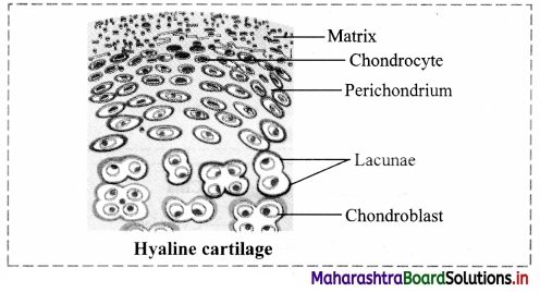
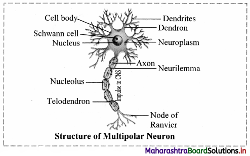
e. The different types of thickening patterns are seen on their walls such as annular (in the form of rings), spiral (in the form of spring/helix), scalariform (ladder like) and pitted (small circular area). Pitted is the most advanced type of thickening which may be simple or bordered.

3. Vessels:
a. Vessels are longer than tracheids with perforated or dissolved ends and formed by union of several vessels end to end.
b. These are involved in conduction of water and minerals.
c. Their lumen is wider than tracheids and the thickening is due to lignin and similar to tracheids.
d. In monocots, vessels are rounded where as they are angular in dicot angiosperms.
e. The first formed xylem vessels (protoxylem) are small and have either annular or spiral thickenings while latter formed xylem vessels are larger (metaxylem) and have reticulate or pitted thickenings.
f. When protoxylem is arranged towards pith and metaxylem towards periphery it is called as endarch
e. g. in stem and when the position is reversed as in the roots is called as exarch.

4. Xylem parenchyma:
a. Xylem parenchyma cells are small associated with tracheids and vessels.
b. This is the only living tissue among this complex tissue.
c. The function is to store food (starch) and sometimes tannins.
d. Xylem parenchyma are involved in lateral or radial conduction of water or sap.

5. Xylem fibres:
a. Xylem fibres are sclerenchymatous cells and serve mainly mechanical support. These are called wood fibres.
b. These are also elongated, narrow and spindle shaped.
c. Cells are tapering at both the ends and their walls are lignified.

Question 11.
Draw neat and labelled diagram of xylem tissue and vascular bundle.
Answer:
Maharashtra Board Class 11 Biology Important Questions Chapter 8 Plant Tissues and Anatomy 7

Question 12.
Match the following.

Column I Column II
1. Protoxylem (a) Xylem with larger vessels
2. Endarch Xylem (b) Protoxylem arranged towards pith
3. Metaxylem (c) Metaxylem arranged towards pith
4. Exarch xylem (d) First formed xylem vessels

Answer:

Column I Column II
1. Protoxylem (d) First formed xylem vessels
2. Endarch Xylem (b) Protoxylem arranged towards pith
3. Metaxylem (a) Xylem with larger vessels
4. Exarch xylem (c) Metaxylem arranged towards pith

Maharashtra Board Class 11 Biology Important Questions Chapter 8 Plant Tissues and Anatomy

Question 13.
Describe the structure of phloem.
Maharashtra Board Class 11 Biology Important Questions Chapter 8 Plant Tissues and Anatomy 8
Answer:
Structure of phloem:
1. Phloem is a living tissue. It is also called as bast.
It is responsible for conduction of organic food material from source (generally leaf) to a sink (other plant parts).
On the basis of origin, it can be protophloem (first formed) and metaphloem (latterly formed).
It is composed of sieve elements (sieve cells and sieve tubes), companion cells, phloem parenchyma and phloem fibres.

2. Sieve elements:
a. Sieve tubes are long tubular conducting channel of phloem.
b. These are placed end to end with bulging at end walls.
c. The sieve tube has sieve plate formed by septa with small pores.
d. The sieve plates connect protoplast of adjacent sieve tube cells.
e. The sieve tube cell is a living cell with a thin layer of cytoplasm, but loses its nucleus at maturity.
f. The sieve tube cell is connected to companion cell through phloem parenchyma by plasmodesmata.
g. Sieve cells are found in lower plants like pteridophytes and gymnosperms and sieve tubes are found in angiosperms.
h. The cells are narrow, elongated with tapering ends and sieve area located laterally.

3. Companion cells:
a. These are narrow elongated and living.
b. Companion cells are laterally associated with sieve tube elements.
c. Companion cells have dense cytoplasm and prominent nucleus.
d. Nucleus of companion cell regulates functions of sieve tube cells through simple pits.
e. From origin point of view, sieve tube cells and companion cell are derived from same cell. Death of the one result in death of the other type.

4. Phloem parenchyma:
a. Cells of phloem parenchyma are living, elongated found associated with sieve tube and companion cells.
b. Their chief function is to store food, latex, resins, mucilage, etc.
c. The cells carry out lateral conduction of food material.
d. These cells are absent in most of the monocots.

5. Phloem fibres (Bast fibres):
a. Phloem fibres are the only dead tissue among this unit.
b. They are sclerenchymatous.
c. They are generally absent in primary phloem, but present in secondary phloem.
d. These cells have with lignified walls and provide mechanical support.
e. They are used in making ropes and rough clothes.

Question 14.
Draw a diagram of phloem.
Answer:
Maharashtra Board Class 11 Biology Important Questions Chapter 8 Plant Tissues and Anatomy 9

Question 15.
Name the types of tissue systems in plants.
Answer:
The types of tissue systems in plants are epidermal tissue system, ground tissue system and vascular tissue system.

Maharashtra Board Class 11 Biology Important Questions Chapter 8 Plant Tissues and Anatomy

Question 16.
Write a short note on Epidermis.
Answer:
Epidermis:

  1. It is the outermost protective cell layer made up of compactly arranged cells without intercellular spaces.
  2. Cells show presence of central large vacuole, thin cytoplasm and a nucleus.
  3. The outer side of the epidermis is often covered with a waxy thick layer called the cuticle which prevents the loss of water.
  4. Root epidermis (Epiblema) has root hairs. These are unicellular, elongated and involved in absorption of sap from the soil.
  5. In stem, epidermal hairs are called trichomes. These are generally multicellular, branched or unbranched, stiff or soft or even secretory. These help in preventing water loss due to transpiration.

Question 17.
Draw a diagram representing epidermal tissue system.
Answer:
Maharashtra Board Class 11 Biology Important Questions Chapter 8 Plant Tissues and Anatomy 10

Question 18.
Write a short note on Structure of stomata.
Answer:
Structure of stomata:

  1. Small gateways in the epidermal cells are called as stomata.
  2. Stoma is controlled or guarded by specially modified cells called guard cells.
  3. These guard cells may be kidney shaped (dicot) or dumbbell shaped (monocot), collectively called as stomata.
  4. Guard cells have chloroplasts to carry out photosynthesis.
  5. Change in turgor pressure of guard cells causes opening and closing of stomata, which enables exchange of gases and water vapour.
  6. Stomata are further covered by subsidiary cells.
  7. Stoma, guard cells and subsidiary cells form a unit called stomatal apparatus.

Question 19.
Write the information related to diagrams given below.
Answer:
1. The given diagram represents stoma in dicot leaf.
[Note: We have given additional label of ‘chloroplast ’for better understanding of students]
2. Structure of stomata:

  1. Small gateways in the epidermal cells are called as stomata.
  2. Stoma is controlled or guarded by specially modified cells called guard cells.
  3. These guard cells may be kidney shaped (dicot) or dumbbell shaped (monocot), collectively called as stomata.
  4. Guard cells have chloroplasts to carry out photosynthesis.
  5. Change in turgor pressure of guard cells causes opening and closing of stomata, which enables exchange of gases and water vapour.
  6. Stomata are further covered by subsidiary cells.
  7. Stoma, guard cells and subsidiary cells form a unit called stomatal apparatus.

Question 20.
Explain the term ground tissue.
Answer:
Ground tissue:

  1. All the plant tissues excluding epidermal and vascular tissue is ground tissue.
  2. It is made up of simple permanent tissue e.g. parenchyma.
  3. It is present in cortex, pericycle, pith and medullary rays in the primary stem and root.
  4. Collenchyma and sclerenchyma in the hypodermis and chloroplasts containing mesophyll tissue in leaves is also ground tissue.

Question 21.
Describe various types of vascular bundles.
Answer:
Vascular bundles occur in the form of distinct patches of the complex tissue viz. Xylem and Phloem. On the basis of their arrangement in the plant body they are classified as follows:
1. Radial vascular bundles:
When the complex tissues (xylem and phloem) are situated separately on separate radius as separate bundle, vascular bundle is called Radial vascular bundle. This is a common feature of roots.

2. Conjoint vascular bundles:
When the complex tissue (xylem and phloem) is collectively present as neighbours of each other on the same radius, vascular bundle is called Conjoint vascular bundle.
They are of two types:
a. Collateral vascular bundle:
In this type of vascular bundle, xylem lies inwards and the phloem lies outwards.
These bundles may be further of open type (secondary growth takes place) containing cambium in between xylem and phloem and closed type if cambium is not present (secondary growth absent).
b. Bicollateral vascular bundle:
When phloem is present in a vascular bundle on both the sides of xylem and intervening cambium tissue, it is called bicollateral vascular bundle. It is a feature of family Cucurbitaceae.

3. Concentric vascular bundle:
a. When one vascular tissue is completely encircling the other, it is called as concentric vascular bundle.
b. When phloem is encircled by xylem, it is called as leptocentric vascular bundle, whereas when xylem is encircled by phloem, it is called as hadrocentric vascular bundle.
c. When xylem is encircled by phloem on both faces, it is called as amphicribral vascular bundle. When phloem is encircled by xylem on both faces it is called as amphivasal vascular bundle.
Maharashtra Board Class 11 Biology Important Questions Chapter 8 Plant Tissues and Anatomy 11

Maharashtra Board Class 11 Biology Important Questions Chapter 8 Plant Tissues and Anatomy

Question 22.
Explain how formation of cambial ring occurs in dicot stem.
Answer:

  1. The cambium present between the primary xylem and primary phloem of a vascular bundle is called intrafascicular cambium.
  2. With the onset of favourable season, meristematic cells of intrafascicular cambium become active.
  3. Simultaneously, the ray parenchyma cells, both fusiform initials and ray initials become meristematic. This is known as dedifferentiation.
  4. These form patch of cambial cells (meristematic cells) in between the adjacent bundles and produce interfascicular cambium.
  5. Both intrafascicular and interfascicular cambium join and form a complete ring, known as the cambial ring. This is possible because they lie in one plane.

Question 23.
‘Secondary growth is observed in most of the dicot and gymnospermic root.’ State whether the given statement is true or false and justify your answer.
Answer:

  1. The given statement is true.
  2. Secondary growth is observed in most of the dicot and gymnospermic root by producing secondary vascular tissue and periderm.
  3. Secondary growth is produced by vascular cambium and cork cambium respectively.
  4. Conjunctive parenchyma cells present on the inner edges of primary phloem bundles become meristematic.
  5. These cells add secondary xylem and secondary phloem on the inner and outer side respectively which results in secondary growth.

Question 24.
Differentiate between heartwood and sap wood.
Answer:

Heartwood Sap wood
1. It is central region of secondary xylem (wood). It is the peripheral region of secondary xylem (wood).
2. It is darker in colour due to deposition of oils, gums, resins, tannins, etc It is lighter in colour and without any depositions.
3. It is non- functional part of secondary xylem. It is functional part of secondary xylem.
4. It is resistant to pathogens. It is more susceptible to pathogens
5. It is not involved in conduction of sap. It is involved in conduction of sap.
6. It is also called as duramen. It is also called as alburnum.

Question 25.
What are tyloses?
Answer:
Tyloses:
1. Tracheary elements of heartwood are plugged by in-growth of adjacent parenchyma cells are known as tyloses.
2. Tyloses are fdled by oils, gums, resins, tannins called as extractives.

Question 26.
Explain how periderm is formed?
Answer:
Formation of periderm:
As the stem increase in diameter due to activity of vascular cambium, the outer cortical and epidermal layer get ruptured. Thus, it becomes necessaiy to replace these cells by new cells.

  1. Phellogen (cork cambium) develops in extrastelar region (cortex region) of the stem.
  2. The outer cortical cells of cortex become meristematic and produce a layer of thin walled, rectangular cells. These cells cut off new cells on both sides.
  3. The cells produced on outer side develop phellem (cork), whereas on the inner side produce phelloderm (secondary cortex).
  4. The cork is impervious in nature and does not allow entry of water due to suberized walls. Secondary cortex is parenchymatous in nature.
  5. Phellogen, phellem and phelloderm constitute periderm.

Question 27.
Explain the given terms:
1. Bark
2. Lenticels
3. Anomalous secondary growth
Answer:
1. Bark:
a. Bark is non-technical term referring to all cell types found external to vascular cambium including secondary phloem.
b. Bark of early season is soft and of the late season is hard.

2. Lenticels:
a. Lenticels are aerating pores present as raised scars on the surface of bark.
b. These are portions of periderm, where phellogen activity is more.
c. Lenticels are meant for gaseous and water vapour exchange.

3. Anomalous secondary growth:
a. Monocot stems lack cambium hence secondary growth does not take place.
b. However, accessory cambium development in plants like, Dracaena, Agave, Palms and root of sweet potato shows presence of secondary growth. This is called as anomalous secondary growth.

Maharashtra Board Class 11 Biology Important Questions Chapter 8 Plant Tissues and Anatomy

Question 28.
With the help of neat and labelled diagram explain the anatomy of dicot root.
Answer:
Maharashtra Board Class 11 Biology Important Questions Chapter 8 Plant Tissues and Anatomy 12
The transverse section of a typical dicotyledonous root shows following anatomical features:
1. Epiblema: It is the outermost single layer of cells without cuticle. Some epidermal cells prolong to form unicellular root hairs.
2. Cortex: It is made up of many layers of thin walled parenchyma cells. Cortical cells store food and water.
3. Exodermis: After the death of epiblema, outer layer of cortex become cutinized and is called Exodermis.

4. Endodermis:
The innermost layer of cortex is called Endodermis.
The cells are barrel-shaped and their radial walls bear Casparian strip or Casparian bands composed of suberin. Near the protoxylem, there are unthickened passage cells.

5. Stele: It consists of pericycle, vascular bundles and pith.
a. Pericycle: Next to the endodermis, there is a single layer of thin walled parenchyma cells called pericycle. It forms outermost layer of stele or vascular cylinder.
b. Vascular bundle: Vascular bundles are radial. Xylem and Phloem occur in separate patches arranged on alternate radii. Xylem is exarch in root that means protoxylem vessels are towards periphery and metaxylem elements are towards centre. Xylem bundles vary from two to six number, i.e. they may be diarch, triarch, tetrarch, etc.
Connective tissue: A parenchymatous tissue is present in between xylem and phloem.
c. Pith: The central part of stele is called pith. It is narrow and made up of parenchymatous cells, with or without intercellular spaces.
6. At a later stage cambium ring develops between the xylem and phloem causing secondary growth.

Question 29.
With the help of neat and labelled diagram explain the anatomy of monocot root.
Answer:
The transverse section of a typical monocotyledonous root shows following anatomical features:
1. Epiblema: It is the outermost single layer of cells without cuticle. Some epidermal cells prolong to form unicellular root hairs.
2. Cortex: It is made up of many layers of thin walled parenchyma cells. Cortical cells store food and water.
Endodermis: It is innermost layer of cortex. The cells of endodermis are thick walled except the passage cells which lie just opposite to the protoxylem.
Stele: It consists of pericycle, vascular bundles and pith.
a. Pericycle: Pericycle is present below the endodermis.
b. Vascular bundle: Vascular bundles are radial. Xylem and Phloem occur in separate patches arranged on alternate radii. Xylem is exarch in root that means protoxylem vessels are towards periphery and metaxylem elements are towards centre. Polyarch condition of xylem is observed, (xylem bundles are more than six).
Pith: Pith is large and well developed.
Secondary growth does not occur due to absence of cambium.

Maharashtra Board Class 11 Biology Important Questions Chapter 8 Plant Tissues and Anatomy

Question 30.
What is polyarch condition of root?
Answer:
Polyarch condition is the one in which roots possess more than six xylem bundles.

Question 31.
Explain in detail anatomy of sunflower stem.
Answer:
A transverse section of sunflower (dicot) stem shows the following structures:
1. Epidermis: It is a single, outermost layer with multicellular outgrowth called trichomes. A layer of cuticle
is usually present towards the outer surface of epidermis.

2. Cortex: Cortex is situated below the epidermis and is usually differentiated into three regions namely, hypodermis, general cortex and endodermis.
a. Hypodermis: It is situated just below the epidermis and is made of 3-5 layers of collenchymatous cells. Intercellular spaces are absent.
b. General cortex: It is made up of several layers of large parenchymatous cells with intercellular spaces.
c. Endodermis: It is an innermost layer of cortex which is made up of barrel shaped cells. It is also called starch sheath, as it is rich in starch grain.

3. Stele: It is differentiated into pericycle, vascular bundles and pith.
a. Pericycle: It is the outermost layer of vascular system situated between the endodermis and vascular bundles. In sunflower, it is multi-layered and also called hard bast.
b. Vascular bundles: Vascular bundles are conjoint, collateral, open, and are arranged in a ring. Each one is composed of xylem, phloem and cambium. Xylem is endarch. A strip of cambium is present between xylem and phloem.
c. Pith: It is situated in the centre of the young stem and is made up of large-sized parenchymatous cells with conspicuous intercellular spaces.

Question 32.
With the help of neat and labelled diagram explain the anatomy of maize stem.
Answer:
Maharashtra Board Class 11 Biology Important Questions Chapter 8 Plant Tissues and Anatomy 13
A transverse section of maize (monocot) stem shows the following structures:

  1. Epidermis: It is single layered and without trichomes.
  2. Hypodermis: It is sclerenchymatous.
  3. Ground tissue: It consists of thin walled parenchyma cells. It extends from hypodermis to the centre. It is not differentiated into cortex, endodermis, pericycle and pith.
  4. Vascular bundles: Vascular bundles are numerous and are scattered in ground tissue. Each vascular bundle is surrounded by a sclerenchymatous bundle sheath. Vascular bundles are conjoint, collateral and closed (without cambium). Xylem is endarch and shows lysigenous cavity.
  5. Pith: Pith is absent.

Question 33.
With the help of a neat and labelled diagram, describe the internal structure of dorsiventral leaf.
Answer:
1. Structure of dorsiventral leaf: The mesophyll tissue is differentiated into palisade and spongy parenchyma in a dorsiventral leaf. This type is very common in dicot leaf. The different parts of this leaf are as follows:
2. Upper epidermis: It consists of a single layer of tightly packed rectangular, barrel shaped, parenchymatous cells which are devoid of chloroplast. A distinct layer of cuticle lies on the outside of the epidermis. Stomata are generally absent.
3. Mesophyll: Between upper and lower epidermis, there is chloroplast-containing photosynthetic tissue called mesophyll It is differentiated into Palisade parenchyma and Spongy parenchyma.
a. Palisade parenchyma:
Palisade parenchyma is present below upper epidermis and consists of closely packed elongated cells. The cells contain abundant chloroplasts and help in photosynthesis.
b. Spongy parenchyma:
Spongy parenchyma is present below palisade tissue and consists of loosely arranged irregularly shaped cells with intercellular spaces. The spongy parenchyma cells contain chloroplast and are in contact with the atmosphere through stomata.
Maharashtra Board Class 11 Biology Important Questions Chapter 8 Plant Tissues and Anatomy 14
4. Vascular system: It is made up of a number of vascular bundles of varying size depending upon the venation. Each one is surrounded by a thin layer of parenchymatous cells called bundle sheath. Vascular bundles are closed. Xylem lies towards upper epidermis and phloem towards lower epidermis. Cambium is absent, hence there is no secondary growth in the leaf.
5. Lower epidermis: It consists of a single layer of compactly arranged rectangular, parenchymatous cells. A thin layer of cuticle is also present. The lower epidermis contains a large number of microscopic pores called stomata. There is an air-space called substomatal chamber at each stoma.

Maharashtra Board Class 11 Biology Important Questions Chapter 8 Plant Tissues and Anatomy

Question 34.
With the help of a neat labelled diagram, describe the anatomy of isobilateral leaf.
Answer:
The parts of isobilateral leaf are as follows:
Maharashtra Board Class 11 Biology Important Questions Chapter 8 Plant Tissues and Anatomy 15
1. Epidermis:
It is single layered, present on both sides of the leaf.
It consists of compactly arranged rectangular transparent parenchymatous cells.
Both the surfaces contain stomata.
Both the surfaces have a distinct layer of cuticle.
2. Mesophyll:
Mesophyll is not differentiated into palisade and spongy tissue.
3. Vascular bundle:
These are conjoint, collateral and closed.

Question 35.
Compare between dorsiventral and isobilateral leaf.
Answer:

Dorsiventral leaf Isobilateral leaf
1. Dorsiventral Leaf is very common in dicotyledonous plants. Isobilateral leaf is very common in monocotyledonous plants.
2. In this mesophyll tissue is differentiated into palisade and spongy parenchyma. In this mesophyll tissue is not differentiated into palisade and spongy parenchyma.
3. The leaves are commonly horizontal in orientation with distinct upper and lower surfaces. The upper surface which faces the sun is darker than the lower surface. In this leaf both the surfaces are equally illuminated as both the surface can face the sun, and show similar structure. The two surfaces are equally green.
4. Stomata is absent on the upper surface of these leaves. Stomata is present on both the upper and lower surfaces of these leaves.

Question 36.
Distinguish between anatomy of dicot root and monocot root.
Answer:

Anatomy of dicot root Anatomy of monocot root
1. Pith is narrow. Pith is large and well developed.
2. Diarch, triarch or tetrarch condition can be observed. (Xylem bundles vary from two to six number) Polyarch condition is observed, (xylem bundles are more than six)
3. Cambium is formed in later stage between xylem and phloem which causes secondary growth. Secondary growth is absent.

Maharashtra Board Class 11 Biology Important Questions Chapter 8 Plant Tissues and Anatomy

Question 37.
Distinguish between anatomy of dicot stem and monocot stem.
Answer:

Anatomy of Dicot stem Anatomy of Monocot stem
1. Epidermis shows presence of multicellular trichomes. Epidermis is without trichomes.
2. Hypodermis is made up of collenchymatous cells. Hypodermis is made up of sclerenchymatous cells.
3. Medullary rays are present between vascular bundles. Medullary rays are absent.
4. Vascular bundles are arranged in the form of a ring. Vascular bundles are scattered in the ground tissue.
5. It is conjoint, collateral and open (Cambium present) They are conjoint, collateral and closed (cambium is absent).
6. Vascular bundle is not surrounded by a sclerenchymatous bundle sheath. Vascular bundle is surrounded by a sclerenchymatous bundle sheath.
7. Secondary growth takes place due to presence of cambium. Secondary growth does not occur due to absence of cambium.
8. Pith is present. Pith is absent.

Question 38.
Apply Your Knowledge

Question 1.
Which plant part would show the following:

  1. Radial vascular bundles.
  2. Large and well-developed pith.
  3. Differentiation of mesophyll into palisade and spongy tissue.
  4. Presence of stomata on both upper and lower epidermis.

Answer:

  1. Root
  2. Monocot root and Dicot stem,
  3. Dicot leaf
  4. Monocot leaf

Question 2.
When a tree is debarked, which tissues are removed?
Answer:
The bark is made up of tissues like cork, cork cambium and secondary cortex, which are removed when a tree is debarked.

Question 3.
While eating fruits like pear or guava, it feels gritty. What gives stiffness to these fruits?
Answer:
Sclereids are found in pulp of fruits like pear and guava which gives them stiffness and thus we feel gritty while eating these fruits.

Maharashtra Board Class 11 Biology Important Questions Chapter 8 Plant Tissues and Anatomy

Question 39.
Quick Review

Maharashtra Board Class 11 Biology Important Questions Chapter 8 Plant Tissues and Anatomy 16
Maharashtra Board Class 11 Biology Important Questions Chapter 8 Plant Tissues and Anatomy 17

Question 40.
Exercise

Question 1.
Define tissue.
Answer:
A group of cells having essentially a common function and origin is called as tissue.

Question 2.
Classify the meristematic tissue based on its origin.
Answer:
Classification of meristematic tissue on the basis of origin:
1. Promeristem / Primordial meristem:
a. It is also called as embryonic meristem.
b. It usually occupies very minute area at the tip of root and shoot.

2. Primary meristem:
a. It originates from the primordial meristem and occurs in the plant body from the beginning, at the root and shoot apices.
b. Cells are always in active state of division and give rise to permanent tissues.

3. Secondary meristem:
a. These tissues develop from living permanent tissues during later stages of plant growth hence are called as secondary meristems.
b. This tissue occurs in the mature regions of root and shoot of many plants.
c. Secondary meristem is always lateral (to the central axis) in position e.g. Fascicular cambium, inter fascicular cambium, cork cambium.

Maharashtra Board Class 11 Biology Important Questions Chapter 8 Plant Tissues and Anatomy

Question 3.
Explain in detail classification of meristematic tissue based on its position.
Answer:
Classification of meristematic tissue based on its position:
1. Apical meristem:
a. It is produced from promeristem and forms growing point of apices of root, shoot and their lateral branches.
b. It brings about increase in length of plant body and is called as apical initials.
c. Shoot apical meristem is terminal in position whereas in root it is subterminal i.e. located behind the root cap.

2. Intercalary meristem:
a. Intercalary meristematic tissue is present in the top or base area of node.
b. Their activity is mainly seen in monocots.
c. These are short lived.

3. Lateral meristem:
a. It is present along the sides of central axis of organs.
b. It takes part in increasing girth of stem or root, e.g. Intrafascicular cambium.
c. It is found in vascular bundles of gymnosperms and dicot angiosperms.

Question 4.
Give any two examples of secondary meristematic tissue.
Answer:
Secondary meristem is always lateral (to the central axis) in position e.g. Fascicular cambium, inter fascicular cambium, cork cambium.

Question 5.
Draw a diagram of meristematic cells.
Answer:
1. Cell wall
2. Nucleus
3. Cytoplasm
Characteristics of meristematic tissue:

  1. It is a group of young, immature cells.
  2. These are living cells with ability to divide in the regions where they are present.
  3. These are polyhedral or isodiametric in shape without intercellular spaces.
  4. Cell wall is thin, elastic and mainly composed of cellulose.
  5. Protoplasm is dense with distinct nucleus at the centre and vacuoles if present, are very small.
  6. Cells show high rate of metabolism.

Maharashtra Board Class 11 Biology Important Questions Chapter 8 Plant Tissues and Anatomy

Question 6.
Write a short note on tracheids.
Answer:
Tracheids:
a. These are elongated, tubular and dead cells (without protoplasm).
b. The ends are oblique and tapering.
c. The cell walls is unevenly thickened and lignified. This provides mechanical strength.
d. Tracheids contribute 95% of wood in gymnosperms and 5% in angiosperms.
e. The different types of thickening patterns are seen on their walls such as annular (in the form of rings), spiral (in the form of spring/helix), scalariform (ladder like) and pitted (small circular area). Pitted is the most advanced type of thickening which may be simple or bordered.

Question 7.
Describe parenchyma in detail.
Answer:
Cell is the component that brings about important processes in the living organisms.

Question 8.
Describe the structure of xylem in detail.
Answer:
1. Xylem is the water conducting tissue in higher plants. It is a dead complex tissue.
It also provides mechanical strength to the plant body.
Components of xylem are tracheids, vessels, xylem parenchyma and xylem fibres.

2. Tracheids:
a. These are elongated, tubular and dead cells (without protoplasm).
b. The ends are oblique and tapering.
c. The cell walls is unevenly thickened and lignified. This provides mechanical strength.
d. Tracheids contribute 95% of wood in gymnosperms and 5% in angiosperms.
e. The different types of thickening patterns are seen on their walls such as annular (in the form of rings), spiral (in the form of spring/helix), scalariform (ladder like) and pitted (small circular area). Pitted is the most advanced type of thickening which may be simple or bordered.

3. Vessels:
a. Vessels are longer than tracheids with perforated or dissolved ends and formed by union of several vessels end to end.
b. These are involved in conduction of water and minerals.
c. Their lumen is wider than tracheids and the thickening is due to lignin and similar to tracheids.
d. In monocots, vessels are rounded where as they are angular in dicot angiosperms.
e. The first formed xylem vessels (protoxylem) are small and have either annular or spiral thickenings while latter formed xylem vessels are larger (metaxylem) and have reticulate or pitted thickenings.
f. When protoxylem is arranged towards pith and metaxylem towards periphery it is called as endarch
e. g. in stem and when the position is reversed as in the roots is called as exarch.

4. Xylem parenchyma:
a. Xylem parenchyma cells are small associated with tracheids and vessels.
b. This is the only living tissue among this complex tissue.
c. The function is to store food (starch) and sometimes tannins.
d. Xylem parenchyma are involved in lateral or radial conduction of water or sap.

5. Xylem fibres:
a. Xylem fibres are sclerenchymatous cells and serve mainly mechanical support. These are called wood fibres.
b. These are also elongated, narrow and spindle shaped.
c. Cells are tapering at both the ends and their walls are lignified.

Question 9.
What are Sclerenchyma fibres?
Answer:
a. Fibres:
Fibres are thread-like, elongated and narrow structures with tapering and interlocking end walls. Fibres are mostly in bundles. Pits are narrow, unbranched and oblique. They provide mechanical strength.

Maharashtra Board Class 11 Biology Important Questions Chapter 8 Plant Tissues and Anatomy

Question 10.
Write the functions of parenchyma cells.
Answer:
Parenchyma, Collenchyma and Sclerenchyma are the simple permanent tissues in plants.

Question 11.
Write function of collenchyma tissue.
Answer:
Function:
Collenchyma is a living mechanical tissue and serves different functions in plants.
a. It gives mechanical strength to young stem and parts like petiole of leaf.
b. It allows bending and pulling action in plant parts and also prevents tearing of leaf.
c. It also allows growth and elongation of organs.

Question 12.
Which are the different types of tracheids based on the types of thickenings on their walls?
Answer:
The different types of thickening patterns are seen on their walls such as annular (in the form of rings), spiral (in the form of spring/helix), scalariform (ladder like) and pitted (small circular area). Pitted is the most advanced type of thickening which may be simple or bordered.

Question 13.
Death of companion cell causes death of sieve tube cells and vice versa. Justify.
Answer:
Companion cells:
a. These are narrow elongated and living.
b. Companion cells are laterally associated with sieve tube elements.
c. Companion cells have dense cytoplasm and prominent nucleus.
d. Nucleus of companion cell regulates functions of sieve tube cells through simple pits.
e. From origin point of view, sieve tube cells and companion cell are derived from same cell. Death of the one result in death of the other type.

Question 14.
Which are the three types of simple permanent tissues?
Answer:
Parenchyma, Collenchyma and Sclerenchyma are the simple permanent tissues in plants.

Maharashtra Board Class 11 Biology Important Questions Chapter 8 Plant Tissues and Anatomy

Question 15.
Write the functions of sclerenchyma tissue.
Answer:
Functions:
a. This tissue functions as the main mechanical tissue.
b. It permits bending, shearing and pulling.
c. It gives rigidity to leaves and prevents it from falling.
d. It also gives rigidity to epicarps and seeds.

Question 16.
What are the components of xylem?
Answer:
1. Xylem is the water conducting tissue in higher plants. It is a dead complex tissue.
It also provides mechanical strength to the plant body.
Components of xylem are tracheids, vessels, xylem parenchyma and xylem fibres.

Question 17.
Name the living component of xylem.
Answer:
This is the only living tissue among this complex tissue.

Question 18.
Name the dead component of phloem.
Answer:
Phloem fibres are the only dead tissue among this unit.

Question 19.
What is closed vascular bundle?
Answer:
When cambium is not present between xylem and phloem, it is known as closed vascular bundle.

Question 20.
Describe two types of conjoint vascular bundles.
Answer:
Conjoint vascular bundles:
When the complex tissue (xylem and phloem) is collectively present as neighbours of each other on the same radius, vascular bundle is called Conjoint vascular bundle.
They are of two types:
a. Collateral vascular bundle:
In this type of vascular bundle, xylem lies inwards and the phloem lies outwards.
These bundles may be further of open type (secondary growth takes place) containing cambium in between xylem and phloem and closed type if cambium is not present (secondary growth absent).
b. Bicollateral vascular bundle:
When phloem is present in a vascular bundle on both the sides of xylem and intervening cambium tissue, it is called bicollateral vascular bundle. It is a feature of family Cucurbitaceae.

Question 21.
Write the function of trichomes.
Answer:
In stem, epidermal hairs are called trichomes. These are generally multicellular, branched or unbranched, stiff or soft or even secretory. These help in preventing water loss due to transpiration.

Question 22.
What are bicollateral vascular bundle?
Answer:
Bicollateral vascular bundle:
When phloem is present in a vascular bundle on both the sides of xylem and intervening cambium tissue, it is called bicollateral vascular bundle. It is a feature of family Cucurbitaceae.

Question 23.
Name the tissue that are not included in ground tissue.
Answer:
All the plant tissues excluding epidermal and vascular tissue is ground tissue.

Maharashtra Board Class 11 Biology Important Questions Chapter 8 Plant Tissues and Anatomy

Question 24.
Which type of conjoint – vascular bundles are found in members of Cucurbitaceae family?
Answer:
Bicollateral vascular bundle:
When phloem is present in a vascular bundle on both the sides of xylem and intervening cambium tissue, it is called bicollateral vascular bundle. It is a feature of family Cucurbitaceae.

Question 25.
What is concentric vascular bundle?
Answer:
Concentric vascular bundle:
a. When one vascular tissue is completely encircling the other, it is called as concentric vascular bundle.
b. When phloem is encircled by xylem, it is called as leptocentric vascular bundle, whereas when xylem is encircled by phloem, it is called as hadrocentric vascular bundle.
c. When xylem is encircled by phloem on both faces, it is called as amphicribral vascular bundle. When phloem is encircled by xylem on both faces it is called as amphivasal vascular bundle.

Question 26.
Define intrafascicular cambium.
Answer:
The cambium present between the primary xylem and primary phloem of a vascular bundle is called intrafascicular cambium.

Question 27.
What is the difference between spring wood and autumn wood?
Answer:
During favourable conditions, spring wood (early wood) is formed which has broader xylem bands, lighter colour, tracheids with thin wall and wide lumen, fibres are less in number, low density. Whereas, during unfavourable conditions, autumn wood (late wood) is formed which has narrow xylem band, darker in colour, lumen is narrow and walls are thick with abundant fibres, high density.

Question 28.
Explain how growth rings are formed in trees?
Answer:
1. Growth rings are formed due cambial activity during favourable and non-favourable climatic conditions.
2. During favourable conditions, spring wood (early wood) is formed which has broader xylem bands, lighter colour, tracheids with thin wall and wide lumen, fibres are less in number, low density. Whereas, during unfavourable conditions, autumn wood (late wood) is formed which has narrow xylem band, darker in colour, lumen is narrow and walls are thick with abundant fibres, high density.
3. Spring wood and autumn wood that appear as alternate light and dark concentric rings, constitute an annual ring or growth ring.

Question 29.
Which tissues are together called as periderm?
Answer:
Phellogen, phellem and phelloderm constitute periderm.

Maharashtra Board Class 11 Biology Important Questions Chapter 8 Plant Tissues and Anatomy

Question 30.
What is bark?
Answer:
Bark:
a. Bark is non-technical term referring to all cell types found external to vascular cambium including secondary phloem.
b. Bark of early season is soft and of the late season is hard.

Question 31.
What is the function of lenticels?
Answer:
Lenticels are meant for gaseous and water vapour exchange.

Question 32.
Explain the term anomalous secondary growth.
Answer:
Anomalous secondary growth:
a. Monocot stems lack cambium hence secondary growth does not take place.
b. However, accessory cambium development in plants like, Dracaena, Agave, Palms and root of sweet potato shows presence of secondary growth. This is called as anomalous secondary growth.

Question 33.
Explain in detail anatomical structure of a dicot stem.
Answer:
A transverse section of sunflower (dicot) stem shows the following structures:
1. Epidermis: It is a single, outermost layer with multicellular outgrowth called trichomes. A layer of cuticle
is usually present towards the outer surface of epidermis.

2. Cortex: Cortex is situated below the epidermis and is usually differentiated into three regions namely, hypodermis, general cortex and endodermis.
a. Hypodermis: It is situated just below the epidermis and is made of 3-5 layers of collenchymatous cells. Intercellular spaces are absent.
b. General cortex: It is made up of several layers of large parenchymatous cells with intercellular spaces.
c. Endodermis: It is an innermost layer of cortex which is made up of barrel shaped cells. It is also called starch sheath, as it is rich in starch grain.

3. Stele: It is differentiated into pericycle, vascular bundles and pith.
a. Pericycle: It is the outermost layer of vascular system situated between the endodermis and vascular bundles. In sunflower, it is multi-layered and also called hard bast.
b. Vascular bundles: Vascular bundles are conjoint, collateral, open, and are arranged in a ring. Each one is composed of xylem, phloem and cambium. Xylem is endarch. A strip of cambium is present between xylem and phloem.
c. Pith: It is situated in the centre of the young stem and is made up of large-sized parenchymatous cells with conspicuous intercellular spaces.

Question 34.
Draw neat and labelled diagrams of dicot and monocot root and differentiate between their anatomical characters.
Answer:
The transverse section of a typical dicotyledonous root shows following anatomical features:
1. Epiblema: It is the outermost single layer of cells without cuticle. Some epidermal cells prolong to form unicellular root hairs.
2. Cortex: It is made up of many layers of thin walled parenchyma cells. Cortical cells store food and water.
3. Exodermis: After the death of epiblema, outer layer of cortex become cutinized and is called Exodermis.

4. Endodermis:
The innermost layer of cortex is called Endodermis.
The cells are barrel-shaped and their radial walls bear Casparian strip or Casparian bands composed of suberin. Near the protoxylem, there are unthickened passage cells.

5. Stele: It consists of pericycle, vascular bundles and pith.
a. Pericycle: Next to the endodermis, there is a single layer of thin walled parenchyma cells called pericycle. It forms outermost layer of stele or vascular cylinder.
b. Vascular bundle: Vascular bundles are radial. Xylem and Phloem occur in separate patches arranged on alternate radii. Xylem is exarch in root that means protoxylem vessels are towards periphery and metaxylem elements are towards centre. Xylem bundles vary from two to six number, i.e. they may be diarch, triarch, tetrarch, etc.
Connective tissue: A parenchymatous tissue is present in between xylem and phloem.
c. Pith: The central part of stele is called pith. It is narrow and made up of parenchymatous cells, with or without intercellular spaces.
6. At a later stage cambium ring develops between the xylem and phloem causing secondary growth.

Maharashtra Board Class 11 Biology Important Questions Chapter 8 Plant Tissues and Anatomy

Question 35.
Which type of vascular bundles are observed in isobilateral leaf?
Answer:
Vascular bundle:
These are conjoint, collateral and closed.

Question 36.
Describe the internal structure of a leaf in which mesophyll is differentiated in palisade and spongy parenchyma.
Answer:
1. Structure of dorsiventral leaf: The mesophyll tissue is differentiated into palisade and spongy parenchyma in a dorsiventral leaf. This type is very common in dicot leaf. The different parts of this leaf are as follows:
2. Upper epidermis: It consists of a single layer of tightly packed rectangular, barrel shaped, parenchymatous cells which are devoid of chloroplast. A distinct layer of cuticle lies on the outside of the epidermis. Stomata are generally absent.
3. Mesophyll: Between upper and lower epidermis, there is chloroplast-containing photosynthetic tissue called mesophyll It is differentiated into Palisade parenchyma and Spongy parenchyma.
a. Palisade parenchyma:
Palisade parenchyma is present below upper epidermis and consists of closely packed elongated cells. The cells contain abundant chloroplasts and help in photosynthesis.
b. Spongy parenchyma:
Spongy parenchyma is present below palisade tissue and consists of loosely arranged irregularly shaped cells with intercellular spaces. The spongy parenchyma cells contain chloroplast and are in contact with the atmosphere through stomata.
4. Vascular system: It is made up of a number of vascular bundles of varying size depending upon the venation. Each one is surrounded by a thin layer of parenchymatous cells called bundle sheath. Vascular bundles are closed. Xylem lies towards upper epidermis and phloem towards lower epidermis. Cambium is absent, hence there is no secondary growth in the leaf.
5. Lower epidermis: It consists of a single layer of compactly arranged rectangular, parenchymatous cells. A thin layer of cuticle is also present. The lower epidermis contains a large number of microscopic pores called stomata. There is an air-space called substomatal chamber at each stoma.

Question 37.
Multiple Choice Questions:

Question 1.
Meristematic tissues are found
(A) only in stems of the plants
(B) in both roots and stems
(C) in all growing tips of the plant body
(D) only in roots of the plants
Answer:
(C) in all growing tips of the plant body

Question 2.
The tissue responsible for translocation of food material is _________
(A) xylem
(B) cambium
(C) parenchyma
(D) phloem
Answer:
(D) phloem

Maharashtra Board Class 11 Biology Important Questions Chapter 8 Plant Tissues and Anatomy

Question 3.
________ are used in making ropes and rough clothes.
(A) Phloem parenchyma
(B) Trachieds
(C) Phloem fibres
(D) Sieve tube elements
Answer:
(C) Phloem fibres

Question 4.
_______ are the only dead tissue among the phloem.
(A) Phloem parenchyma
(B) Sieve tubes
(C) Companion cells
(D) Phloem fibres
Answer:
(D) Phloem fibres

Question 5.
Phloem was named as _______ by Haberlandt as similar to xylem.
(A) Bast
(B) Leptome
(C) Wood fibres
(D) Casparian
Answer:
(B) Leptome

Question 6.
The sieve tube cell is connected to companion cell through phloem parenchyma by
(A) Plasmodesmata
(B) Interfascicular cambium
(C) Pericycle
(D) Hypodermis
Answer:
(A) Plasmodesmata

Maharashtra Board Class 11 Biology Important Questions Chapter 8 Plant Tissues and Anatomy

Question 7.
Which of the following tissues is with dead thick-walled cells without intercellular spaces?
(A) parenchyma
(B) collenchyma
(C) sclerenchyma
(D) phloem
Answer:
(C) sclerenchyma

Question 8.
The tissue which is present in between xylem and phloem of stem is called
(A) apical meristem
(B) pericycle
(C) vascular cambium
(D) cork cambium
Answer:
(C) vascular cambium

Question 9.
In stem, epidermal hairs are called as
(A) Cuticles
(B) Casparian strip
(C) Trichomes
(D) Companion cells
Answer:
(C) Trichomes

Question 10.
_______ forms the outer covering of plant body and is derived from protodenn or dermatogen.
(A) Ground tissue system
(B) Interfascicular cambium
(C) Vascular tissue system
(D) Epidermal tissue system
Answer:
(D) Epidermal tissue system

Question 11.
_______ play a vital role in exchange of gases and water vapour.
(A) Vascular bundles
(B) Stomata
(C) Ground tissues
(D) Trichomes
Answer:
(B) Stomata

Maharashtra Board Class 11 Biology Important Questions Chapter 8 Plant Tissues and Anatomy

Question 12.
In which of the following leaf possesses dumbbell shaped guard cell’?
(A) Pisum sativum
(B) Wheat
(C) Datura
(D) Sunflower
Answer:
(B) Wheat

Question 13.
Which of the following is NOT a characteristic of spring wood?
(A) Tracheids with wide lumen
(B) Less number of fibres
(C) Narrow xylem band
(D) Lighter colour
Answer:
(C) Narrow xylem band

Question 14.
Periderm consists of
(A) Phellogen
(B) Phellem
(C) Phelloderm
(D) All of these
Answer:
(D) All of these

Question 15.
Which of the following is essential for secondary growth?
(A) Xylem
(B) Pith
(C) Phloem
(D) Cambium
Answer:
(D) Cambium

Question 16.
Vascular bundles of dicot root are
(A) radial exarch
(B) radial endarch
(C) conjoint exarch
(D) conjoint endarch
Answer:
(A) radial exarch

Question 17.
In which of the following characters, a monocot root differs from dicot root?
(A) Open vascular bundle
(B) Large pith
(C) Radial vascular bundles
(D) Scattered vascular bundles
Answer:
(B) Large pith

Question 18.
Which of the following plant shows isobilateral leaves?
(A) Hibiscus
(B) Maize
(C) Mangifera indica
(D) Sunflower
Answer:
(B) Maize

Maharashtra Board Class 11 Biology Important Questions Chapter 8 Plant Tissues and Anatomy

Question 19.
Secondary growth does not occur in
(A) Maize stem
(B) Mango leaf
(C) Carina root
(D) All of these
Answer:
(D) All of these

Question 20.
Stele of a dicot stem consists of all the given below, EXCEPT
(A) Pericycle
(B) Cortex
(C) Vascular bundles
(D) Pith
Answer:
(B) Cortex

Question 21.
Hypodermis is collenchymatous in
(A) monocot stem
(B) dicot stem
(C) monocot root
(D) both (A) and (B)
Answer:
(B) dicot stem

Question 22.
Lysigenous cavity filled with water is present in
(A) dicot stem
(B) monocot stem
(C) monocot root
(D) dicot root
Answer:
(B) monocot stem

Question 23.
The vascular bundles in a dicot stem are
(A) collateral and open
(B) radial
(C) bicollateral and open
(D) collateral and closed
Answer:
(A) collateral and open

Question 38.
Competitive Corner

Question 1.
Phloem in gymnosperms lacks:
(A) companion cells only
(B) both sieve tubes and companion cells
(C) albuminous cells and sieve cells
(D) sieve tubes only
Answer:
(B) both sieve tubes and companion cells

Question 2.
Grass leaves curl inwards during very dry weather. Select the most appropriate reason from the following:
(A) Shrinkage of air spaces in spongy mesophyll
(B) Tyloses in vessels
(C) Closure of stomata
(D) Flaccidity of bulliform cells
Hint: Grass leaves curl inwards to the minimize water loss.
Answer:
(D) Flaccidity of bulliform cells

Question 3.
Which of the statements given below is NOT true about formation of ‘annual rings’ in trees?
(A) Activity of cambium depends upon variation in climate.
(B) Annual rings are not prominent in trees of temperate region.
(C) Annual ring is a combination of spring wood and autumn wood produced in a year.
(D) Differential activity of cambium causes light and dark bands of tissue – early and late wood respectively.
Hint: Annual rings are formed due to activity of cambium. The activity of cambium is under the control of many physiological and environmental factors. In temperate regions, the climatic conditions are not uniform throughout the year due to this, annual rings are formed.
Answer:
(B) Annual rings are not prominent in trees of temperate region.

Question 4.
Regeneration of damaged growing grass following grazing is largely due to:
(A) secondary meristem
(B) lateral meristem
(C) apical meristem
(D) intercalary meristem
Hint: Intercalary meristems occur in grasses at the base of intemode, which regenerates the grass damaged due to grazing.
Answer:
(D) intercalary meristem

Question 5.
In the dicot root, the vascular cambium originates from:
(A) intrafascicular and interfascicular tissue in a ring
(B) tissue located below the phloem bundles and a portion of pericycle tissue above protoxylem
(C) cortical region
(D) parenchyma between endodermis and pericycle
Answer:
(B) tissue located below the phloem bundles and a portion of pericycle tissue above protoxylem

Maharashtra Board Class 11 Biology Important Questions Chapter 8 Plant Tissues and Anatomy

Question 6.
Casparian strips occur in
(A) Cortex
(B) Pericycle
(C) Epidermis
(D) Endodermis
Answer:
(D) Endodermis

Question 7.
Plants having little or no secondary growth are
(A) Conifers
(B) Deciduous angiosperms
(C) Grasses
(D) Cycads
Hint: Secondary growth takes place in stems and roots of dicotyledons and gymnosperms, but does not occur in monocotyledons.
Answer:
(C) Grasses

Question 8.
Secondary xylem and phloem in dicot stem are produced by
(A) Phellogen
(B) Vascular cambium
(C) Apical meristems
(D) Axillary meristems
Answer:
(B) Vascular cambium

Question 9.
The vascular cambium normally gives rise to
(A) Phelloderm
(B) Primary phloem
(C) Secondary xylem
(D) Periderm
Answer:
(C) Secondary xylem

Maharashtra Board Class 11 Biology Important Questions Chapter 8 Plant Tissues and Anatomy

Question 10.
Identify the wrong statement in context of heartwood.
(A) Organic compounds are deposited in it
(B) It is highly durable
(C) It conducts water and minerals efficiently
(D) It comprises dead elements with highly lignified walls
Hint: In old trees, secondary xylem (wood) becomes physiologically non active. It does not conduct water and becomes dark due to organic deposits (tannins, resins, oils, aromatic substances, etc.) It comprises of dead elements and called as heart wood. It is non conductive, hard, durable and resistant to microbes and insects.
Answer:
(C) It conducts water and minerals efficiently

Question 11.
Which of the following is made up of dead cell?
(A) Xylem parenchyma
(B) Collenchyma
(C) Phellem
(D) Phloem
Hint: Cork cambium (phellogen) cuts off cells on both the sides. The outer cells differentiate into cork or phellem. The cork is impervious to water due to suberin deposition in the cell wall.
Answer:
(C) Phellem

Maharashtra Board Class 11 Biology Important Questions Chapter 7 Cell Division

Balbharti Maharashtra State Board 11th Biology Important Questions Chapter 7 Cell Division Important Questions and Answers.

Maharashtra State Board 11th Biology Important Questions Chapter 7 Cell Division

Question 1.
Why interphase is known as the preparatory phase.
Answer:
1. During interphase, the cell is metabolically very active.
2. In this phase, a cell grows to its maximum size, chromosomal material (DNA and histone proteins) duplicates and the cell prepares itself for the next mitotic division. Hence, the interphase is known as the preparatory phase.

Maharashtra Board Class 11 Biology Important Questions Chapter 7 Cell Division

Question 2.
Name the following.
1. In which phase does the amount of DNA per cell doubles?
2. Which types of RNA are synthesized during first growth phase?
Answer:
1. S-phase.
2. m-RNA, t-RNA and r-RNA

Question 3.
Match the Column I (Phases of Cell cycle) with Column II (Approximate time for completion).

Column I Column II
1. G: Phase (a) 1-3 Hours
2. Gi Phase (b) 2-5 Hours
3. M Phase (c) 6-8 Hours
4. S Phase (d) 8 Hours

Answer:

Column I Column II
1. G: Phase (b) 2-5 Hours
2. Gi Phase (d) 8 Hours
3. M Phase (a) 1-3 Hours
4. S Phase (c) 6-8 Hours

Question 4.
What is cell division? Mention the types of cell division.
Answer:
The division of cells into two (or more) daughter cells with same (or different) genetic material is called cell division. There are three types of cell divisions:
1. Amitosis:
a. It is the simplest form of cell division. The nucleus elongates and a constriction appears along its length.
b. This constriction deepens and divides nucleus into two daughter nuclei followed by division of cytoplasm resulting in formation of two daughter cells.
c. This type of division is observed in unicellular organisms, abnormal cells, old cells and in foetal membrane cells.

2. Mitosis:
a. In this type of cell division, the cell divides and forms two similar daughter cells which are identical to the parent cell.
b. It is completed in two steps as karyokinesis and cytokinesis.

3. Meiosis:
a. In this type of cell division, the number of chromosomes is reduced to half. Hence, this type of cell division is also called reductional division.
b. Meiosis produces four haploid daughter cells from a diploid parent cell.

Maharashtra Board Class 11 Biology Important Questions Chapter 7 Cell Division

Question 5.
With the help of suitable diagrams, explain karyokinesis in brief.
Answer:
Karyokinesis is the nuclear division which is divided into prophase, metaphase, anaphase and telophase.
1. Prophase:
a. In this phase, condensation of chromatin material, migration of centrioles, appearance of mitotic apparatus and disappearance of nuclear membrane takes place.
b. Due to condensation, each chromosome with its sister chromatids connected by centromere is clearly visible under light microscope.
c. The nucleolus starts to disappear.
d. Centrosome start moving towards the opposite poles of the cell.
e. Mitotic apparatus is almost completely formed.

2. Metaphase:
a. Chromosomes are completely condensed and appear short.
b. Centromere and sister chromatids become very prominent.
c. All the chromosomes are arranged at equatorial plane of cell. This is called metaphase plate.
d. Mitotic spindle is fully formed in this phase.
e. Centromere of each chromosome divides horizontally into two, each being associated with a chromatid. [Note: The centromeres divide at the beginning of anaphase so that the two chromatids of each chromosome become separated from each other.
Source: Cell Division, Donald B. McMillan, Richard J. Harris, in An Atlas of Comparative Vertebrate Histology, 2018.]

3. Anaphase:
a. In this phase, chromatids of each chromosome separate and form two chromosomes called daughter chromosomes.
b. The chromosomes which are formed are pulled away in opposite direction by spindle apparatus.
c. Anaphase ends when each set of chromosomes reach at opposite poles of the cell.

4. Telophase:
a. This is the final stage of karyokinesis.
b. The chromosomes with their centromeres begin to uncoil at the poles.
c. The chromosomes lengthen and lose their individuality.
d. The nucleolus reappears and the nuclear membrane appear around the chromosomes.
e. Spindle fibres breakdown and get absorbed in the cytoplasm. Thus, two daughter nuclei are formed.

These are small disc-shaped structures at the surface of the centromeres which serve as the sites of attachment of spindle fibres to the chromosomes.

Question 6.
Draw neat and labelled diagram of Anaphase.
Answer:
Anaphase:
a. In this phase, chromatids of each chromosome separate and form two chromosomes called daughter chromosomes.
b. The chromosomes which are formed are pulled away in opposite direction by spindle apparatus.
c. Anaphase ends when each set of chromosomes reach at opposite poles of the cell.

Maharashtra Board Class 11 Biology Important Questions Chapter 7 Cell Division

Question 7.
Match the following.

Column I Column II
1. Prophase (a) Chromatids moving to opposite poles.
2. Metaphase (b) Nuclear membrane starts disappearing.
3. Anaphase (c) Chromosomes at equatorial plane of the cell.
4. Telophase (d) Nuclear membrane reappears

Answer:

Column I Column II
1. Prophase (b) Nuclear membrane starts disappearing.
2. Metaphase (c) Chromosomes at equatorial plane of the cell.
3. Anaphase (a) Chromatids moving to opposite poles.
4. Telophase (d) Nuclear membrane reappears

Question 8.
Observe the given diagram and explain the depicted process in your own words.
Maharashtra Board Class 11 Biology Important Questions Chapter 7 Cell Division 1
Answer:

  1. The process depicted in the given diagram is cytokinesis in animal cell.
  2. This step takes place at the end of karyokinesis (nuclear division) of mitosis.
  3. It depicts the division of the cytoplasmic material in order to form two daughter cells that resemble each other.
  4. The division starts with a constriction generally at the equator. This constriction gradually deepens and ultimately joins in the centre dividing into two cells.
  5. At the time of cytoplasmic division, organelles like mitochondria and plastids get distributed between the two daughter cells.

Maharashtra Board Class 11 Biology Important Questions Chapter 7 Cell Division

Question 9.
Diagrammatically differentiate between cytokinesis in animal cell and plant cell.
Answer:
Maharashtra Board Class 11 Biology Important Questions Chapter 7 Cell Division 2

Question 10.
How cell wall is formed in plant cell?
Answer:
The formation of the new cell wall begins with the formation of a simple precursor, called the ‘cell-plate’ that represents the middle lamella between the walls of two adjacent cells.

Question 11.
What is necrosis?
Answer:
Necrosis is a form of cell injury which leads to the premature death of cells. For example: due to scrape or a harmful chemical.

Question 12.
What is apoptosis? Write its significance.
Answer:

  1. Apoptosis also known as programmed cell death or cellular suicide. In apoptosis cells die in controlled way.
  2. For example: during embryonic development the cells between the embryonic fingers die in a normal process called apoptosis to give a definite shape to the fingers.
  3. Apoptosis also helps in eliminating potential cancer cells.

Maharashtra Board Class 11 Biology Important Questions Chapter 7 Cell Division

Question 13.
Which type of cell division is known as reductional division? Why?
Answer:
1. Meiosis is known as reductional division.
2. The number of chromosome is reduced to half, hence, meiosis is known as reductional division.

Question 14.
Describe the various phases of heterotypic division.
Answer:
Heterotypic division is first meiotic division, during which a diploid cell is divided into two haploid cells. The daughter cells resulting from this division are different from the parent cell in chromosome number. Hence the division is called heterotypic division.
It consists of following phases:
1. Prophase -I:
It is the most complicated and longest phase of meiotic division.
It is further divided into five sub-phases viz. leptotene, zygotene, pachytene, diplotene and diakinesis.

a. Leptotene:

  1. The volume of the nucleus increases.
  2. The chromosomes become long distinct and coiled.
  3. They orient themselves in a specific fonn known as bouquet stage. This is characterized with the ends of chromosomes converged towards the side of nucleus where the centrosome lies.
  4. The centriole duplicates into two and migrates to opposite poles. [Note: Centrioles divide during Gj phase of interphase.]

b. Zygotene:

  1. Pairing of non-sister chromatids of homologous chromosomes takes place by formation of synaptonemal complex. This pairing is called synapsis.
  2. Each pair consists of a maternal chromosome and a paternal chromosome. Chromosomal pairs are called bivalents or tetrads.

c. Pachytene:

  1. Each individual chromosome begins to split longitudinally into two similar chromatids. Therefore, each bivalent now appears as a tetrad consisting of four chromatids.
  2. The homologous chromosomes begin to separate but they do not separate completely and remain attached to one or more points. These points are called chiasmata (Appear like a cross-X).
  3. Chromatids break at these points and broken segments are exchanged between non-sister chromatids of homologous chromosomes resulting in recombination.

d. Diplotene:
The chiasma becomes clearly visible in diplotene due to beginning of repulsion between synapsed homologous chromosomes. This is known as desynapsis. Synaptonemal complex also starts to disappear.

e. Diakinesis:

  1. The chiasmata begin to move along the length of chromosomes from the centromere towards the ends of chromosomes. The displacement of chiasmata is termed as terminalization.
  2. The terminal chiasmata exist till the metaphase.
  3. The nucleolus and nuclear membrane completely disappear and spindle fibres begin to appear.

2. Metaphase -1:
a. The spindle fibres are well developed.
b. The tetrads orient themselves on equator in such a way that centromeres of homologous tetrads lie towards the poles and arms towards the equator.
c. They are ready to separate as repulsive force increases.
a. Homologous chromosomes are carried towards the opposite poles by spindle apparatus. This is known as disjunction.
b. The two sister chromatids of each chromosome do not separate in meiosis -I. This is reductional division.
c. The sister chromatids of each chromosome are connected by a common centromere.
d. Both sister chromatids of each chromosome are now different in genetic content as one of them has undergone recombination.
Maharashtra Board Class 11 Biology Important Questions Chapter 7 Cell Division 3

3. Anaphase – I:
1. Homologous chromosomes are carried towards the opposite poles by spindle apparatus. This is known as disjunction.
2. The two sister chromatids of each chromosome do not separate in meiosis -I. This is reductional division.
3. The sister chromatids of each chromosome are connected by a common centromere.
4. Both sister chromatids of each chromosome are now different in genetic content as one of them has undergone recombination.
Maharashtra Board Class 11 Biology Important Questions Chapter 7 Cell Division 4

4. Telophase-I:
a. The haploid number of chromosomes becomes uncoiled and elongated after reaching their respective poles.
b. The nuclear membrane and nucleolus reappear and thus two daughter nuclei are formed.
Maharashtra Board Class 11 Biology Important Questions Chapter 7 Cell Division 5

Cytokinesis -1:
Cytokinesis occurs after karyokinesis and two haploid cells are formed. In many cases, these daughter cells pass through interkinesis.
Maharashtra Board Class 11 Biology Important Questions Chapter 7 Cell Division 6
[Note: The association between the homologous chromosomes i.e. chiasmata remain till metaphase I. During metaphase /, the paired homologous chromosomes move to the metaphase plate. In anaphase [ the spindle fibers begin to shorten. As these spindle fibres shorten, the association between homologous chromosomes (chiasmata) are broken, allowing homologous chromosomes to be pulled to opposite poles.]

Maharashtra Board Class 11 Biology Important Questions Chapter 7 Cell Division

Question 15.
What is Homotypic Division? Explain its phases.
Answer:
Two haploid cells formed during first meiotic division divide further into four haploid cells this division is called homotypic division. It consists of five phases: prophase – II, metaphase – II, anaphase – II, telophase – II, and Cytokinesis – II.
Maharashtra Board Class 11 Biology Important Questions Chapter 7 Cell Division 7

1. Prophase-II:
a. The chromosomes are distinct with two chromatids.
b. Each centriole divides into two resulting in formation of two centrioles which migrate to opposite poles and form asters.
c. Spindle fibres are formed between the centrioles.
d. The nuclear membrane and nucleolus disappears in this phase.

2. Metaphase -II:
a. Chromosomes are arranged at the equator.
b. The two chromatids of each chromosome are separated by division of the centromere.
c. Some of the spindle fibres are attached to the centromeres and some are arranged end to end between two opposite centrioles.

3. Anaphase – II:
In this phase, the separated chromatids become daughter chromosomes and move to opposite poles due to the contraction of the spindle fibres attached to centromeres.

4. Telophase – II:
a. In this stage daughter chromosomes starts to uncoil.
b. The nuclear membrane surrounds each group of chromosomes.
c. Nucleolus reappears in this phase.

5. Cytokinesis – II
a. Cytokinesis takes place after the nuclear division.
b. Two haploid cells are formed from each haploid cell.
c. Thus, four haploid daughter cells are formed.
d. These cells then undergo changes to form gametes.

Maharashtra Board Class 11 Biology Important Questions Chapter 7 Cell Division

Question 16.
Why meiosis is important?
Answer:

  1. Meiotic division produces gametes or spores.
  2. If it is absent, the number of chromosomes would double or quadruple resulting in the formation of monstrosities (abnormal gametes).
  3. The constant number of chromosomes in a given species across generations is maintained by meiosis and fertilization.
  4. Because of crossing over, exchange of genetic material takes place leading to genetic variations, which are the raw materials for evolution.

Question 17.
Observe the diagram and answer the questions given below it.
Maharashtra Board Class 11 Biology Important Questions Chapter 7 Cell Division 8
1. Identify the type of cell division shown in the diagram.
2. Write its significance of meiosis.
Answer:
1. The type of cell division shown in diagram is meiosis II.
2. Meiotic division produces gametes or spores.

  1. If it is absent, the number of chromosomes would double or quadruple resulting in the formation of monstrosities (abnormal gametes).
  2. The constant number of chromosomes in a given species across generations is maintained by meiosis and fertilization.
  3. Because of crossing over, exchange of genetic material takes place leading to genetic variations, which are the raw materials for evolution.

Question 18.
Explain Anaphase-I with a neat labelled diagram.
Answer:
Maharashtra Board Class 11 Biology Important Questions Chapter 7 Cell Division 9
Anaphase:
a. In this phase, chromatids of each chromosome separate and form two chromosomes called daughter chromosomes.
b. The chromosomes which are formed are pulled away in opposite direction by spindle apparatus.
c. Anaphase ends when each set of chromosomes reach at opposite poles of the cell.

Question 19.
What is crossing over? Give its significance.
Answer:
Crossing over:
The process of exchange of genetic material between non-sister chromatids of homologous chromosomes is known as crossing over.
Significance of crossing over:
Crossing over results in genetic recombination of parental characters that leads to variations.

Maharashtra Board Class 11 Biology Important Questions Chapter 7 Cell Division

Question 20.
What happens during diakinesis?
Answer:

  1. In diakinesis, the chiasmata begin to move along the length of chromosomes from the centromere towards the ends of chromosomes.
  2. The displacement of chiasmata is termed as terminalization. The terminal chiasmata exist till the metaphase.
  3. The nucleolus disappears and the nuclear membrane also begins to disappear.
  4. Spindle fibres starts to appear in the cytoplasm.

Question 21.
Differentiate between anaphase of mitosis and anaphase – I of meiosis.
Answer:

Anaphase of mitosis Anaphase – I of meiosis
1. Centromere divides into two, resulting in the separation of chromatids. Centromere does not divide.
2. Homologous chromosomes are not involved. Homologous chromosomes are involved.
3. Disjunction does not occur. Disjunction occurs.
4. Same number of chromosomes gather at each pole. Half the chromosome number gather at respective pole.

Question 22.
Give reasons: Meiosis is known as reductional division.
Answer:
Meiosis is known as reductional division because the parent cell produces four daughter cells each having half the number of chromosomes present in the parent cell.

Question 23.
Fill in the blanks:

  1. The process of mitosis maintains the _______.
  2. ________ involves the cell death, but it benefits the organism as a whole.
  3. Crossing over takes place in _______ phase of Prophase-I.

Answer:

  1. The process of mitosis maintains the nucleo-cytoplasmic ratio.
  2. Apoptosis involves the cell death, but it benefits the organism as a whole.
  3. Crossing over takes place in pachytene phase of Prophase-I.

Question 24.
1. Complete the following flowchart.
2. Explain the type of cell division in which chromosome number remain the same as that of the parent cell.
Maharashtra Board Class 11 Biology Important Questions Chapter 7 Cell Division 10
Answer:
Maharashtra Board Class 11 Biology Important Questions Chapter 7 Cell Division 11Karyokinesis is the nuclear division which is divided into prophase, metaphase, anaphase and telophase.
1. Prophase:
a. In this phase, condensation of chromatin material, migration of centrioles, appearance of mitotic apparatus and disappearance of nuclear membrane takes place.
b. Due to condensation, each chromosome with its sister chromatids connected by centromere is clearly visible under light microscope.
c. The nucleolus starts to disappear.
d. Centrosome start moving towards the opposite poles of the cell.
e. Mitotic apparatus is almost completely formed.

2. Metaphase:
a. Chromosomes are completely condensed and appear short.
b. Centromere and sister chromatids become very prominent.
c. All the chromosomes are arranged at equatorial plane of cell. This is called metaphase plate.
d. Mitotic spindle is fully formed in this phase.
e. Centromere of each chromosome divides horizontally into two, each being associated with a chromatid. [Note: The centromeres divide at the beginning of anaphase so that the two chromatids of each chromosome become separated from each other.
Source: Cell Division, Donald B. McMillan, Richard J. Harris, in An Atlas of Comparative Vertebrate Histology, 2018.]

3. Anaphase:
a. In this phase, chromatids of each chromosome separate and form two chromosomes called daughter chromosomes.
b. The chromosomes which are formed are pulled away in opposite direction by spindle apparatus.
c. Anaphase ends when each set of chromosomes reach at opposite poles of the cell.

4. Telophase:
a. This is the final stage of karyokinesis.
b. The chromosomes with their centromeres begin to uncoil at the poles.
c. The chromosomes lengthen and lose their individuality.
d. The nucleolus reappears and the nuclear membrane appear around the chromosomes.
e. Spindle fibres breakdown and get absorbed in the cytoplasm. Thus, two daughter nuclei are formed.

These are small disc-shaped structures at the surface of the centromeres which serve as the sites of attachment of spindle fibres to the chromosomes.

Maharashtra Board Class 11 Biology Important Questions Chapter 7 Cell Division

Question 25.
While studying mitosis, different teams of students made following observations in the cells focused under microscope.
1. In certain cells chromosomes were arranged at equatorial plane with fibres originating from cylindrical structures at both the poles.
2. Few cells showed chromatids moving towards opposite poles.
a. In first observation which stage of mitosis is seen by students and what is the scientific term used to represent cylindrical structures?
b. Which stage is seen in the second observation?
Answer:
a. The stage observed in the first case is metaphase. The scientific term used to represent the cylindrical structures are centrioles.
b. The other stage seen in second observation is anaphase.

Question 26.
During biology practical students were asked to see the slide mounted under microscope and note down their observations. Few students noted that the stage observed is anaphase of mitosis and others said that it is anaphase I of meiosis. Later while explaining about experiments teacher said that it is anaphase I of meiosis. On what basis teacher confirmed that it is anaphase I of meiosis?
Answer:
Chromosomes moving towards opposite poles during anaphase I do not separate at the centromeres.

Question 27.
Colchicine is an alkaloid extracted from plants. It prevents the formation of spindle fibres. In the presence colchicine, if a cell enters mitosis what would be the outcome?
Answer:
The spindle fibres are necessary for segregating the sister chromatids to opposite poles of the cell during anaphase. In the presence of colchicine, no spindle fibres will form to attach to the kinetochores (small disc¬like structures present on chromosomes). Therefore, the cell will be stuck in mitosis with the condensed pairs of sister chromatids in an unorganized array.

Question 28.
Read the following statements and mention whether they are TRUE or FALSE in respective boxes.
1. Life of all multicellular organisms starts from single cell which is known as zygote.
2. Spindle fibres present between centriole and centromere are known as polar fibres which can contract.
3. Growth of every living organism depends on cell division.
4. Spindle fibres present between opposite centrioles are called as kinetochore fibres which can elongate.

(i) (ii) (iii) (iv)
(A) T T F T
(B) F F T F
(C) T F T F
(D) 1 T F F ‘

Answer:
(C)

Maharashtra Board Class 11 Biology Important Questions Chapter 7 Cell Division

Question 29.
Quick Review

Maharashtra Board Class 11 Biology Important Questions Chapter 7 Cell Division 12

Question 30.
Exercise:

Question 1.
Define cell cycle.
Answer:

  1. Sequential events occurring in the life of a cell is called cell cycle.
  2. Interphase and M – phase are the two phases of cell cycle.
  3. Cell undergoes growth or rest during interphase and divides during M – phase.

Question 2.
Observe the following diagram and the questions based on it.
Maharashtra Board Class 11 Biology Important Questions Chapter 7 Cell Division 13
1. If the initial amount of DNA in a cell is 2C then in which phase of cell cycle the amount of this DNA would become 4C? Also name the process.
2. Which sub-phase of the interphase is of short duration?
3. Enlist the phases of karyokinesis in proper order.
Answer:
S – phase (Synthesis phase):
In this phase DNA is synthesized (replicated), so that amount of DNA per cell doubles.
Synthesis of histone proteins takes place in this phase.
Karyokinesis is the nuclear division which is divided into prophase, metaphase, anaphase and telophase.
1. Prophase:
a. In this phase, condensation of chromatin material, migration of centrioles, appearance of mitotic apparatus and disappearance of nuclear membrane takes place.
b. Due to condensation, each chromosome with its sister chromatids connected by centromere is clearly visible under light microscope.
c. The nucleolus starts to disappear.
d. Centrosome start moving towards the opposite poles of the cell.
e. Mitotic apparatus is almost completely formed.

2. Metaphase:
a. Chromosomes are completely condensed and appear short.
b. Centromere and sister chromatids become very prominent.
c. All the chromosomes are arranged at equatorial plane of cell. This is called metaphase plate.
d. Mitotic spindle is fully formed in this phase.
e. Centromere of each chromosome divides horizontally into two, each being associated with a chromatid. [Note: The centromeres divide at the beginning of anaphase so that the two chromatids of each chromosome become separated from each other.
Source: Cell Division, Donald B. McMillan, Richard J. Harris, in An Atlas of Comparative Vertebrate Histology, 2018.]

3. Anaphase:
a. In this phase, chromatids of each chromosome separate and form two chromosomes called daughter chromosomes.
b. The chromosomes which are formed are pulled away in opposite direction by spindle apparatus.
c. Anaphase ends when each set of chromosomes reach at opposite poles of the cell.

4. Telophase:
a. This is the final stage of karyokinesis.
b. The chromosomes with their centromeres begin to uncoil at the poles.
c. The chromosomes lengthen and lose their individuality.
d. The nucleolus reappears and the nuclear membrane appear around the chromosomes.
e. Spindle fibres breakdown and get absorbed in the cytoplasm. Thus, two daughter nuclei are formed.

These are small disc-shaped structures at the surface of the centromeres which serve as the sites of attachment of spindle fibres to the chromosomes.

Maharashtra Board Class 11 Biology Important Questions Chapter 7 Cell Division

Question 3.
During which stage of Prophase-I synapsis occurs?
Answer:
b. Zygotene:
Pairing of non-sister chromatids of homologous chromosomes takes place by formation of synaptonemal complex. This pairing is called synapsis.
Each pair consists of a maternal chromosome and a paternal chromosome. Chromosomal pairs are called bivalents or tetrads.

Question 4.
During which stage disjunction takes place?
Answer:
Telophase-I:
a. The haploid number of chromosomes becomes uncoiled and elongated after reaching their respective poles.
b. The nuclear membrane and nucleolus reappear and thus two daughter nuclei are formed.

Question 5.
What is disjunction?
Answer:
Telophase-I:
a. The haploid number of chromosomes becomes uncoiled and elongated after reaching their respective poles.
b. The nuclear membrane and nucleolus reappear and thus two daughter nuclei are formed.

Question 6.
Why meiosis is known as reductional division?
Answer:
In this type of cell division, the number of chromosomes is reduced to half. Hence, this type of cell division is also called reductional division.

Question 7.
Sketch and label the phase of cell division in which synaptonemal complex is formed?
Answer:
Zygotene:
Pairing of non-sister chromatids of homologous chromosomes takes place by formation of synaptonemal complex. This pairing is called synapsis.
Each pair consists of a maternal chromosome and a paternal chromosome. Chromosomal pairs are called bivalents or tetrads.

Maharashtra Board Class 11 Biology Important Questions Chapter 7 Cell Division

Question 8.
Make a schematic representation of a type of cell division in which chromosome number is reduced to half.
Answer:
Karyokinesis is the nuclear division which is divided into prophase, metaphase, anaphase and telophase.
1. Prophase:
a. In this phase, condensation of chromatin material, migration of centrioles, appearance of mitotic apparatus and disappearance of nuclear membrane takes place.
b. Due to condensation, each chromosome with its sister chromatids connected by centromere is clearly visible under light microscope.
c. The nucleolus starts to disappear.
d. Centrosome start moving towards the opposite poles of the cell.
e. Mitotic apparatus is almost completely formed.

2. Metaphase:
a. Chromosomes are completely condensed and appear short.
b. Centromere and sister chromatids become very prominent.
c. All the chromosomes are arranged at equatorial plane of cell. This is called metaphase plate.
d. Mitotic spindle is fully formed in this phase.
e. Centromere of each chromosome divides horizontally into two, each being associated with a chromatid. [Note: The centromeres divide at the beginning of anaphase so that the two chromatids of each chromosome become separated from each other.
Source: Cell Division, Donald B. McMillan, Richard J. Harris, in An Atlas of Comparative Vertebrate Histology, 2018.]

3. Anaphase:
a. In this phase, chromatids of each chromosome separate and form two chromosomes called daughter chromosomes.
b. The chromosomes which are formed are pulled away in opposite direction by spindle apparatus.
c. Anaphase ends when each set of chromosomes reach at opposite poles of the cell.

4. Telophase:
a. This is the final stage of karyokinesis.
b. The chromosomes with their centromeres begin to uncoil at the poles.
c. The chromosomes lengthen and lose their individuality.
d. The nucleolus reappears and the nuclear membrane appear around the chromosomes.
e. Spindle fibres breakdown and get absorbed in the cytoplasm. Thus, two daughter nuclei are formed.

These are small disc-shaped structures at the surface of the centromeres which serve as the sites of attachment of spindle fibres to the chromosomes.

Question 9.
Describe mitosis and its stages in brief.
Answer:
Karyokinesis is the nuclear division which is divided into prophase, metaphase, anaphase and telophase.
1. Prophase:
a. In this phase, condensation of chromatin material, migration of centrioles, appearance of mitotic apparatus and disappearance of nuclear membrane takes place.
b. Due to condensation, each chromosome with its sister chromatids connected by centromere is clearly visible under light microscope.
c. The nucleolus starts to disappear.
d. Centrosome start moving towards the opposite poles of the cell.
e. Mitotic apparatus is almost completely formed.

2. Metaphase:
a. Chromosomes are completely condensed and appear short.
b. Centromere and sister chromatids become very prominent.
c. All the chromosomes are arranged at equatorial plane of cell. This is called metaphase plate.
d. Mitotic spindle is fully formed in this phase.
e. Centromere of each chromosome divides horizontally into two, each being associated with a chromatid. [Note: The centromeres divide at the beginning of anaphase so that the two chromatids of each chromosome become separated from each other.
Source: Cell Division, Donald B. McMillan, Richard J. Harris, in An Atlas of Comparative Vertebrate Histology, 2018.]

3. Anaphase:
a. In this phase, chromatids of each chromosome separate and form two chromosomes called daughter chromosomes.
b. The chromosomes which are formed are pulled away in opposite direction by spindle apparatus.
c. Anaphase ends when each set of chromosomes reach at opposite poles of the cell.

4. Telophase:
a. This is the final stage of karyokinesis.
b. The chromosomes with their centromeres begin to uncoil at the poles.
c. The chromosomes lengthen and lose their individuality.
d. The nucleolus reappears and the nuclear membrane appear around the chromosomes.
e. Spindle fibres breakdown and get absorbed in the cytoplasm. Thus, two daughter nuclei are formed.

These are small disc-shaped structures at the surface of the centromeres which serve as the sites of attachment of spindle fibres to the chromosomes.

Maharashtra Board Class 11 Biology Important Questions Chapter 7 Cell Division

Question 10.
Describe chiasmata. Draw diagram to illustrate your answer.
Answer:
Pachytene:
Each individual chromosome begins to split longitudinally into two similar chromatids. Therefore, each bivalent now appears as a tetrad consisting of four chromatids.
The homologous chromosomes begin to separate but they do not separate completely and remain attached to one or more points. These points are called chiasmata (Appear like a cross-X).
Chromatids break at these points and broken segments are exchanged between non-sister chromatids of homologous chromosomes resulting in recombination.

Diplotene:
The chiasma becomes clearly visible in diplotene due to beginning of repulsion between synapsed homologous chromosomes. This is known as desynapsis. Synaptonemal complex also starts to disappear.

Question 11.
Correct the following diagram and write a short note on it.
Maharashtra Board Class 11 Biology Important Questions Chapter 7 Cell Division 14
Answer:
b. Zygotene:
Pairing of non-sister chromatids of homologous chromosomes takes place by formation of synaptonemal complex. This pairing is called synapsis.
Each pair consists of a maternal chromosome and a paternal chromosome. Chromosomal pairs are called bivalents or tetrads.

Question 12.
Explain prophase I in your own words.
Answer:
Prophase -I:
It is the most complicated and longest phase of meiotic division.
It is further divided into five sub-phases viz. leptotene, zygotene, pachytene, diplotene and diakinesis.

Question 13.
Explain homotypic division.
Answer:
Two haploid cells formed during first meiotic division divide further into four haploid cells this division is called homotypic division. It consists of five phases: prophase – II, metaphase – II, anaphase – II, telophase – II, and Cytokinesis – II.

1. Prophase-II:
a. The chromosomes are distinct with two chromatids.
b. Each centriole divides into two resulting in formation of two centrioles which migrate to opposite poles and form asters.
c. Spindle fibres are formed between the centrioles.
d. The nuclear membrane and nucleolus disappears in this phase.

2. Metaphase -II:
a. Chromosomes are arranged at the equator.
b. The two chromatids of each chromosome are separated by division of the centromere.
c. Some of the spindle fibres are attached to the centromeres and some are arranged end to end between two opposite centrioles.

3. Anaphase – II:
In this phase, the separated chromatids become daughter chromosomes and move to opposite poles due to the contraction of the spindle fibres attached to centromeres.

4. Telophase – II:
a. In this stage daughter chromosomes starts to uncoil.
b. The nuclear membrane surrounds each group of chromosomes.
c. Nucleolus reappears in this phase.

5. Cytokinesis – II
a. Cytokinesis takes place after the nuclear division.
b. Two haploid cells are formed from each haploid cell.
c. Thus, four haploid daughter cells are formed.
d. These cells then undergo changes to form gametes.

Maharashtra Board Class 11 Biology Important Questions Chapter 7 Cell Division

Question 14.
How does cytokinesis in plant cells differ from animal cells?
Answer:
The formation of the new cell wall begins with the formation of a simple precursor, called the ‘cell-plate’ that represents the middle lamella between the walls of two adjacent cells.

Question 15.
What is the significance of meiosis in sexually reproducing animals?
Answer:

  1. Meiotic division produces gametes or spores.
  2. If it is absent, the number of chromosomes would double or quadruple resulting in the formation of monstrosities (abnormal gametes).
  3. The constant number of chromosomes in a given species across generations is maintained by meiosis and fertilization.
  4. Because of crossing over, exchange of genetic material takes place leading to genetic variations, which are the raw materials for evolution.
  5. Gametes are produced by the process of meiosis which are essential for sexual reproduction.
  6. Diploid organisms have two set of chromosomes (one paternal and one maternal).
  7. For a diploid organism to undergo sexual reproduction it needs to create gametes that contain only one set of chromosomes so the number of chromosomes remains same in the next generation.
  8. In absence of meiosis, the chromosome number of parents and their offsprings will differ in every generation; hence no species will hold its characters.
  9. Also, there will be no crossing over of homologous chromosomes. Thus, there will be no variations with respect to the changing environment in progeny to maintain their existence, which may lead to extinction of species.

Question 16.
Explain the first three stages of Meiosis II.
Answer:
Two haploid cells formed during first meiotic division divide further into four haploid cells this division is called homotypic division. It consists of five phases: prophase – II, metaphase – II, anaphase – II, telophase – II, and Cytokinesis – II.

1. Prophase-II:
a. The chromosomes are distinct with two chromatids.
b. Each centriole divides into two resulting in formation of two centrioles which migrate to opposite poles and form asters.
c. Spindle fibres are formed between the centrioles.
d. The nuclear membrane and nucleolus disappears in this phase.

2. Metaphase -II:
a. Chromosomes are arranged at the equator.
b. The two chromatids of each chromosome are separated by division of the centromere.
c. Some of the spindle fibres are attached to the centromeres and some are arranged end to end between two opposite centrioles.

3. Anaphase – II:
In this phase, the separated chromatids become daughter chromosomes and move to opposite poles due to the contraction of the spindle fibres attached to centromeres.

4. Telophase – II:
a. In this stage daughter chromosomes starts to uncoil.
b. The nuclear membrane surrounds each group of chromosomes.
c. Nucleolus reappears in this phase.

5. Cytokinesis – II
a. Cytokinesis takes place after the nuclear division.
b. Two haploid cells are formed from each haploid cell.
c. Thus, four haploid daughter cells are formed.
d. These cells then undergo changes to form gametes.

Maharashtra Board Class 11 Biology Important Questions Chapter 7 Cell Division

Question 17.
Sketch, label and describe telophase in mitosis.
Answer:
Telophase:
a. This is the final stage of karyokinesis.
b. The chromosomes with their centromeres begin to uncoil at the poles.
c. The chromosomes lengthen and lose their individuality.
d. The nucleolus reappears and the nuclear membrane appear around the chromosomes.
e. Spindle fibres breakdown and get absorbed in the cytoplasm. Thus, two daughter nuclei are formed.

These are small disc-shaped structures at the surface of the centromeres which serve as the sites of attachment of spindle fibres to the chromosomes.

Question 18.
Explain the process recombination.
Answer:
a. Recombination is exchange of genetic material between paternal and maternal chromosomes during gamete formation.
b. The points where crossing over takes place is known as chiasmata.
c. Chromatids acquire new combinations of alleles by physically exchanging segments in crossing-over.
d. The exchange of genetic material between homologous chromosomes involves accurate breakage and joining of DNA molecules through a complex mechanism.
e. It is catalyzed by enzymes.

Question 19.
1. What is necrosis?
2. What is apoptosis? Mention its significance.
Answer:
1. Necrosis is a form of cell injury which leads to the premature death of cells. For example: due to scrape or a harmful chemical.
(2) 1. Apoptosis also known as programmed cell death or cellular suicide. In apoptosis cells die in controlled way.
2. For example: during embryonic development the cells between the embryonic fingers die in a normal process called apoptosis to give a definite shape to the fingers.
3. Apoptosis also helps in eliminating potential cancer cells.

Question 20.
Multiple Choice Questions:

Question 1.
Replication of DNA takes place during
(A) prophase
(B) S-phase
(C) G2 phase
(D) Interkinesis
Answer:
(B) S-phase

Question 2.
During cell division, spindle fibers are attached to
(A) telomere
(B) centromere
(C) chromomeres
(D) chromosome
Answer:
(B) centromere

Question 3.
Which of the following is the shortest phase?
(A) metaphase
(B) anaphase
(C) interphase
(D) S-phase
Answer:
(B) anaphase

Question 4.
Reappearance of nucleolus is during
(A) telophase
(B) prophase
(C) cytokinesis
(D) inter-kinesis
Answer:
(A) telophase

Maharashtra Board Class 11 Biology Important Questions Chapter 7 Cell Division

Question 5.
During telophase,
(A) nuclear membrane is formed.
(B) nucleolus appears.
(C) astral rays disappear.
(D) all the above
Answer:
(D) all the above

Question 6.
Cytokinesis in plant cell takes place by
(A) furrowing
(B) cell plate formation
(C) any one of (A) or (B)
(D) none of these
Answer:
(B) cell plate formation

Question 7.
Meiosis is a
(A) homotypic division
(B) equatorial division
(C) reductional division
(D) none of the above
Answer:
(C) reductional division

Question 8.
Formation of Synaptonemal complex during meiosis occurs at
(A) Leptotene
(B) Zygotene
(C) Diplotene
(D) Pachytene
Answer:
(B) Zygotene

Question 9.
Crossing over takes place in the ________ stage.
(A) leptotene
(B) zygotene
(C) pachytene
(D) diplotene
Answer:
(C) pachytene

Maharashtra Board Class 11 Biology Important Questions Chapter 7 Cell Division

Question 10.
Crossing over takes place between
(A) sister chromatids
(B) non-homologous chromosomes
(C) non-sister chromatids of homologues
(D) any two chromatids
Answer:
(C) non-sister chromatids of homologues

Question 11.
Crossing over of chromosomes during meiosis leads to
(A) mutation
(B) sex determination
(C) new gene combination
(D) loss of chromosomes
Answer:
(C) new gene combination

Question 12.
Points at which crossing over has taken place between homologous chromosomes are called
(A) chiasmata
(B) synaptonemal complexes
(C) centromeres
(D) telomere
Answer:
(A) chiasmata

Question 13.
Which of the following events take place during diplotene stage of prophase I of meiosis?
(A) Compaction of chromosomes
(B) Formation of synapsis
(C) Process of crossing over
(D) Repulsion of homologues
Answer:
(D) Repulsion of homologues

Question 14.
The correct sequence of stages in prophase I of meiosis is
(A) Leptotene, Pachytene, Zygotene, Diakinesis, Diplotene
(B) Zygotene, Leptotene, Pachytene, Diakinesis, Diplotene
(C) Leptotene, Zygotene, Pachytene, Diplotene, Diakinesis
(D) Diplotene, Diakinesis, Pachytene, Zygotene, Leptotene
Answer:
(C) Leptotene, Zygotene, Pachytene, Diplotene, Diakinesis

Question 15.
In which phase of meiosis are homologous chromosomes separated?
(A) Anaphase I
(B) Prophase II
(C) Anaphase II
(D) Prophase I
Answer:
(A) Anaphase I

Maharashtra Board Class 11 Biology Important Questions Chapter 7 Cell Division

Question 16.
Mitosis differs from meiosis in not having
(A) Long prophase
(B) duplication of DNA
(C) Synapsis and crossing over
(D) interphase
Answer:
(C) Synapsis and crossing over

Question 17.
How many divisions are required to produce 128 gametes?
(A) 64
(B) 16
(C) 32
(D) 12
Answer:
(C) 32

Question 18.
Number of cells undergoing meiotic divisions to produce 124 microspores in angiosperm is
(A) 62
(B) 31
(C) 124
(D) 8
Answer:
(B) 31

Question 19.
How many haploid daughter cells are produced at the end of meiosis-II?
(A) 2
(B) 4
(C) 6
(D) 8
Answer:
(B) 4

Question 21.
Competitive Corner:

Question 1.
Crossing over takes place between which chromatids and in which stage of the cell cycle?
(A) Non-sister chromatids of nonhomologous chromosomes at Pachytene stage of prophase I
(B) Non-sister chromatids of nonhomologous chromosomes at Zygotene stage of prophase I
(C) Non-sister chromatids of homologous chromosomes at Pachytene stage of prophase I
(D) Non-sister chromatids of homologous chromosomes at Zygotene stage of prophase I
Answer:
(C) Non-sister chromatids of homologous chromosomes at Pachytene stage of prophase I

Maharashtra Board Class 11 Biology Important Questions Chapter 7 Cell Division

Question 2.
After meiosis I, the resultant daughter cells have
(A) four times the amount of DNA in comparison to haploid gamete.
(B) same amount of DNA as in the parent cell in S phase.
(C) twice the amount of DNA in comparison to haploid gamete.
(D) same amount of DNA in comparison to haploid gamete.
Answer:
(C) twice the amount of DNA in comparison to haploid gamete.

Question 3.
Cells in G0 phase
(A) suspend the cell cycle
(B) terminate the cell cycle
(C) exit the cell cycle
(D) enter the cell cycle
Answer:
(C) exit the cell cycle

Question 4.
The CORRECT sequence of phases of cell cycle is: [NEET (UG) 2019]
(A) S → G1 → G2 → M
(B) G1 → S → G2 → M
(C) M → G1 → G2 → S
(D) G1 → G2 → S → M
Answer:
(B) G1 → S → G2 → M

Question 5.
The stage during which separation of the paired homologous chromosomes begins is
(A) Diakinesis
(B) Diplotene
(C) Pachytene
(D) Zygotene
Answer:
(B) Diplotene

Question 6.
Which of the following options gives the correct sequence of events during mitosis?
(A) Condensation → nuclear membrane disassembly → crossing over – segregation → telophase
(B) Condensation → nuclear membrane disassembly → arrangement at equator → centromere division → segregation → telophase
(C) Condensation → crossing over → nuclear membrane disassembly → segregation → telophase
(D) Condensation → arrangement at equator → centromere division → segregation → telophase
Answer:
(B) Condensation → nuclear membrane disassembly → arrangement at equator → centromere division → segregation → telophase

Question 7.
Which of the following is not a characteristic feature during mitosis in somatic cells?
(A) Chromosome movement
(B) Synapsis
(C) Spindle fibres
(D) Disappearance of nucleolus
Answer:
(B) Synapsis

Maharashtra Board Class 11 Biology Important Questions Chapter 7 Cell Division

Question 8.
Arrange the following events of meiosis in correct sequence. [AIPMT Retest 2015]
(a) Crossing over
(b) Synapsis
(c) Terminalisation of chiasmata
(d) Disappearance of nucleolus
(A) (b), (c), (d), (a)
(B) (b), (a), (d), (c)
(C) (b),(a), (c), (d)
(D) (a), (b), (c), (d)
Answer:
(C) (b),(a), (c), (d)

Maharashtra Board Class 11 Biology Important Questions Chapter 6 Biomolecules

Balbharti Maharashtra State Board 11th Biology Important Questions Chapter 6 Biomolecules Important Questions and Answers.

Maharashtra State Board 11th Biology Important Questions Chapter 6 Biomolecules

Question 1.
How are living organisms classified? Give examples of each.
Answer:
1. Living organisms are classified as unicellular (consisting of single-cell) and multicellular (having many cells).
2. Example of unicellular organisms: bacteria, yeast.
Examples of multicellular organisms: plants, animals.

Maharashtra Board Class 11 Biology Important Questions Chapter 6 Biomolecules

Question 2.
What is biochemistry?
Answer:
1. Biochemistry is biological chemistry that provides us the idea of the chemistry of living organisms and molecular basis for changes taking place in plants, animals and microbial cells.
2. It develops the foundation for understanding all biological processes and communication within and between cells as well as chemical basis of inheritance and diseases in animals and plants.

Question 3.
What does chemical analysis of living organisms indicate?
Answer:
Chemical analysis of all living organisms indicates the presence of the most common elements like carbon, hydrogen, nitrogen, oxygen, sulphur, calcium, phosphorus, magnesium and others with their respective content per unit mass of a living tissue.

Question 4.
Name the basic macromolecules present in the living organisms.
Answer:
Polysaccharides (carbohydrate) polymer of monosaccharide, polypeptides (proteins) polymer of amino acids and polynucleotides (nucleic acids) polymer of nucleotides are the three basic macromolecule present in the living organisms.

Question 5.
Draw a flowchart showing classification of carbohydrates.
Answer:
Maharashtra Board Class 11 Biology Important Questions Chapter 6 Biomolecules 1

Maharashtra Board Class 11 Biology Important Questions Chapter 6 Biomolecules

Question 6.
Write a short note on
1. Glucose
2. Galactose and
3. Fructose.
Answer:
1. Glucose:
a. It is the most important fuel in living cells.
b. Its concentration in the human blood is about 90mg per 100ml of blood.
c. The small size and solubility in water of glucose molecules allows them to pass through the cell membrane into the cell.
d. Energy is released when the molecules are metabolized by cellular respiration.

2. Galactose:
a. It looks very similar to glucose molecules.
b. They can also exist in a and p forms.
c. Galactose react with glucose to form the disaccharide lactose.
d. However, glucose and galactose cannot be easily converted into one another.
e. Galactose cannot play the same role in respiration as glucose.

3. Fructose:
a. It is the fruit sugar and chemically it is ketohexose but it has a five-atom ring rather than a six-atom ring.
b. Fructose reacts with glucose to form the sucrose, a disaccharide.

Question 7.
How are disaccharides absorbed through the cell membrane?
Answer:
1. Disaccharides are soluble in water but they are too big to pass through the cell membrane by diffusion.
2. They are broken down in the small intestine during digestion.
3. Thus, formed monosaccharides then pas into the blood and through cell membranes into the cells.
Maharashtra Board Class 11 Biology Important Questions Chapter 6 Biomolecules 2

Question 8.
Identify the X and Y in the following structure of a disaccharide.
Answer:
Maharashtra Board Class 11 Biology Important Questions Chapter 6 Biomolecules 3
X: Glycosidic bond Y: Glucose

Question 9.
Distinguish between monosaccharides and disaccharides.
Answer:

Monosaccharides Disaccharides
1. They are composed of 3-6 carbon atoms. They are composed of two monosaccharide units covalently linked to each other.
2. They cannot be hydrolyzed into smaller units. They can be hydrolysed into monosaccharides.
3. Glucose, Fructose Sucrose and Lactose

Question 10.
Which macromolecules are too big to escape from the cell?
Answer:
Polysaccharides are too big to escape from the cell.

Maharashtra Board Class 11 Biology Important Questions Chapter 6 Biomolecules

Question 11.
Write a short note on
1. Starch
2. Glycogen
3. Cellulose.
Answer:
1. Starch:
a. Starch is a stored food in the plants.
b. Starch contains two types of glucose polymer: amylose and amylopectin.
c. Both are made from a-glucose.
d. Amylose is an unbranched polymer of a-glucose.
e. The molecules coil into a helical structure.
f. It foims a colloidal suspension in hot water.
g. Amylopectin is a branched polymer of a-glucose.
h. It is completely insoluble in water.
Maharashtra Board Class 11 Biology Important Questions Chapter 6 Biomolecules 4

2. Glycogen:
a. It is amylopectin with very short distances between the branching side-chains.
b. Glycogen is stored in animal body particularly in liver and muscles from where it is hydrolyzed as per need to produce glucose.

3. Cellulose:
a. It is a polymer made from P-glucosc molecules and the polymer molecules are ‘straight’.
b. Cellulose serves to form the cell walls in plant cells.
c. These are much tougher than cell membranes.
d. This toughness is due to the arrangement of glucose units in the polymer chain and the hydrogen-bonding between neighbouring chains.

Maharashtra Board Class 11 Biology Important Questions Chapter 6 Biomolecules

Question 12.
Why plant fats are liquid at room temperature while animal fats are solid?
Answer:

  1. Plant fats are unsaturated fatty acids, whereas animal fats are saturated fatty acids.
  2. Fats having unsaturated fatty acids are liquid at room temperature.
  3. Saturated fatty acids are solid at room temperature. Hence, plant fats are liquid at room temperature, while animal fats are solid.

Question 13.
Draw the structure of triglyceride.
Answer:
Maharashtra Board Class 11 Biology Important Questions Chapter 6 Biomolecules 5

Question 14.
Observe the following diagram and answer the questions based on it.
1. Identify the part ‘X’ in the given diagram.
2. What is the chemical property of the part ‘X’.
Answer:
1. The part labelled as ‘X’ is non-polar tail.
2. Non-polar tails are hydrophobic in nature.

Question 15.
Give two examples of unsaturated fatty acids.
Answer:
Oleic acid and linoleic acid are the examples of unsaturated fatty acids.

Maharashtra Board Class 11 Biology Important Questions Chapter 6 Biomolecules

Question 16.
Explain primary structure of protein.
Answer:
The linear sequence of amino acids in polypeptide chain of a protein forms the primary structure of a protein.

Question 17.
Explain the secondary structure of protein with examples.
Answer:

  1. There are two types of secondary structure of protein: a-helix and P-pleated sheets.
  2. The polypeptide chain is arranged in a spiral helix. These spiral helices are of two types: a-helix (right handed) and P-helix (left handed).
  3. This spiral configuration is held together by hydrogen bonds.
  4. The sequence of amino acids in the polypeptide chain determines the location of its bend or fold and the position of formation of hydrogen bonds between different portions of the chain or between different chains. Thus, peptide chains form an a-helix structure.
  5. Example of a-helix structure is keratin.
  6. In some proteins two or more peptide chains are linked together by intermolecular hydrogen bonds. Such structures are called P-pleated sheets.
  7. Example of P-pleated sheet is silk fibres.
  8. Due to formation of hydrogen bonds peptide chains assume a secondary structure.

Question 18.
Explain the tertiary and quaternary structure of protein with example.
Answer:
Tertiary structure:

  1. In tertiary structure the peptide chains are much looped, twisted and folded back on themselves due to formation of disulphide bonds.
  2. Such loops and bends give the protein a tertiary structure.
  3. E.g. Myoglobin, enzymes.

Quaternary structure:
1. When a protein has more than two polypeptide subunits their arrangement in space is called quaternary structure.
2. E.g. Haemoglobin.

Question 19.
Write a note on properties of protein.
Answer:
Properties of proteins are as follows:

  1. Proteins are extremely reactive and highly specific in behaviour.
  2. Proteins are amphoteric in nature i.e. they act as both acids and bases.
  3. The behaviour of proteins is strongly influenced by pH.
  4. Like amino acids, proteins are dipolar ions at the isoelectric point i.e. the sum of the positive charges is equal to the sum of the negative charges and the net charge is zero.
  5. The ionic groups of a protein are contributed by the side chains of the polyfunctional amino acids.
  6. A protein consists of more basic amino acids such as lysine and arginine exist as a cation at the physiological pH of 7.4. Such proteins are called basic proteins.
  7. Histones of nucleoproteins are basic proteins.
  8. A protein rich in acidic amino acids exists as an anion at the physiological pH. Such proteins are called acidic proteins.
  9. Most of the blood proteins are acidic proteins.

Question 20.
Mention the findings of Feulgen.
Answer:
1. In 1924, Feulgen showed that chromosomes contain DNA.
2. He found that nucleic acids contain two pyrimidine (cytosine and thymine) and two purine (adenine and guanine) bases.

Maharashtra Board Class 11 Biology Important Questions Chapter 6 Biomolecules

Question 21.
What were the findings of Wilkins and coworkers?
Answer:
The findings of Wilkins and coworkers were as follows:

  1. Purine and pyrimidine bases are placed regularly along the DNA molecules at a distance of 3.4 A.
  2. DNA (Deoxyribonucleotide) is composed of sugar molecule (a pentose sugar of deoxyribose type), phosphoric acid (phosphates when in chemical combination), nitrogen containing bases (nitrogen containing organic ring compounds).
  3. Bases are of two types: Pyrimidine bases and purine bases.
  4. Pyrimidine bases are single ring (monocyclic) nitrogenous bases. Cytosine, Thymine and uracil are pyrimidines.
  5. Purine are double ring (dicyclic) nitrogenous bases. Adenine and guanine are purines.

Question 22.
Chargaff analyzed the composition of DNA from various sources. Mention what were his implications from all his experiments.
Answer:
Implications proposed by Erwin Chargaff:
1. Purine and pyrimidine always occur in equal amount in DNA.
2. The base ratio i.e. A+T/G+C may vary in the DNA of different groups of animals and plants but the ratio remains constant for particular species.

Question 23.
Describe the structure of DNA.
Answer:
Maharashtra Board Class 11 Biology Important Questions Chapter 6 Biomolecules 6

  1. DNA is a long chain made up of alternate sugar and phosphate groups. The sugar present in DNA is always a deoxyribose attached to a phosphate group. So, it forms a regular, repeating phosphate sugar sequence.
  2. A base is attached to sugar -phosphate chain. Together this unit which consist of sugar, phosphate and a base is called nucleotide.
  3. The nitrogenous base and a sugar of a nucleotide form a molecule called nucleoside. It lacks phosphate group. Four types of nucleoside are found in DNA molecule.
  4. In a nucleoside, nitrogenous base is attached to the first carbon atom (C-1) of the sugar and when a phosphate group gets attached with that of the carbon (C-5) atom of the sugar molecule a nucleotide molecule is formed.
  5. A single strand of DNA consists of several thousands of nucleotides one above the other.
  6. The phosphate group of the lower nucleotide attached with the 5th carbon atom of the deoxyribose sugar forms phospho-di-ester bond with that of the 3rd carbon atom of the deoxyribose sugar of the nucleotide placed just above it.
  7. Single long chain of polynucleotides of DNA consists of one end with sugar molecules not connected with another nucleotide having C-3 carbon which is not connected with phosphate group, similarly the other end having C-5 of the sugar is not connected with any phosphate group. These two ends of the polynucleotide chain are called as 3′ and 5′ ends respectively.
  8. The single polynucleotide strand of DNA is not straight but helical in shape.
  9. The DNA molecule consists of such two helical polynucleotide chains which are complementary to each other.
  10. The two complementary polynucleotide chains of DNA are held together by the weak hydrogen bonds.
  11. Adenine always pairs with thymine, and guanine with cytosine (a pyrimidine with a purine).
  12. Adenine-thymine pair consists of two hydrogen bonds and guanine-cytosine pair consists of three hydrogen bonds (Thus, if the sequence of bases of a polynucleotide chain is known, that of the other can be determined).

Question 24.
Draw the structures of nitrogen bases in nucleic acid.
Answer:
Maharashtra Board Class 11 Biology Important Questions Chapter 6 Biomolecules 7

Maharashtra Board Class 11 Biology Important Questions Chapter 6 Biomolecules

Question 25.
Describe the structure of RNA.
Answer:
Maharashtra Board Class 11 Biology Important Questions Chapter 6 Biomolecules 8

  1. The other nucleic acid found in living organisms is Ribose nucleic acid.
  2. In most of the organisms it is not found to be hereditary material but in certain organisms like tobacco mosaic virus, it is the hereditary material.
  3. Like DNA, ribose nucleic acid also consists of polynucleotide chain with the difference that it consists of single strand. Exceptions are Reovirus and wound tumor virus where RNA is double stranded.
  4. The nucleotides of RNA have ribose sugar instead of the deoxyribose sugar as in the case of DNA.
  5. In case of RNA, Uracil substitutes thymine of DNA.
  6. Purine, pyrimidine equality is not found in RNA molecule because of its single stranded structure.
  7. RNA strand is usually found folded upon itself in certain regions or entirely. These folding helps in stability of the RNA molecule.
  8. Most of the RNA polynucleotide chains start either with adenine or guanine.
  9. Three types of cellular RNAs have been distinguished:
    1. messenger RNA (mRNA) or template RNA,
    2. ribosomal RNA (rRNA),
    3. transfer RNA (tRNA) or soluble RNA.

Question 26.
Observe the following figure and name the type of bond shown by arrow in the structure.
Maharashtra Board Class 11 Biology Important Questions Chapter 6 Biomolecules 9
Answer:
The type of bond shown in the diagram is hydrogen bond.

Maharashtra Board Class 11 Biology Important Questions Chapter 6 Biomolecules

Question 27.
What would have happened if there were no enzymes in the body?
Answer:
If enzymes were absent in the body, either the reactions would not occur or if they occur they would occur at a very slow rate.

Question 28.
How many reactions are catalyzed by an enzyme?
Answer:
Each enzyme catalyzes only one reaction.

Question 29.
What is a substrate?
Answer:
The substance upon which an enzyme acts is termed as the substrate.

Question 30.
What is endo-enzyines? Give examples.
Answer:
The enzymes which act within the cell in which they are synthesized are known as endo-enzymes E.g., enzymes produced in the chloroplast and mitochondria.

Question 31.
What are exo-enzymes?
Answer:
1. The enzymes which act outside the cell of which they are synthesized are known as exo-enzymes. E.g. enzymes released by many fungi.
2. These enzymes, synthesized by living cell, retain their catalytic property even when extracted from cells.

Question 32.
How are enzymes categorised?
Answer:
On basis of chemical composition enzymes are categorised:
1. Purely proteinaceous enzymes: e.g. Proteases that spilt protein
2. Conjugated enzymes: enzymes are made up of a protein to which a non-protein prosthetic group is attached.

Maharashtra Board Class 11 Biology Important Questions Chapter 6 Biomolecules

Question 33.
What is a prosthetic group ? What w ill happen if it is removed?
Answer:
1. Prosthetic group is non-protein in nature and is attached to the protein component of enzyme by chemical bonds.
2. It is not removed by hydrolysis.
3. If the prosthetic group is removed the protein part of the enzyme becomes inactive.

Question 34.
What are coenzymes?
Answer:
1. Enzymes require certain organic compounds for their activity.
2. The organic compounds that are tightly attached to the protein part are called coenzymes.
3. E.g. Nicotinamaide adenine dinucleotide (NAD), Flavin mononucleotide (FMN).

Question 35.
What are co-factors?
Answer:
1. Enzymes require certain inorganic ions for their activity.
2. The inorganic ions which are loosely attached to the protein part are called co-factors.
E.g. Magnesium, copper, zinc, iron, manganese etc.
[Note: Apoenzyme (protein part) and co-factor together form a complete catalytically active enzyme which is known as holoenzyme. The co-factors are of two types; metal ions and coenzymes. A coenzyme or metal ion that is very tightly or even covalently bound to the enzyme protein is known as a prosthetic group.]

Question 36.
Complete the analogy.
Iron (Fe): Catalase: Manganese (Mn):
Answer:
Peptidase

Question 37.
Give examples of coenzymes and cofactors.
Answer:
1. Nicotinamaide adenine dinucleotide (NAD), Flavin mononucleotide (FMN).
2. Magnesium, copper, zinc, iron, manganese etc.

Maharashtra Board Class 11 Biology Important Questions Chapter 6 Biomolecules

Question 38.
How are enzymes named?
Answer:
  1. Enzymes are named by adding the suffix- ‘ase’ to the name of the substrate on which they act e.g. protease, sucrase, nuclease etc. which break up proteins, sucrose and nucleic acids respectively.
  2. The enzymes can be named according to the type of function they perform.
    For e.g., dehydrogenase remove hydrogen, carboxylase add CO; decarboxylases remove C02, oxidases helping in oxidation.
  3. Some enzymes are named according to the source from which they are obtained.
    For e.g., papain from papaya, bromelain from the member of Bromeliaceae family, pineapple.
  4. According to international code of enzyme nomenclature, the name of each enzyme ends with an -ase and consists of double name!
  5. The first name indicates the nature of substrate upon which the enzyme acts and the second name indicates the reaction catalyzed.

For e.g., pyruvic decarboxylase catalyses the removal of C02 from the substrate pyruvic acid.
Similarly, the enzyme glutamate pyruvate transaminase catalyses the transfer of an amino group from the substrate glutamate to another substrate pyruvate.

Question 39.
Explain in detail the mechanism of enzyme action. Write a note on model proposed by Emil Fischer for mechanism of enzyme action.
Answer:
1. The basic mechanism by which enzymes catalyze chemical reactions begins with the binding of the substrate (or substrates) to the active site on the enzyme.
2. The active site is the specific region of the enzyme which combines with the substrate.
3. The binding of the substrate to the enzyme causes changes in the distribution of electrons in the chemical bonds of the substrate and ultimately causes the reactions that lead to the formation of products.
4. The products are released from the enzyme surface to regenerate the enzyme for another reaction cycle.
Maharashtra Board Class 11 Biology Important Questions Chapter 6 Biomolecules 10
5. Lock and Key model proposed by Emil Fischer: i. Proteinaceous Nature:
All enzymes are basically made up of protein.

Maharashtra Board Class 11 Biology Important Questions Chapter 6 Biomolecules

Question 40.
Describe the concept of metabolism.
Answer:
  1. Metabolism is the sum of the chemical reactions that take place within each cell of a living organism and provide energy for vital processes and for synthesizing new organic material.
  2. It involves continuous process of breakdown and synthesis of biomolecules through chemical reactions.
  3. Each of the metabolic reaction results in a transformation of biomolecules.
  4. Most of these metabolic reactions do not occur in isolation but are always linked with some other reactions.
  5. In cells, metabolism involves two following types of pathways:

a. Catabolic pathways
This involves formation of simpler structure from a complex biomolecule.
For e.g. when we eat wheat, bread or chapati, our gastrointestinal tract digests (hydrolyses) the starch to glucose units with help of enzymes and releases energy in form of ATP (Adenosine triphosphate).

b. Anabolic pathway
It is also called as biosynthetic pathway that involves formation of a more complex biomolecules from a simpler structure
Fone.g., synthesis of glycogen from glucose and protein from amino acids. These pathways consume energy.

Question 41.
Draw a flowchart showing catabolic and anabolic reactions.
Answer:
Maharashtra Board Class 11 Biology Important Questions Chapter 6 Biomolecules 11

Question 42.
Write a short note on secondary metabolites.
Answer:
  1. Secondary metabolites are small organic molecules produced by organisms that are not essential for their growth, development and reproduction.
  2. Several types of bacteria, fungi and plants produce secondary metabolites.
  3. Secondary metabolites can be classified on the basis of chemical structure (e.g. SMs containing rings, sugar), composition (with or without nitrogen), their solubility in various solvents, or the pathway by which they are synthesized (e.g. phenylpropanoid produces tannins).
  4. A simple way of classifying secondary metabolites includes three main groups such as:
    • Terpenes: Made from mevalonic acid that is composed mainly of carbon and hydrogen
    • Phenolics: Made from simple sugars containing benzene rings, hydrogen and oxygen.
    • Nitrogen-containing compounds: Extremely diverse class may also contain sulphur.

Maharashtra Board Class 11 Biology Important Questions Chapter 6 Biomolecules

Question 43.
Fill in the blanks.

  1. Living organism have _________ as the basic structural and functional unit.
  2. The cells have _______ containing numerous chemical molecules, the biomolecules.
  3. ________ are used very quickly by cells but if a cell is not in need of all the energy released immediately then it may get stored.
  4. By ________ reaction monosaccharide is converted to disaccharide.
  5. The balance between catabolism and anabolism maintain _______ in the cell as well as in the whole body.

Answer:
  1. Cell
  2. Protoplasm
  3. monosaccharides
  4. Condensation reaction
  5. Homeostasis

Question 44.
Apply Your Knowledge:

Question 1.
While performing an experiment, to understand effect of pH on enzyme activity, a student prepared
solution of varied pH. He observed that enzyme activity is maximum at a particular range of pH.
What is the reason for its maximum activity at a particular range of pH? What would be the effect on enzyme activity if strong acid or strong base is added?
Answer:
The enzymes are highly specific to pH and remain active within particular range of pH only. Hence, exhibit maximum activity only at particular range of pH. When strong acid or strong base is added in the reaction the enzyme activity is inhibited as most of the enzymes are denatured.

Question 2.
When a compound ‘x’ is added to a chemical solution containing enzyme and substrate, the enzymatic activity stops. What could be the nature of compound ‘x’?
Answer:
Compound ‘x’ could be either competitive or non-competitive inhibitor.

Question 45.
Quick Review

Maharashtra Board Class 11 Biology Important Questions Chapter 6 Biomolecules 12
Maharashtra Board Class 11 Biology Important Questions Chapter 6 Biomolecules 13

Question 46.
Exercise:

Question 1.
Draw a flow chart of biomolecules in living system.
Answer:
Refer Quick Review

Maharashtra Board Class 11 Biology Important Questions Chapter 6 Biomolecules

Question 2.
Explain what is biochemistry?
Answer:
1. Biochemistry is biological chemistry that provides us the idea of the chemistry of living organisms and molecular basis for changes taking place in plants, animals and microbial cells.
2. It develops the foundation for understanding all biological processes and communication within and between cells as well as chemical basis of inheritance and diseases in animals and plants.

Question 3.
Mention the basic macromolecules present in the living organism.
Answer:
Polysaccharides (carbohydrate) polymer of monosaccharide, polypeptides (proteins) polymer of amino acids and polynucleotides (nucleic acids) polymer of nucleotides are the three basic macromolecule present in the living organisms.

Question 4.
Write a note on monosaccharides.
Answer:
Monosaccharides:
a. Monosaccharides are the simplest sugars having crystalline structure, sweet taste and soluble in water.
b. They cannot be further hydrolyzed into smaller molecules.
c. They are the building blocks or monomers of complex carbohydrates.
d. They have the general molecular formula (CH20)n, where n can be 3, 4, 5, 6 and 7.
e. They can be classified as triose, tetrose, pentose, etc.
f. Monosaccharides containing the aldehyde (-CHO) group are classified as aldoses e.g. glucose, xylose, and those with a ketone(-C=0) group are classified as ketoses. E.g. ribulose, fructose.

Question 5.
Explain the absorption of disaccharides through the cell membrane.
Answer:
1. Disaccharides are soluble in water but they are too big to pass through the cell membrane by diffusion.
2. They are broken down in the small intestine during digestion.
3. Thus, formed monosaccharides then pass into the blood and through cell membranes into the cells.

Question 6.
Draw the structure of amylose.
Answer:
Starch:
a. Starch is a stored food in the plants.
b. Starch contains two types of glucose polymer: amylose and amylopectin.
c. Both are made from a-glucose.
d. Amylose is an unbranched polymer of a-glucose.
e. The molecules coil into a helical structure.
f. It foims a colloidal suspension in hot water.
g. Amylopectin is a branched polymer of a-glucose.
h. It is completely insoluble in water.

Question 7.
Write the significance of carbohydrates.
Answer:
Significances of carbohydrates are as follows:

  1. Carbohydrates provide energy for metabolism.
  2. Glucose is the main substrate for ATP synthesis.
  3. Lactose, a disaccharide present in the milk provides energy to babies.
  4. Polysaccharide serves as a structural component of cell membrane, cell wall and reserved food as starch and glycogen.

Maharashtra Board Class 11 Biology Important Questions Chapter 6 Biomolecules

Question 8.
What is glycosidic bond?
Answer:
Oligosaccharides:
a. A carbohydrate polymer comprising of two to six monosaccharide molecules is called oligosaccharide.
b. They are linked together by glycosidic bond.
c. They are classified on the basis of monosaccharide units:
Disaccharides: These are the sugars containing two monosaccharide units and can be further hydrolysed into smaller components. E.g.: Sucrose, maltose, lactose, etc.
Trisaccharides: These contain three monomers. E.g. Raffmose.
Tetrasaccharides: These contain four monomers. E.g.: Stachyose.

Glycosidic bond:
a. Glycosidic bond is a covalent bond that forms a linkage between two monosaccharides by a dehydration reaction.
b. It is formed when a hydroxyl group of one sugar reacts with the anomeric carbon of the other.
c. Glycosidic bonds are readily hydrolyzed by acid but resist cleavage by base.
d. There are two types of glycosidic bonds: a-glycosidic bond and P-glycosidic bond.

Question 9.
What are saturated fatty acids?
Answer:
1. Saturated fatty acids: They contain single chain of carbon atoms with single bonds.
E.g. Palmitic acid, stearic acid
2. Unsaturated fatty acids: They contain one or more double bonds between the carbon atoms of the hydrocarbon chain.
a. Simple lipids: These are esters of fatty acids with various alcohols.
E.g. Fats, wax.
b. Compound lipids: These are ester of fatty acids containing other groups like phosphate (Phospholipids), sugar (glycolipids), etc.
E.g. Lecithin
c. Sterols: They are derived lipids. They are composed of fused hydrocarbon rings (steroid nucleus) and a long hydrocarbon side chain.
E.g. Cholesterol, phytosterols.

Question 10.
Write a note on simple lipids.
Answer:
Lipids are classified into three main types:
Simple lipids:
a. These are esters of fatty acids with various alcohols. Fats and waxes are simple lipids.
b. Fats are esters of fatty acids with glycerol (CH2OH-CHOH-CH2OH).
c. Triglycerides are three molecules of fatty acids and one molecule of glycerol.
d. Unsaturated fats are liquid at room temperature and are called oils. Unsaturated fatty acids are hydrogenated to produce fats e.g. Vanaspati ghee.

Question 11.
Write a note on derived lipids.
Answer:
Derived Lipids:
a. They are composed of fused hydrocarbon rings (steroid nucleus) and a long hydrocarbon side chain.
b. One of the most common sterols is cholesterol.
Biological significance:
a. It is widely distributed in all cells of the animal body, but particularly in nervous tissue.
b. Cholesterol exists either free or as cholesterol ester.
c. Adrenocorticoids, sex hormones (progesterone, testosterone) and vitamin D are synthesized from cholesterol.
d. Cholesterol is not found in plants.
e. Sterols exist as phytosterols in plants.
f. Yam Plant (Dioscorea) produces a steroid compound called diosgenin. It is used in the manufacture of antifertility pills, i.e. birth control pills.

Maharashtra Board Class 11 Biology Important Questions Chapter 6 Biomolecules

Question 12.
What are compound lipids? Mention their biological significance.
Answer:
Compound lipids:
a. These are ester of fatty acids containing other groups like phosphate (Phospholipids), sugar (glycolipids), etc.
b. They contain a molecule of glycerol, two molecules of fatty acids and a phosphate group or simple sugar.
c. Some phospholipids such as lecithin also have a nitrogenous compound attached to the phosphate group.
d. Phospholipids have both hydrophilic polar groups (phosphate and nitrogenous group) and hydrophobic non-polar groups (hydrocarbon chains of fatty acids).
e. Glycolipids contain glycerol, fatty acids, simple sugars such as galactose. They are also called cerebrosides.
Biological significance:
a. Phospholipids contribute in the formation of cell membrane.
b. Large amounts of glycolipids are found in the brain white matter and myelin sheath.

Question 13.
Explain the classification of proteins based on their chemical composition.
Answer:
On the basis of structure, proteins are classified into three categories:
1. Simple proteins:
a. Simple proteins on hydrolysis yield only amino acids.
b. These are soluble in one or more solvents.
c. Simple proteins may be soluble in water.
d. Histones of nucleoproteins are soluble in water.
e. Globular molecules of histones are not coagulated by heat.
f. Albumins are also soluble in water but they get coagulated on heating.
g. Albumins are widely distributed e.g. egg albumin, serum albumin and legumelin of pulses are albumins.
Importance: They are involved in structural components; they also act as a storage kind of protein.
Some are associated with nucleic acids in nucleoproteins of cell.

2. Conjugated proteins:
a. Conjugated proteins consist of a simple protein united with some non-protein substance.
b. The non-protein group is called prosthetic group e.g. haemoglobin.
c. Globin is the protein and the iron containing pigment haem is the prosthetic group.
d. Similarly, nucleoproteins have nucleic acids.
e. Proteins are classified as glycoproteins and mucoproteins.
f. Mucoproteins are carbohydrate-protein complexes e.g. mucin of saliva and heparin of blood.
g. Lipoproteins are lipid-protein complexes e.g. conjugate protein found in brain, plasma membrane, milk etc. Importance: They are involved in structural components of cell membranes and organelles.
They also act as a transporter.
Some conjugated proteins are important in electron transport chain in respiration.

3. Derived proteins:
a. These proteins are not found in nature as such.
b. These proteins are derived from native protein molecules on hydrolysis.
c. Metaproteins, peptones are derived proteins.
Importance: They act as a precursor for many molecules which are essential for life.

Question 14.
What is peptide bond? Explain its formation.
Answer:
1. The covalent bond that links the two amino acids is called a peptide bond.
2. Peptide bond is formed by condensation reaction.

Question 15.
Mention the examples of simple proteins and write their significance.
Answer:
Examples of simple proteins are: E.g.: Albumins and histones.
Significance:
1. Albumin:
a. % It is the main protein in the blood.
b. It maintains the pressure in the blood vessels.
c. It helps in transportation of substances like hormone and drugs in the body.
2. Histones:
a. It is the chief protein of chromatin.
b. They are involved in packaging of DNA into structural units called nucleosomes.

Maharashtra Board Class 11 Biology Important Questions Chapter 6 Biomolecules

Question 16.
What is nucleotide?
Answer:
Nucleotide is a unit which consists of a sugar, phosphate and a base. Nucleotides are basic units of nucleic acids.

Question 17.
Write a note on structure of DNA molecule proposed by Watson and Crick.
Answer:
1. DNA is a long chain made up of alternate sugar and phosphate groups. The sugar present in DNA is always a deoxyribose attached to a phosphate group. So, it forms a regular, repeating phosphate sugar sequence.
2. A base is attached to sugar -phosphate chain. Together this unit which consist of sugar, phosphate and a base is called nucleotide.
3. The nitrogenous base and a sugar of a nucleotide form a molecule called nucleoside. It lacks phosphate group. Four types of nucleoside are found in DNA molecule.
4. In a nucleoside, nitrogenous base is attached to the first carbon atom (C-1) of the sugar and when a phosphate group gets attached with that of the carbon (C-5) atom of the sugar molecule a nucleotide molecule is formed.
5. A single strand of DNA consists of several thousands of nucleotides one above the other.
6. The phosphate group of the lower nucleotide attached with the 5th carbon atom of the deoxyribose sugar forms phospho-di-ester bond with that of the 3rd carbon atom of the deoxyribose sugar of the nucleotide placed just above it.
7. Single long chain of polynucleotides of DNA consists of one end with sugar molecules not connected with another nucleotide having C-3 carbon which is not connected with phosphate group, similarly the other end having C-5 of the sugar is not connected with any phosphate group. These two ends of the polynucleotide chain are called as 3′ and 5′ ends respectively.
8. The single polynucleotide strand of DNA is not straight but helical in shape.
9. The DNA molecule consists of such two helical polynucleotide chains which are complementary to each other.
10. The two complementary polynucleotide chains of DNA are held together by the weak hydrogen bonds.
11. Adenine always pairs with thymine, and guanine with cytosine (a pyrimidine with a purine).
12. Adenine-thymine pair consists of two hydrogen bonds and guanine-cytosine pair consists of three hydrogen bonds (Thus, if the sequence of bases of a polynucleotide chain is known, that of the other can be determined).

  1. According to Watson and Crick, DNA molecule consists of two strands twisted around each other in the form of a double helix.
  2. The two strands i.e. polynucleotide chains are supposed to be in opposite direction so end of one chain having 3′ lies beside the 5′ end of the other.
  3. One turn of the double helix of the DNA measures about 34A.
  4. It consists paired nucleotides and the distance between two neighboring pair nucleotides is 3.4A.
  5. The diameter of the DNA molecule has been found be 20A.

Question 18.
What is the function of ribosomal RNA?
Answer:
Ribosomal RNA (rRNA):
a. rRNA was discovered by Kurland in 1960.
b. It forms 50-60% part of ribosomes.
c. It accounts 80-90% of the cellular RNA.
d. It is synthesized in nucleus.
e. It gets coiled at various places due to intrachain complementary base pairing.
Role of ribosomal RNA: It provides proper binding site for m-RNA during protein synthesis.

Question 19.
Write a short note on m-RNA.
Answer:
Messenger RNA (mRNA):
a. It is a linear polynucleotide.
b. It accounts 3% of cellular RNA.
c. Its molecular weight is several million. , d. mRNA molecule carrying information to form a complete polypeptide chain is called cistron.
e. Size of mRNA is related to the size of message it contains.
f. Synthesis of mRNA begins at 5’ end of DNA strand and terminates at 3’ end.

Role of messenger RNA:
It carries genetic information from DNA to ribosomes, which are the sites of protein synthesis.
Maharashtra Board Class 11 Biology Important Questions Chapter 6 Biomolecules 14

Question 20.
Write a note on types of non-genetic RNA.
Answer:
There are three types of cellular RNAs:
1. messenger RNA (mRNA),
2. ribosomal RNA (rRNA),
3. transfer RNA (tRNA). ‘

1. Messenger RNA (mRNA):
a. It is a linear polynucleotide.
b. It accounts 3% of cellular RNA.
c. Its molecular weight is several million. , d. mRNA molecule carrying information to form a complete polypeptide chain is called cistron.
e. Size of mRNA is related to the size of message it contains.
f. Synthesis of mRNA begins at 5’ end of DNA strand and terminates at 3’ end.

Role of messenger RNA:
It carries genetic information from DNA to ribosomes, which are the sites of protein synthesis.

2. Ribosomal RNA (rRNA):
a. rRNA was discovered by Kurland in 1960.
b. It forms 50-60% part of ribosomes.
c. It accounts 80-90% of the cellular RNA.
d. It is synthesized in nucleus.
e. It gets coiled at various places due to intrachain complementary base pairing.
Role of ribosomal RNA: It provides proper binding site for m-RNA during protein synthesis.

3. Transfer RNA (tRNA):
a. These molecules are much smaller consisting of 70-80 nucleotides.
b. Due to presence of complementary base pairing at various places, it is shaped like clover-leaf.
c. Each tRNA can pick up particular amino acid.
d. Following four parts can be recognized on tRNA
1. DHU arm (Dihydroxyuracil loop/ amino acid recognition site
2. Amino acid binding site
3. Anticodon loop / codon recognition site
4. Ribosome recognition site.
e. In the anticodon loop of tRNA, three unpaired nucleotides are present called as anticodon which pair with codon present on mRNA.
f. The specific amino acids are attached at the 3’ end in acceptor stem of clover leaf of tRNA.
Role of transfer RNA: It helps in elongation of polypeptide chain during the process called translation.

Maharashtra Board Class 11 Biology Important Questions Chapter 6 Biomolecules

Question 21.
What are co-factors? Give examples.
Answer:

  1. Enzymes require certain inorganic ions for their activity.
  2. The inorganic ions which are loosely attached to the protein part are called co-factors.
  3. E.g. Magnesium, copper, zinc, iron, manganese etc.

[Note: Apoenzyme (protein part) and co-factor together form a complete catalytically active enzyme which is known as holoenzyme. The co-factors are of two types; metal ions and coenzymes. A coenzyme or metal ion that is very tightly or even covalently bound to the enzyme protein is known as a prosthetic group.]

Question 22.
Describe the important properties of enzymes.
Answer:
1. Proteinaceous Nature:
All enzymes are basically made up of protein.

2. Three-Dimensional conformation:
a. All enzymes have specific 3-dimensional conformation.
b. They have one or more active sites to which substrate (reactant) combines.
c. The points of active site where the substrate joins with the enzyme is called substrate binding site.

3. Catalytic property:
a. Enzymes are like inorganic catalysts and influence the speed of biochemical reactions but themselves remain unchanged.
b. After completion of the reaction and release of the product they remain active to catalyze again.
c. A small quantity of enzymes can catalyze the transformation of a very large quantity of the substrate
into an end product.
d. For example, sucrase can hydrolyze 100000 times of sucrose as compared with its own weight.

4. Specificity of action:
a. The ability of an enzyme to catalyze one specific reaction and essentially no other is perhaps its most significant property. Each enzyme acts upon a specific substrate or a specific group of substrates.
b. Enzymes are very sensitive to temperature and pH.
c. Each enzyme exhibits its highest activity at a specific pH i.e. optimum pH.
d. Any increase or decrease in pH causes decline in enzyme activity e.g. enzyme pepsin (secreted in stomach)shows highest activity at an optimum pH of 2 (acidic)

5. Temperature:
a. Enzymes are destroyed at higher temperature of 60-70°C or below, they are not destroyed but become inactive.
b. This inactive state is temporary and the enzyme can become active at suitable temperature.
c. Most of the enzymes work at an optimum temperature between 20°C and 35°C.

There are two types of models:
1. Lock and Key model:
a. Lock and Key model was first postulated in 1894 by Emil Fischer.
b. This model explains the specific action of an enzyme with a single substrate.
c. In this model, lock is the enzyme and key is the substrate.
d. The correctly sized key (substrate) fits into the key hole (active site) of the lock (enzyme).

2. Induced Fit model (Flexible Model):
a. Induced Fit model was first proposed in 1959 by Koshland.
b. This model states that approach of a substrate induces a conformational change in the enzyme.
c. It is the more accepted model to understand mode of action of enzyme.
d. The induced fit model shows that enzymes are rather flexible structures in which the active site continually reshapes by its interactions with the substrate until the time the substrate is completely bound to it.
e. It is also the point at which the final form and shape of the enzyme is determined.
[Note: Temperature is a factor affecting enzyme activity and not a property of enzyme.]

Maharashtra Board Class 11 Biology Important Questions Chapter 6 Biomolecules

Question 23.
Explain the classification enzymes and mention the example of each class.
Answer:
1. Enzymes are biological macromolecules which act as a catalyst and accelerates the reaction in the body.
2. Enzymes are classified into six classes:
a. Oxidoreductases: These enzymes catalyze oxidation and reduction reactions by the transfer of hydrogen and/or oxygen, e.g. alcohol dehydrogenase
b. Transferases: These enzymes catalyse the transfer of certain groups between two molecules, e.g. glucokinase
c. Hydrolases: These enzymes catalyse hydrolytic reactions. This class includes amylases, proteases, lipases etc. e.g. Sucrase
d. Lyases: These enzymes are involved in elimination reactions resulting in the removal of a group of atoms from substrate molecule to leave a double bond. It includes aldolases, decarboxylases, and dehydratases, e.g. fumarate hydratase.
e. Isomerases: These enzymes catalyze structural rearrangements within a molecule. Their nomenclature is based on the type of isomerism. Thus, these enzymes are identified as racemases, epimerases, isomerases, mutases, e.g. xylose isomerase.
f. Ligases or Synthetases: These are the enzymes which catalyze the covalent linkage of the molecules utilizing the energy obtained from hydrolysis of an energy-rich compound like ATP, GTP e.g. glutathione synthetase, Pyruvate carboxylase.

Question 24.
Enlist the factors affecting the activity of enzymes.
Answer:
The factors affecting the enzyme activity are as follows:
1. Concentration of substrate:
a. Increase in the substrate concentration gradually increases the velocity of enzyme activity within the limited range of substrate levels.
b. A rectangular hyperbola is obtained when velocity is plotted against the substrate concentration.
c. Three distinct phases (A, B and C) of the reaction are observed in the graph.
Where V = Measured velocity, Vmax = Maximum velocity, S = Substrate concentration,
Km = Michaelis-Menten constant.
d. Km or the Michaelis-Menten constant is defined as the substrate concentration (expressed in moles/lit) to produce half of maximum velocity in an enzyme catalyzed reaction.
e. It indicates that half of the enzyme molecules (i.e. 50%) are bound with the substrate molecules when the substrate concentration equals the Km value.
f. Km value is a constant and a characteristic feature of a given enzyme.
g. It is a representative for measuring the strength of ES complex.
h. A low Km value indicates a strong affinity between enzyme and substrate, whereas a high Km value reflects a weak affinity between them.
i. For majority of enzymes, the Km values are in the range of 10-5 to 10-2 moles.

2. Enzyme Concentration:
a. The rate of an enzymatic reaction is directly proportional to the concentration of the substrate.
b. The rate of reaction is also directly proportional to the square root of the concentration of enzymes.
c. It means that the rate of reaction also increases with the increasing concentration of enzyme and the rate of reaction can also decrease by decreasing the concentration of enzyme.

3. Temperature:
a. The temperature at which the enzymes show maximum activity is called Optimum temperature.
b. The rate of chemical reaction is increased by a rise in temperature but this is true only over a limited range of temperature.
c. Enzymes rapidly denature at temperature above 40°C.
d. The activity of enzymes is reduced at low temperature.
e. The enzymatic reaction occurs best at or around 37°C which is the average normal body temperature in homeotherms.

4. Effect of pH:
a. The pH at which an enzyme catalyzes the reaction at the maximum rate is known as optimum pH.
b. The enzyme cannot perform its function beyond the range of its pH value.

5. Other substances:
a. The enzyme action is also increased or decreased in the presence of some other substances such as co-enzymes, activators and inhibitors.
b. Most of the enzymes are combination of a co-enzyme and an apo-enzyme.
c. Activators are the inorganic substances which increase the enzyme activity.
d. Inhibitor is the substance which reduces the enzyme activity.

Maharashtra Board Class 11 Biology Important Questions Chapter 6 Biomolecules

Question 25.
With the help of lock and key theory explain the mechanism of enzyme action.
Answer:
1. Proteinaceous Nature:
All enzymes are basically made up of protein.

2. Three-Dimensional conformation:
a. All enzymes have specific 3-dimensional conformation.
b. They have one or more active sites to which substrate (reactant) combines.
c. The points of active site where the substrate joins with the enzyme is called substrate binding site.

3. Catalytic property:
a. Enzymes are like inorganic catalysts and influence the speed of biochemical reactions but themselves remain unchanged.
b. After completion of the reaction and release of the product they remain active to catalyze again.
c. A small quantity of enzymes can catalyze the transformation of a very large quantity of the substrate
into an end product.
d. For example, sucrase can hydrolyze 100000 times of sucrose as compared with its own weight.

4. Specificity of action:
a. The ability of an enzyme to catalyze one specific reaction and essentially no other is perhaps its most significant property. Each enzyme acts upon a specific substrate or a specific group of substrates.
b. Enzymes are very sensitive to temperature and pH.
c. Each enzyme exhibits its highest activity at a specific pH i.e. optimum pH.
d. Any increase or decrease in pH causes decline in enzyme activity e.g. enzyme pepsin (secreted in stomach)shows highest activity at an optimum pH of 2 (acidic)

5. Temperature:
a. Enzymes are destroyed at higher temperature of 60-70°C or below, they are not destroyed but become inactive.
b. This inactive state is temporary and the enzyme can become active at suitable temperature.
c. Most of the enzymes work at an optimum temperature between 20°C and 35°C.

There are two types of models:
1. Lock and Key model:
a. Lock and Key model was first postulated in 1894 by Emil Fischer.
b. This model explains the specific action of an enzyme with a single substrate.
c. In this model, lock is the enzyme and key is the substrate.
d. The correctly sized key (substrate) fits into the key hole (active site) of the lock (enzyme).

Question 26.
With the help of suitable examples give any three classes of enzymes.
Answer:
a. Oxidoreductases: These enzymes catalyze oxidation and reduction reactions by the transfer of hydrogen and/or oxygen, e.g. alcohol dehydrogenase
b. Transferases: These enzymes catalyse the transfer of certain groups between two molecules, e.g. glucokinase
c. Hydrolases: These enzymes catalyse hydrolytic reactions. This class includes amylases, proteases, lipases etc. e.g. Sucrase

Question 27.
Following graph represents the effect of substrate concentration on enzyme activity. Identify ‘X’ and ‘Y’ Write proper explanation of the process.
Maharashtra Board Class 11 Biology Important Questions Chapter 6 Biomolecules 17
Answer:
The factors affecting the enzyme activity are as follows:
1. Concentration of substrate:
a. Increase in the substrate concentration gradually increases the velocity of enzyme activity within the limited range of substrate levels.
b. A rectangular hyperbola is obtained when velocity is plotted against the substrate concentration.
c. Three distinct phases (A, B and C) of the reaction are observed in the graph.
Where V = Measured velocity, Vmax = Maximum velocity, S = Substrate concentration,
Km = Michaelis-Menten constant.
d. Km or the Michaelis-Menten constant is defined as the substrate concentration (expressed in moles/lit) to produce half of maximum velocity in an enzyme catalyzed reaction.
e. It indicates that half of the enzyme molecules (i.e. 50%) are bound with the substrate molecules when the substrate concentration equals the Km value.
f. Km value is a constant and a characteristic feature of a given enzyme.
g. It is a representative for measuring the strength of ES complex.
h. A low Km value indicates a strong affinity between enzyme and substrate, whereas a high Km value reflects a weak affinity between them.
i. For majority of enzymes, the Km values are in the range of 10-5 to 10-2 moles.

Maharashtra Board Class 11 Biology Important Questions Chapter 6 Biomolecules

Question 28.
Explain the concept of metabolism.
Answer:

  1. Metabolism is the sum of the chemical reactions that take place within each cell of a living organism and provide energy for vital processes and for synthesizing new organic material.
  2. It involves continuous process of breakdown and synthesis of biomolecules through chemical reactions.
  3. Each of the metabolic reaction results in a transformation of biomolecules.
  4. Most of these metabolic reactions do not occur in isolation but are always linked with some other reactions.
  5. In cells, metabolism involves two following types of pathways:

a. Catabolic pathways
This involves formation of simpler structure from a complex biomolecule.
For e.g. when we eat wheat, bread or chapati, our gastrointestinal tract digests (hydrolyses) the starch to glucose units with help of enzymes and releases energy in form of ATP (Adenosine triphosphate).

b. Anabolic pathway
It is also called as biosynthetic pathway that involves formation of a more complex biomolecules from a simpler structure
Fone.g., synthesis of glycogen from glucose and protein from amino acids. These pathways consume energy.

Question 29.
Distinguish between Catabolic pathways and anabolic pathways.
Answer:
In cells, metabolism involves two following types of pathways:

a. Catabolic pathways
This involves formation of simpler structure from a complex biomolecule.
For e.g. when we eat wheat, bread or chapati, our gastrointestinal tract digests (hydrolyses) the starch to glucose units with help of enzymes and releases energy in form of ATP (Adenosine triphosphate).

b. Anabolic pathway
It is also called as biosynthetic pathway that involves formation of a more complex biomolecules from a simpler structure
Fone.g., synthesis of glycogen from glucose and protein from amino acids. These pathways consume energy.

Question 30.
Write the application of secondary metabolites.
Answer:

  1. Drugs developed from secondary metabolites have been used to treat infectious diseases, cancer, hypertension and inflammation.
  2. Morphine, the first alkaloid isolated from Papaver somniferum is used as pain reliver and cough suppressant.
  3. Secondary metabolites like alkaloids, nicotine, cocaine and the terpenes, cannabinol are widely used for recreation and stimulation.
  4. Flavours of secondary metabolites improve our food preferences.
  5. Tannins are added to wines and chocolate for improving astringency.
  6. Since most secondary metabolites have antibiotic property, they are also used as food preservatives.
  7. Glucosinolates is a secondary metabolite which is naturally present in cabbage imparts a characteristic flavour and aroma because of nitrogen and sulphur-containing chemicals. It also offers protection to these plants from many pests.

Question 31.
Explain the formation of metabolic pool.
Answer:

  1. Metabolism is the sum of the chemical reactions that take place within each cell of a living organism and provide energy for vital processes and for synthesizing new’ organic material.
  2. Metabolic pool in the cell is formed due to glycolysis and Krebs cycle.
  3. The catabolic chemical reaction of glycolysis and Krebs cycle provides ATP and biomolecules. These biomolecules form the metabolic pool of the cell.
  4. These biomolecules can be utilized for synthesis of many important cellular components.
  5. The metabolites can be added or withdrawn from the pool according to the need of the cell.

Question 32.
Explain the concept of metabolic pool.
Answer:
1. Metabolic pool is the reservoir of biomolecules in the cell on which enzymes can act to produce useful products as per the need of the cell.
2. The concept of metabolic pool is significant in cell biology because it allows one type of molecule to change into another type E.g. Carbohydrates can be converted to fats and vice-versa.

Maharashtra Board Class 11 Biology Important Questions Chapter 6 Biomolecules

Question 33.
Multiple Choice Questions:

Question 1.
Most common constituents of organic compounds found in organims are
(A) C, H, O, P
(B) C, H, O
(C) C, H, N, P
(D) C, H, O, N, P
Answer:
(B) C, H, O

Question 2.
Carbohydrates are composed of
(A) carbon
(B) hydrogen
(C) oxygen
(D) all of these
Answer:
(D) all of these

Question 3.
In which of the following, the ratio of hydrogen and oxygen atoms is 2:1?
(A) proteins
(B) fats
(C) oil
(D) carbohydrates
Answer:
(D) carbohydrates

Question 4.
Which of the following do not give smaller sugar units on hydrolysis?
(A) Monosaccharides
(B) Disaccharides
(C) Polysaccharides
(D) Glycogen
Answer:
(A) Monosaccharides

Question 5.
The simplest monosaccharide made up of three carbons amongst the following is
(A) erythrose
(B) glucose
(C) glyceraldehyde
(D) ribose
Answer:
(C) glyceraldehyde

Question 6.
Deoxyribose sugar is an example of
(A) monosaccharide
(B) disaccharide
(C) polysaccharide
(D) simple protein
Answer:
(A) monosaccharide

Question 7.
Common examples of hexose sugar is/are
(A) glucose
(B) fructose
(C) erythrose
(D) both (A) and (B)
Answer:
(D) both (A) and (B)

Maharashtra Board Class 11 Biology Important Questions Chapter 6 Biomolecules

Question 8.
If a compound contains 2 monosaccharides, then it is described as
(A) derived monosaccharide
(B) disaccharide
(C) polysaccharide
(D) pentose sugar
Answer:
(B) disaccharide

Question 9.
In a disaccharide, monomers are linked with each other through ________ bonds.
(A) peptide
(B) hydrogen
(C) glycosidic
(D) ester
Answer:
(C) glycosidic

Question 10.
A disaccharide that gives two molecules of glucose on hydrolysis is
(A) sucrose
(B) maltose
(C) lactose
(D) none of these
Answer:
(B) maltose

Question 11.
Sugar present in milk is
(A) fructose
(B) lactose
(C) galactose
(D) sucrose
Answer:
(B) lactose

Question 12.
Polysaccharides consist of
(A) two monosaccharide units
(B) eight monosaccharide units
(C) many monosaccharide units
(D) amino acids
Answer:
(C) many monosaccharide units

Maharashtra Board Class 11 Biology Important Questions Chapter 6 Biomolecules

Question 13.
________ are water insoluble and small molecular weight compounds as compared to macromolecules.
(A) Lipids
(B) proteins
(C) carbohydrates
(D) nucleic acids.
Answer:
(A) Lipids

Question 14.
Simple lipids are esters of
(A) amino acids
(B) proteins
(C) phosphorus
(D) fatty acids with glycerol
Answer:
(D) fatty acids with glycerol

Question 15.
Fatty acids which do not contain double bond between carbon atoms are
(A) saturated fatty acids
(B) unsaturated fatty acids
(C) oleic and linoleic acids
(D) linoleic and linolenic acids
Answer:
(A) saturated fatty acids

Question 16.
Proteins are linear polymers of
(A) amino acids
(B) fatty acids
(C) monosaccharides
(D) nucleic acids
Answer:
(A) amino acids

Question 17.
Proteins are formed by the condensation of
(A) nucleic acids
(B) amino acids
(C) fatty acids
(D) carbohydrates
Answer:
(B) amino acids

Question 18.
Protein is
(A) micromolecule
(B) macromolecule
(C) soluble
(D) specific
Answer:
(B) macromolecule

Maharashtra Board Class 11 Biology Important Questions Chapter 6 Biomolecules

Question 19.
Keratin is a ________ protein.
(A) transport
(B) protective
(C) structural
(D) storage
Answer:
(C) structural

Question 20.
A nucleotide contains
(A) sugar + phosphate
(B) N-base + phosphate
(C) sugar + nitrogenous base
(D) sugar + N-base + phosphate
Answer:
(D) sugar + N-base + phosphate

Question 21.
Nucleotides, the polymers of nucleic acid are joined together by __________ bond.
(A) Peptide
(B) Ester
(C) Phosphodiester
(D) Glycosidic
Answer:
(C) Phosphodiester

Question 22.
Find the odd one.
(A) Adenine
(B) Cytosine
(C) Thymine
(D) Uracil
Answer:
(D) Uracil

Question 23.
The two strands of DNA are
(A) similar in nature and complementary
(B) anti-parallel and complementary
(C) parallel and complementary
(D) basically, different in nature
Answer:
(B) anti-parallel and complementary

Question 24.
RNA is genetic material in
(A) bacteria
(B) cyanobacteria
(C) bacteriophages
(D) plant viruses
Answer:
(D) plant viruses

Maharashtra Board Class 11 Biology Important Questions Chapter 6 Biomolecules

Question 25.
Which RNA is present in more amount in the cell?
(A) m-RNA
(B) t-RNA
(C) r-RNA
(D) not certain
Answer:
(C) r-RNA

Question 26.
Smallest RNA is
(A) t-RNA
(B) m-RNA
(C) r-RNA
(D) not specific
Answer:
(A) t-RNA

Question 27.
________ catalyze hydrolysis of ester, ether etc.
(A) Lyases
(B) Ligases
(C) Hydrolases
(D) Transferases
Answer:
(C) Hydrolases

Question 28.
_______ catalyze interconversions of geometric, optical and positional isomers.
(A) Transferases
(B) Ligases
(C) Oxidoreductase
(D) Isomerases
Answer:
(D) Isomerases

Question 29.
Metal cofactors are also known as?
(A) prosthetic group
(B) coenzyme
(C) activators
(D) inhibitors
Answer:
(C) activators

Question 30.
________ are also known as dehydrogenases.
(A) Oxidoreductases
(B) Ligases
(C) Lyases
(D) Transferases
Answer:
(A) Oxidoreductases

Question 31.
The enzyme functions best at temperature
(A) 30°C to 50°C
(B) 15°C to 25°C
(C) 20°C to 35°C
(D) 40°C to 50°C
Answer:
(C) 20°C to 35°C

Maharashtra Board Class 11 Biology Important Questions Chapter 6 Biomolecules

Question 32.
As temperature changes from 30° to 45° C, the rate of enzyme activity will
(A) decrease
(B) increase
(C) first increase and then decrease
(D) first decrease and then increase
Answer:
(C) first increase and then decrease

Question 33.
Out of the following, which is not a property of enzymes?
(A) Specific in nature
(B) Proteinaceous
(C) Used up in reaction
(D) Increased rate of biochemical reaction
Answer:
(C) Used up in reaction

Question 34.
Majority of cellular enzymes function best at _______ PH.
(A) acidic
(B) basic
(C) neutral
(D) strong base
Answer:
(B) basic

Question 35.
The _______ action of enzyme with a substrate is explained by lock and key theory.
(A) relative
(B) specific
(C) random
(D) abstract
Answer:
(B) specific

Question 36.
Morphine, the first alkaloid isolated from ________
(A) Pisum sativum
(B) Hibiscus rosa sinensis
(C) Papaver somniferum
(D) Azadirachta indica
Answer:
(C) Papaver somniferum

Maharashtra Board Class 11 Biology Important Questions Chapter 6 Biomolecules

Question 34.
Competitive Corner:

Question 1.
Prosthetic groups differ from co-enzymes in that –
(A) They can serve as co-factors in a number of enzyme – catalyzed reactions
(B) They require metal ions for their activity
(C) They (prosthetic groups) are tightly bound to apoenzymes
(D) Their association with apoenzymes is transient
Hint: Apoenzyme (protein part) and co-factor together form a complete catalytically active enzyme which is known as holoenzyme. The co-factors are of two types; metal ions and coenzymes. A coenzyme or metal ion that is very tightly or even covalently bound to the enzyme protein is known as a prosthetic group.
Answer:
(C) They (prosthetic groups) are tightly bound to apoenzymes

Question 2.
Consider the following statements:
1. Coenzyme or metal ion that is tightly bound to enzyme protein is called prosthetic group.
2. A complete catalytic active enzyme with its bound prosthetic group is called apoenzyme. Select the correct option.
(A) Both (i) and (ii) are false.
(B) (i) is false but (ii) is true.
(C) Both (i) and (ii) are true.
(D) (i) is true but (ii) is false.
Answer:
(D) (i) is true but (ii) is false.

Question 3.
Concanavalin A is:
(A) a lectin
(B) a pigment
(C) an alkaloid
(D) an essential oil
Answer:
(A) a lectin

Question 4.
Which one of the following carbohydrates is a heteropolysaccharide?
(A) Cellulose
(B) Starch
(C) Glycogen
(D) Hyaluronic acid
Answer:
(D) Hyaluronic acid

Question 5.
The two functional groups characteristic of sugars are
(A) Carbonyl and phosphate
(B) Carbonyl and methyl
(C) Hydroxyl and methyl
(D) Carbonyl and hydroxyl
Answer:
(D) Carbonyl and hydroxyl

Maharashtra Board Class 11 Biology Important Questions Chapter 6 Biomolecules

Question 6.
Which one of the following statements is correct with reference to enzymes?
(A) Apoenzyme = Holoenzyme + coenzyme
(B) Holoenzyme = Apoenzyme + Coenzyme
(C) Coenzyme = Apoenzyme + Holoenzyme
(D) Holoenzyme = Coenzyme + Co-factor
Answer:
(B) Holoenzyme = Apoenzyme + Coenzyme

Question 7.
Which of the following are NOT polymeric?
(A) Nucleic acids
(B) Proteins
(C) Polysaccharides
(D) Lipids
Answer:
(D) Lipids

Maharashtra Board Class 11 Biology Important Questions Chapter 5 Cell Structure and Organization

Balbharti Maharashtra State Board 11th Biology Important Questions Chapter 5 Cell Structure and Organization Important Questions and Answers.

Maharashtra State Board 11th Biology Important Questions Chapter 5 Cell Structure and Organization

Question 1.
Define cell.
Answer:
The cell is defined as a structural and functional unit of life of all living organisms capable of independent existence and can perform all functions of life.

Maharashtra Board Class 11 Biology Important Questions Chapter 5 Cell Structure and Organization

Question 2.
Write information about the instrument which is used for observing smaller organisms or cells.
Answer:

  1. To observe cells or organisms of smaller size we use a microscope.
  2. Larger cells can be seen through simple microscope but to observe smaller cells we require a compound microscope.
  3. Simple microscope can magnify image 50 to 100 times but a compound microscope can do so 1000 times or more.
  4. In the microscope a beam of light is used to make things visible hence it is a light microscope.
  5. To observe interior of cell we need electron microscope which can magnify the image 500000 times.

Question 3.
Write the shapes of the cells that can be observed.
Answer:
There is no typical shape of a cell. Cells may be spherical, rectangular, flattened, polygonal, oval, triangular, conical, columnar, etc.

Question 4.
1. Smallest cell
2. Longest cell in animals
3. Largest cell
Answer:
1. Mycoplasma (0.3 µm)
2. Nerve cell
3. Ostrich egg

Maharashtra Board Class 11 Biology Important Questions Chapter 5 Cell Structure and Organization

Question 5.
Explain the term totipotency.
Answer:

  1. Totipotency (totus – entire, potential – power) is the capacity or the potential of living nucleated cell, to differentiate into any other type of cell and thus, can form a complete new organism.
  2. A cell is totipotent as it has the entire genetic information of the organism stored in its nucleus.
  3. Embryonic animal cells are totipotent and are termed as stem cells.
  4. Stem cells are used in curing many diseases. Therefore, they have great potential for medical applications.

Question 6.
Who proposed the cell theory?
Answer:
Schwann and Schleiden proposed the cell theory.

Question 7.
Give the postulates of modern cell theory.
Answer:
Postulates of modern cell theory:

  1. All living organisms are made up of cells.
  2. Cell is the basic structural and functional unit of life.
  3. All cells arise from pre-existing cells. (Rudolf Virchow 1858 – “Omnis cellula-e-cellula”).
  4. Total activities of cells are responsible for activity of an organism.
  5. Cells show transformation of energy.
  6. Cells contain nucleic acids; DNA and RNA in the nucleus and cytoplasm.

Question 8.
State the two general categories on which living organisms are grouped.
Answer:
Living organisms are grouped into two main categories the Prokaryotes and Eukaryotes.

Question 9.
State the general characteristics of prokaryotic cell.
Answer:
General characteristics of prokaryotic cell:
1. Prokaryotic cells are primitive type of cells.
2. It does not have membrane bound cell organelles (like endoplasmic reticulum, Golgi complex, mitochondria, etc.) and well-defined nucleus (nuclear membrane is absent).
3. Genetic material is in the form of nucleoid.

4. Cell envelope:
a. Prokaryotic cell has chemically complex protective cell envelope having glycocalyx, cell wall and plasma membrane.
b. In some bacteria, glycocalyx occurs in the form of a slime layer (loose sheath). Other bacteria may have a thick and tough covering called capsule. It helps in protection of bacterial cell.

5. Cell wall:
The Gram-positive bacteria show presence of peptidoglycan layer in the cell wall and Gram-negative bacteria show presence of murein in the cell wall. It gives mechanical strength to the cell.
[Note: In Gram-negative bacteria, cell wall is made up of two layers; inner layer of Murein or peptidoglycan and outer layer of Lipopolysaccharides.]

6. Cell membrane:
a. It is the innermost covering of the cell envelope, chemically composed of lipids and proteins.
b. It helps in intercellular communication.
c. Cell membrane shows infoldings called mesosomes which help in cell wall formation, cellular respiration and DNA replication.
d. The cyanobacteria show longer extensions called as chromatophores which carry photosynthetic pigments.

7. In motile bacteria either cilia or flagella are found. Both are driven by rotatory movement produced by basal body (which works as motor) of flagellum. Other parts of flagellum are filament and hook.

8. Some other surface projections are the tubular pili (which help in inter-cellular communication) and fimbriae (for clinging to support).

9. Ribosomes:
Bacterial cell cytoplasm contains dense particles called ribosomes which help in protein synthesis. Ribosomes are 70S type (composed of a larger sub-unit 50S and + smaller sub-unit 30S).

Maharashtra Board Class 11 Biology Important Questions Chapter 5 Cell Structure and Organization

Question 10.
What is the difference between Gram-positive and Gram-negative bacterial cells? Name the technique used for differentiating such bacterial cells.
Answer:
The Gram-positive bacteria show presence of peptidoglycan layer in the cell wall and Gram-negative bacteria show presence of murein in the cell wall. The technique used for differentiating bacterial cells is Gram staining.
[Note: Murein is similar to peptidoglycan in structure and function. It is present in the cell walls of archaebacteria.

Question 11.
Write the constituents of prokaryotic cytoplasm.
Answer:
1. Cytoplasm of prokaryotes is a pool of all necessary materials like water, enzymes, elements, amino acids, etc.
2. Some inclusion bodies in form of organic (cyanophycean starch and glycogen) and inorganic granules (phosphate and sulphur) are also found.

Question 12.
Fill in the blanks.

  1. Genetic material in bacterium is a single chromosome made up of circular and coiled _______.
  2. The bacterial chromosome remains attached to _________.
  3. The _________ model of replication is observed in bacterial cells.
  4. _________ present in the bacterial cells are known as extrachromosomal self-replicating DNA.

Answer:

  1. DNA
  2. Mesosomes
  3. Theta
  4. Plasmids

Question 13.
What are eukaryotic cells?
Answer:
1. Eukaryotic cells are the cells possessing well-defined nucleus and membrane bound organelles (like mitochondria, endoplasmic reticulum, ribosomes, Golgi complex etc.).
2. Eukaryotes include protists, plants, animals and fungi.

Maharashtra Board Class 11 Biology Important Questions Chapter 5 Cell Structure and Organization

Question 14.
Write a note on cell wall in Eukaryotic cells.
Answer:

  • The rigid, protective and supportive covering, outside the cell membrane is called cell wall. It is present in plant cells, fungi and some protists.
  • Algae show presence of cellulose, galactans, mannans and minerals like calcium carbonate in cell wall.
  • In other plants, it is made up of hemicelluloses, pectin, lipids and protein.
  • Microfibrils of plant cell wall show presence of cellulose which is responsible for rigidity.
  • Some of the depositions of cell wall are silica (grass stem), cutin (epidermal walls of land plants), suberin (endodermal cells of root), wax, lignin.
  • Function:
    • Provides support, rigidity and shape to the cell.
    • Protects the protoplasm against mechanical injury and infections.

Question 15.
Explain the structure of plant cell wall.
Answer:
In plants, cell wall shows middle lamella, primary wall and secondary wall
Maharashtra Board Class 11 Biology Important Questions Chapter 5 Cell Structure and Organization 1
1. Middle lamella:
It is thin and present between two adjacent r cells. It is the first structure formed from cell plate during cytokinesis. It is mainly made up of pectin, calcium and magnesium pectate. Softening of ripe fruit is due to solubilization of pectin.
2. Primary wall:
In young plant cell, it is capable of growth. It is laid inside to middle lamella.
It is the only wall seen in meristematic tissue, mesophyll, pith, etc.
3. Secondary wall:
It is present inner to primary wall. Once the growth of primary wall stops, secondary wall is laid. At some places thickening is absent which leads to formation of pits.

Question 16.
Draw a well labelled diagram of a plant cell.
Answer:
Maharashtra Board Class 11 Biology Important Questions Chapter 5 Cell Structure and Organization 2

Maharashtra Board Class 11 Biology Important Questions Chapter 5 Cell Structure and Organization

Question 17.
Give an account of eukaryotic plasma membrane.
Answer:
Eukaryotic plasma membrane/ Cell membrane/ Biomembrane:

  1. It is thin, quasi-fluid structure present both extracellularly and intracellularly.
  2. Extracellularly, it is present around protoplast and intracellularly, it is present around most of the cell organelles in eukaryotic cell. It separates cell organelles from cytosol.
  3. Thickness of bio-membrane is about 75A.
  4. Cell membrane appears trilaminar (made up of three layers) when observed under electron microscope. It shows presence of lipids (mostly phospholipids) arranged in bilayer.
  5. Lipids possess one hydrophilic polar head and two hydrophobic non-polar tails. Therefore, phospholipids are amphipathic.
  6. Lipid molecules are arranged in two layers (bilayer) in such a way that their tails are sandwiched in between heads. Due to this, tails never come in direct contact with aqueous surrounding.
  7. Cell membrane also shows presence of proteins and carbohydrates.
  8. Ratio of proteins and lipids varies in different cells. For example, in human beings, RBCs show approximately 52% protein and 40% lipids.

Question 18.
Explain the structure of plasma membrane on the basis of Fluid mosaic model.
Answer:
Fluid mosaic model:

  1. Fluid mosaic model was proposed by Singer and Nicholson (1972).
  2. This model states that plasma membrane is made up of phospholipid bilayer and proteins.
  3. Proteins are embedded in the lipid membrane like icebergs in the sea of lipids.
  4. Phospholipid bilayer is fluid in nature.
  5. Quasi-fluid nature of lipid enables lateral movement of proteins. This ability to move within the membrane is measured as fluidity.
  6. Based on organization of membrane proteins they are of two types, as:

a. The intrinsic proteins occur at different depths of bilayer i.e. they are tightly bound to the phospholipid bilayer and are embedded in it. They span the entire thickness of the membrane. Therefore, they are known as transmembrane proteins. They form channels for passage of water.
b. The extrinsic or peripheral proteins are found on two surfaces of the membrane i.e. are loosely held to the phospholipid layer and can be easily removed.

Question 19.
Draw neat and labelled diagram of structure of plasma membrane proposed by Singer and Nicolson.
Answer:
Fluid mosaic model:

  1. Fluid mosaic model was proposed by Singer and Nicholson (1972).
  2. This model states that plasma membrane is made up of phospholipid bilayer and proteins.
  3. Proteins are embedded in the lipid membrane like icebergs in the sea of lipids.
  4. Phospholipid bilayer is fluid in nature.
  5. Quasi-fluid nature of lipid enables lateral movement of proteins. This ability to move within the membrane is measured as fluidity.
  6. Based on organization of membrane proteins they are of two types, as:

a. The intrinsic proteins occur at different depths of bilayer i.e. they are tightly bound to the phospholipid bilayer and are embedded in it. They span the entire thickness of the membrane. Therefore, they are known as transmembrane proteins. They form channels for passage of water.
b. The extrinsic or peripheral proteins are found on two surfaces of the membrane i.e. are loosely held to the phospholipid layer and can be easily removed.

Maharashtra Board Class 11 Biology Important Questions Chapter 5 Cell Structure and Organization

Question 20.
Give the functions of plasma membrane.
Answer:
1. The significant function of plasma membrane is transport of molecules across it. Plasma membrane is selectively permeable.

2. Passive transport:
a. Many molecules move across the membrane without spending energy.
b. Some molecules move by simple diffusion along the concentration gradient i.e. from higher to lower concentration.
c. Neutral molecules may move across the membrane by the process of simple diffusion.
d. Water may also move by osmosis.

3. Active transport:
a. Few ions or molecules are transported against concentration gradient i.e. from lower to higher concentration.
b. This requires energy, hence ATP is utilized. As such a transport is an energy dependent process in which ATP is utilized, it is called Active transport e.g. Na+ /K+ pump.
c. Polar molecules cannot pass through non-polar lipid bilayer. Therefore, they require carrier proteins to facilitate their transport across the membrane.

Question 21.
Write a note on cytoplasm in Eukaryotic cell.
Answer:
Cytoplasm in Eukaryotic cell:

  1. The cell contains ground substance called cytoplasmic matrix or cytosol.
  2. This colloidal jelly like material shows streaming movements called cyclosis.
  3. The cytoplasm contains water as major component along with organic and inorganic molecules like sugars, amino acids, vitamins, enzymes, nucleotides, minerals and waste products.
  4. It also contains various membrane-bound cell organelles like endoplasmic reticulum, Golgi complex, mitochondria, plastids, nucleus, microbodies and cytoskeletal elements like microtubules.
  5. Cytoplasm acts as a source of raw materials as well as seat for various metabolic activities taking place in the cell.
  6. It helps in distribution and exchange of materials between various cell organelles.

Maharashtra Board Class 11 Biology Important Questions Chapter 5 Cell Structure and Organization

Question 22.
Explain the endomembrane system of the cell.
Answer:

  1. Cell organelles are compartments in the cell that carry out specific functions.
  2. Some of these organelles coordinate with each other and complete the specific function of the cell.
  3. Nuclear membrane, endoplasmic reticulum, Golgi complex, lysosomes and various types of vesicles and vacuoles form such a group and are together considered as endomembrane system of the cell.

Question 23.
Why mitochondria and chloroplasts are not considered as a part of endomembrane system?
Answer:
1. Organelles having distinct functions are not included in endomembrane system.
2. Mitochondria or chloroplast carry out specific type of energy conversions in the cell. Therefore, mitochondria and chloroplasts are not considered as a part of endomembrane system.

Question 24.
Describe the structure of Endoplasmic Reticulum.
Answer:

  1. Endoplasmic reticulum is a network present within the cytosol.
  2. It is present in all a cells except ova and mature red blood corpuscles.
  3. Under the electron microscope, it appears like network of membranous tubules and sacs called cisternae.
  4. This network of ER divides the cytoplasm in two parts viz. one within the lumen of ER called laminal cytoplasm and non-laminal cytoplasm that lies outside ER.
  5. Membrane of ER is continuous with nuclear envelope at one end and extends till cell membrane. It thus acts as intracellular supporting framework and helps in maintaining position of various cell organelles in the cytoplasm.
  6. Depending upon the presence or absence of ribosomes, endoplasmic reticulum is called rough endoplasmic reticulum (RER) or smooth endoplasmic reticulum (SER) respectively.

Question 25.
Label the diagram
Answer:

  1. Endoplasmic reticulum is a network present within the cytosol.
  2. It is present in all a cells except ova and mature red blood corpuscles.
  3. Under the electron microscope, it appears like network of membranous tubules and sacs called cisternae.
  4. This network of ER divides the cytoplasm in two parts viz. one within the lumen of ER called laminal cytoplasm and non-laminal cytoplasm that lies outside ER.
  5. embrane of ER is continuous with nuclear envelope at one end and extends till cell membrane. It thus acts as intracellular supporting framework and helps in maintaining position of various cell organelles in the cytoplasm.
  6. Depending upon the presence or absence of ribosomes, endoplasmic reticulum is called rough endoplasmic reticulum (RER) or smooth endoplasmic reticulum (SER) respectively.

Maharashtra Board Class 11 Biology Important Questions Chapter 5 Cell Structure and Organization

Question 26.
Explain the structure, location and functions of Golgi complex.
Answer:
Golgi complex or Golgi apparatus or Golgi body act as a assembly, manufacturing cum packaging and transport unit of cell.
1. Structure of Golgi complex:
a. Golgi complex consists of stacks of membranous sacs called cistemae.
b. Diameter of cistemae varies from 0.5 to 1pm.
c. A Golgi complex may have few to several cistemae depending on its function.
d. The thickness and molecular composition of membranes at one end of the stack of a Golgi sac differ from those at the other end.
e. The Golgi sacs show specific orientation in the cell.
f. Each cistema has a forming or ‘cis’ face (cis: on the same side) and maturing or ‘trans’ face (trAnswer:the opposite side).
g. Transport vesicles that pinch off from transitional ER merge with cis face of Golgi cistema and add its contents into the lumen.

2. Location of Golgi complex:
Golgi bodies are usually located near endoplasmic reticulum.

3. Functions of Golgi complex:
a. Golgi body carries out two types of functions, modification of secretions of ER and production of its own secretions.
b. Cistemae contain specific enzymes for specific functions.
c. Refining (modification) of product takes place in a sequential manner.
d. For example, certain sugar component is added or removed from glycolipids and glycoproteins that are brought from ER, thus forming a variety of products.
e. Golgi bodies also manufacture their own products. Golgi bodies in many plant cells produce non-cellulose polysaccharides like pectin.
f. Manufactured or modified, all products of Golgi complex leave cistemae from trans face as transport vesicles.

Question 27.
How transport vesicles identify their target cell or cell membrane?
Answer:
While transport vesicles are leaving from the trans face of the Golgi, certain markers get impregnated on their membrane. These markers help them to identify their specific target cell or cell organelle.

Question 28.
Label the diagrams and write down the details of concept in your words.
Answer:
Golgi complex or Golgi apparatus or Golgi body act as a assembly, manufacturing cum packaging and transport unit of cell.
1. Structure of Golgi complex:
a. Golgi complex consists of stacks of membranous sacs called cistemae.
b. Diameter of cistemae varies from 0.5 to 1pm.
c. A Golgi complex may have few to several cistemae depending on its function.
d. The thickness and molecular composition of membranes at one end of the stack of a Golgi sac differ from those at the other end.
e. The Golgi sacs show specific orientation in the cell.
f. Each cistema has a forming or ‘cis’ face (cis: on the same side) and maturing or ‘trans’ face (trAnswer:the opposite side).
g. Transport vesicles that pinch off from transitional ER merge with cis face of Golgi cistema and add its contents into the lumen.

2. Location of Golgi complex:
Golgi bodies are usually located near endoplasmic reticulum.

3. Functions of Golgi complex:
a. Golgi body carries out two types of functions, modification of secretions of ER and production of its own secretions.
b. Cistemae contain specific enzymes for specific functions.
c. Refining (modification) of product takes place in a sequential manner.
d. For example, certain sugar component is added or removed from glycolipids and glycoproteins that are brought from ER, thus forming a variety of products.
e. Golgi bodies also manufacture their own products. Golgi bodies in many plant cells produce non-cellulose polysaccharides like pectin.
f. Manufactured or modified, all products of Golgi complex leave cistemae from trans face as transport vesicles.

Maharashtra Board Class 11 Biology Important Questions Chapter 5 Cell Structure and Organization

Question 29.
Write a note on lysosomes and make a list of lysosomal enzymes.
Answer:
Lysosomes:

  1. Lysosomes are considered as dismantling and restructuring units of a cell.
  2. These are membrane bound vesicles containing hydrolytic enzymes. The enzymes in lysosomes are used by most eukaryotic cells to digest (hydrolyse) macromolecules.
  3. The lysosomal enzymes show optimal activity in acidic pH.
  4. Lysosomes arise from Golgi associated endoplasmic reticulum.
  5. Lysosomes are polymorphic in nature and are classified as primary lysosomes, secondary or hybrid lysosomes, residual body and autophagic vesicle.
  6. The list of lysosomal enzymes includes:
    All types of hydrolases viz, amylases, proteases and lipases.

Question 30.
“Lysosomes are polymorphic in nature.” Justify the statement.
Answer:

  1. Lysosomes are classified as, Primary lysosomes; which are nothing but membrane bound vesicles in which enzymes are in inactive state.
  2. Secondary lysosomes or hybrid lysosomes, which are formed by fusion of lysosome with endocytic vesicle containing materials to be digested, represented as heterophagic vesicle. This is larger in size than primary lysosome.
  3. When organic molecules or membrane bound old cell organelle to be recycled fuses with primary lysosome, autophagic vesicles are formed.
  4. Residual body is the vesicle containing undigested remains left over in the heterophagic vesicle after releasing the products of digestion in the cytosol. Hence, lysosomes are polymorphic in nature.

Question 31.
“Lysosomes are called suicide bags of the cells”. Why?
Answer:

  1. Lysosomes which bring about digestion of cell’s own organic material like a damaged cell organelle are called autophagic vesicle (suicide bags).
  2. An autophagic vesicle essentially consists of lysosome fused with membrane bound old cell organelle or organic molecules to be recycled.
  3. Thus, lysosomes are capable of destructing all kinds of material in the cell. Therefore, can digest its own cell organelles due to presence of lysosome. Hence, lysosomes are also called as suicide bags.

Maharashtra Board Class 11 Biology Important Questions Chapter 5 Cell Structure and Organization

Question 32.
Write a note on vacuoles.
Answer:
The organelle which helps in maintaining turgidity of the cell and a proper internal balance of cellular contents is known as vacuole.

  1. The vacuoles are bound by semipermeable membrane, called tonoplast membrane. This membrane helps in maintaining the composition of vacuolar fluid (cell sap), different from that of the cytosol.
  2. Composition of cell sap differs in different types of cells.
  3. In vacuoles along with excretory products other compounds are stored that are harmful or unpalatable to herbivores, thereby protecting the plants.
  4. Attractive colours of the petals are due to storage of such pigments in vacuoles.
  5. Generally, there are two or three permanent vacuoles in a plant cell.
  6. In some large plant cells, a single large vacuole occupies the central part of the cell. It is called central vacuole. In such cells, vacuole can occupy about 90% of the total volume of the cell.
  7. The cell sap of central vacuole is a store house of various ions and thus is hypertonic to cytosol.
  8. Small vacuoles in seeds of certain plants store organic materials like proteins.
  9. In animal cells, they are few in number and smaller in size.
  10. Intake of food or foreign particle by phagocytosis involves formation of food vacuole.

Question 33.
What is the function of contractile vacuole in Paramoecium?
Answer:
Contractile vacuole performs excretion and osmoregulation in fresh water unicellular forms like Paramoecium.

Question 34.
What are microbodies? Mention their types and functions.
Answer:
Microbodies are minute membrane bound sacs found in both plant and animal cells. Microbodies contain various types of enzymes based on which they are classified into following types:
1. Sphaerosomes:
a. These are found mainly in cells involved in synthesis and storage of fats. For e. g. endosperm of oil seeds.
b. The membrane of sphaerosome is half unit membrane i.e. this membrane has only one phospholipid layer.

2. Peroxisomes:
a. Peroxisomes contain enzymes that remove hydrogen atoms from substrate and produce toxic hydrogen peroxide by utilisation of oxygen.
b. At the same time peroxisome also contains enzymes that convert toxic H202 to water. Conversion of toxic substances like alcohol takes place in liver cells by peroxisomes.

Question 35.
Draw a neat and labelled diagram and explain the functions of glyoxysomes.
Answer:
Maharashtra Board Class 11 Biology Important Questions Chapter 5 Cell Structure and Organization 3
Glyoxysomes are membrane bound organelles containing enzymes that convert fatty acids to sugar. They are observed in cells of germinating seeds where the cells utilize sugar (formed by conversion of stored fatty acids) till it starts photosynthesising on its own.

Question 36.
Describe the structure of mitochondria.
Answer:
Mitochondrion is known as the power house of the cell. It plays significant role in aerobic respiration. Mitochondria are absent in prokaryotic cells and red blood corpuscles (RBCs).

The structure of mitochondrion:

  1. Shape of the mitochondria may be oval or spherical or like spiral strip.
  2. It is a double membrane bound organelle.
  3. Outer membrane is permeable to various metabolites due to presence of a protein-Porin or Parson’s particles.
  4. Inner membrane is selectively permeable to few substances only.
  5. Both membranes are separated by intermembrane space.
  6. Inner membrane shows several finger like or plate like folds called as cristae which bears numerous particles oxysomes and cytochromes / electron carriers.
  7. Inner membrane encloses a cavity called inner chamber, containing a fluid-matrix.
  8. Matrix contains few coils of circular DNA, RNA, 70S types of ribosomes, lipids and various enzymes of Krebs’ cycle and other pathways.

Maharashtra Board Class 11 Biology Important Questions Chapter 5 Cell Structure and Organization

Question 37.
Label the diagram and write down the details of concept in your words.
Maharashtra Board Class 11 Biology Important Questions Chapter 5 Cell Structure and Organization 4
Answer:
Mitochondrion is known as the power house of the cell. It plays significant role in aerobic respiration. Mitochondria are absent in prokaryotic cells and red blood corpuscles (RBCs).

The structure of mitochondrion:

  1. Shape of the mitochondria may be oval or spherical or like spiral strip.
  2. It is a double membrane bound organelle.
  3. Outer membrane is permeable to various metabolites due to presence of a protein-Porin or Parson’s particles.
  4. Inner membrane is selectively permeable to few substances only.
  5. Both membranes are separated by intermembrane space.
  6. Inner membrane shows several finger like or plate like folds called as cristae which bears numerous particles oxysomes and cytochromes / electron carriers.
  7. Inner membrane encloses a cavity called inner chamber, containing a fluid-matrix.
  8. Matrix contains few coils of circular DNA, RNA, 70S types of ribosomes, lipids and various enzymes of Krebs’ cycle and other pathways.

Question 38.
Identify and label the following structure. Write a note on it.
Maharashtra Board Class 11 Biology Important Questions Chapter 5 Cell Structure and Organization 5
Answer:
1. The given structure is of Oxysome/ F1 Particle.
2. A: Head (F1)
B: Pedicel
C: Foot (Base / F0)
3. Structure of Oxysome:
a. Inner membrane of mitochondria bears numerous particles called as Oxysomes (F1 – Fo / Fernandez – Moran Elementary particles / Mitochondrial particles).
b. Each particle consists of head, stalk (Pedicel) and base.
c. Head (F1) / lollipop head faces towards matrix and foot (F0) is embedded in inner membrane.
d. Head acts as an enzyme ATP synthase and foot (base) as proton channel. Oxysomes are involved in proton pumping and ATP synthesis.

Maharashtra Board Class 11 Biology Important Questions Chapter 5 Cell Structure and Organization

Question 39.
What are plastids?
Answer:
Plastids are double membraned organelles containing DNA, RNA and 70S ribosomes.

Question 40.
Draw a labelled diagram of the organelle which plays a significant role in synthesis of starch in plants. Write a note on its structure.
Answer:
Chloroplast plays a significant role in synthesis of starch in plants.
Structure of chloroplast:

  1. In plants, chloroplast is found mainly in mesophyll of leaf.
  2. Chloroplast is lens shaped but it can also be oval, spherical, discoid or ribbon like.
  3. A cell may contain single large chloroplast as in Chlamydomonas or there can be 20 to 40 chloroplasts per cell as seen in mesophyll cells.
  4. Chloroplasts contain green pigment called chlorophyll along with other enzymes that help in production of sugar by photosynthesis.
  5. Inner membrane of double membraned chloroplast is comparatively less permeable.
  6. Inside the cavity of inner membrane, there is another set of membranous sacs called thylakoids.
  7. Thylakoids are arranged in the form of stacks called grana (singular: granum).
  8. The grana are connected to each other by means of membranous tubules called stroma lamellae.
  9. Space outside thylakoids is filled with stroma.
  10. The stroma and the space inside thylakoids contain various enzymes essential for photosynthesis.
  11. Stroma of chloroplast contains DNA and ribosomes (70S).

Question 41.
Insulin is the protein hormone synthesized by pancreatic cells. Name the component that performs the role of protein factory and draw their labelled structure as seen in prokaryotes and eukaryotes.
Answer:
Ribosomes are the protein factories that synthesize proteins using genetic information.
Maharashtra Board Class 11 Biology Important Questions Chapter 5 Cell Structure and Organization 6

Question 42.
Give the detailed information on ribosomes found in eukaryotic cell.
Answer:

  1. Ribosomes are protein factories of cell and were first observed as dense particles in electron micrograph of a cell by scientist Palade in 1953.
  2. Ribosomes lack membranous covering around them and are made up of Ribosomal RNA and proteins.
  3. In a eukaryotic cell, ribosomes are present in mitochondria, plastids (in plant cells) and in cytosol.
  4. Ribosomes are either found attached to outer surface of Rough Endoplasmic Reticulum and nuclear membrane or freely suspended in cytoplasm.
  5. Both are of 80S type. Each ribosome is made up of two subunits- a large (60S) and a small (40S) subunit.
  6. Bound ribosomes generally produce proteins that are transported outside the cell after processing in ER and Golgi body. e.g. Bound ribosomes of acinar cells of pancreas produce pancreatic digestive enzymes.
  7. Free ribosomes come together and form chains called polyribosomes for protein synthesis.
  8. Free ribosomes generally produce enzymatic proteins that are used up in cytoplasm, like enzymes required for breakdown of sugar.
  9. Both types of ribosomes (bound and free) can interchange position and function.
  10. Number of ribosomes is high in cells actively engaged in protein synthesis.

Maharashtra Board Class 11 Biology Important Questions Chapter 5 Cell Structure and Organization

Question 43.
What is Svedberg unit?
Answer:
The particle size of ribosomes is measured in terms of Svedberg unit (S). It is a measure of sedimentation rate of a particle in ultracentrifuge. It is thus a measure of density and size of a particle. 1S = 10-13 sec.

Question 44.
Describe the structure of nucleus.
Answer:
Nucleus is known as the master cell organelle as it regulates various metabolic activities through synthesis of various proteins and enzymes.
The nucleus in eukaryotic cell is made up of nuclear envelope, nucleoplasm, nucleolus and chromatin network.
1. Nuclear envelope:
a. Nuclear envelope is a double layered delimiting membrane of nucleus.
b. Two membranes are separated from each other by perinuclear space (10 to 50nm).
c. Outer membrane is connected with endoplasmic reticulum at places and harbours ribosomes on it.
d. The inner membrane is lined by nuclear lamina- a network of protein fibres that helps in maintaining shape of the nucleus.
e. The two membranes along with perinuclear space help in separating nucleoplasm from cytoplasm. However, nuclear membrane is not continuous.
f. There are small openings called nucleopores on the nuclear membrane.
g. The nucleopores are guarded by pore complexes which regulate flow of substances from nucleus to cytoplasm and in reverse direction.

2. Nucleoplasm or karyolymph:
a. The nucleoplasm or karyolymph contains various substances like nucleic acids, protein molecules, minerals and salts.
b. It contains chromatin network and nucleolus.

3. Nucleolus:
a. Nucleolus is made up of rRNA and ribosomal proteins and it is known as the site of ribosome biogenesis.
b. The rRNA and ribosomal proteins are transported to cytoplasm and are assembled together to form ribosomes.
c. Depending on synthetic activity of a cell, there are one or more nucleoli present in the nucleoplasm. For e.g. cells of oocyte contain large nucleolus whereas sperm cells contain small inconspicuous one.
d. Nucleolus appear as dense spherical body present near chromatin network.

Question 45.
Write the functions of the controlling unit of the cell.
Answer:
Nucleus is known as the controlling unit of the cell.
Functions of the nucleus:
1. The nucleus contains entire genetic information; hence play important role in heredity and variation.
2. It is the site for synthesis of DNA, RNA and ribosomes.
3. It plays important role in protein synthesis.

Maharashtra Board Class 11 Biology Important Questions Chapter 5 Cell Structure and Organization

Question 46.
Write a note on chromatin material.
Answer:

  1. Nucleus contains genetic information in the form of chromosomes which are DNA molecules associated with proteins.
  2. In a non-dividing cell, the chromosomes appear as thread like network and cannot be identified individually. This network is called chromatin material.
  3. The chromatin material contains DNA, histone and non-histone proteins and RNA.
  4. In some regions of chromatin, DNA is more and is genetically active called euchromatin.
  5. Some regions that contain more of proteins and less DNA and are genetically inert, are called
    heterochromatin.

[Note: Heterochromatin is a region in chromatin that is highly compacted during interphase and is generally not accessible for transcription of genes.]

Question 47.
What is the significance of having constant chromosome number in a species?
Answer:
Constant chromosome number in a species is important in phylogenetic studies.

Question 48.
Explain the cytoskeletal system of a cell.
Answer:

  1. The cytoskeleton is a supportive structure built from microtubules, intermediate filaments, and
    microfilaments.
  2. Microtubules are made up of protein- tubulin.
  3. Microfilaments are made up of actin.
  4. Intermediate filaments are composed of fibrous proteins.

Question 49.
Compete the following concept map representing the functions of cytoskeleton.
Answer:
Maharashtra Board Class 11 Biology Important Questions Chapter 5 Cell Structure and Organization 7

Question 50.
Explain in detail the structures of components that help in locomotion of unicellular organisms.
Answer:

  1. Cilium or flagellum helps in locomotion of unicellular organisms.
  2. They consist of basal body, basal plate and shaft.
  3. Basal body is placed in outer part of cytoplasm. It is derived from centriole. It has nine peripheral triplets of fibrils.
  4. Shaft is exposed part of cilia or flagella. It consists of two parts- sheath and axoneme.
  5. Sheath is covering membrane of cilium or flagellum.
  6. Core called axoneme possesses 11 fibrils (microtubules) running parallel to long axis.
  7. It shows 9 peripheral doublet microtubules and two single central microtubules (9+2).
  8. The central tubules are enclosed by central sheath.
  9. This sheath is connected to one of the tubules of peripheral doublets by a radial spoke.
  10. Central tubules are connected to each other by bridges.
  11. The peripheral doublet microtubules are connected to each other through linkers or inter-doublet bridge.

Maharashtra Board Class 11 Biology Important Questions Chapter 5 Cell Structure and Organization

Question 51.
Draw a labelled diagram of the structure of cilia.
Answer:
Cilia act as oars causing movement of cell.
Maharashtra Board Class 11 Biology Important Questions Chapter 5 Cell Structure and Organization 8

Question 52.
Spindle apparatus is formed during cell division. Write the information on the components of cell which help in formation of this.
Answer:

  1. Centrioles and centrosomes play significant role in formation of spindle apparatus during cell division.
  2. Centrosome is usually found near the nucleus of an animal cell.
  3. It contains a pair of cylindrical structures called centrioles.
  4. The cylinder (centriole) are perpendicular to each other and are surrounded by amorphous substance called pericentriolar material.
  5. Each cylinder of centriole is made up of nine sets of triplet microtubules made up of tubulin.
  6. Evenly spaced triplets are connected to each other by means of non-tubulin proteins.
  7. At the proximal end of centriole, there is a set of tubules called hub.
  8. The peripheral triplets are connected to hub by means of radial spokes. Due to this proximal end of centriole looks like a cartwheel.
  9. Centriole forms basal body of cilia and flagella.

Question 53.
Draw a labelled diagram of the structure of centriole.
Answer:
Maharashtra Board Class 11 Biology Important Questions Chapter 5 Cell Structure and Organization 9

Question 54.
Match the column I with column II.

Column I Column II
1. Mitochondria (a) Synthesis of protein
2. Nucleus (b) Photosynthesis
3. Chloroplast (c) Respiration
4. Ribosomes (d) Nucleoplasm

Answer:

Column I Column II
1. Mitochondria (c) Respiration
2. Nucleus (d) Nucleoplasm
3. Chloroplast (b) Photosynthesis
4. Ribosomes (a) Synthesis of protein

Question 55.
Distinguish between Plant cell and Animal cell.
Answer:

Plant cell Animal cell
(a) Cell wall is present. Cell wall is absent.
(b) Plastids present. Plastids absent.
(c) Chloroplast present. Chloroplast absent.
(d) Centrioles are present only in lower plant forms. Centrioles are present in all animal cells.
(e) Lysosomes absent. Lysosomes present in all animal cells.
(f) Two or three large and permanent vacuoles. Small and temporary vacuoles are present.
(g) Carbohydrates stored as starch. Carbohydrates stored as glycogen.

Maharashtra Board Class 11 Biology Important Questions Chapter 5 Cell Structure and Organization

Question 56.
Label the A, B, C, and D in above diagram and write the functions of organelles A and B.
Maharashtra Board Class 11 Biology Important Questions Chapter 5 Cell Structure and Organization 10Answer:
1. A: Mitochondria B: Endoplasmic Reticulum
C: Golgi complex D: Amyloplast
2. Functions of Mitochondria: Mitochondrion is known as the powerhouse of the cell. It plays a significant role in aerobic respiration. Mitochondria are absent in prokaryotic cells and red blood corpuscles (RBCs).
3. Functions of Endoplasmic Reticulum: Refer Q.33.

Question 57.
Draw a labelled diagram of an animal cell.
Answer:
Maharashtra Board Class 11 Biology Important Questions Chapter 5 Cell Structure and Organization 11

Question 58.
Classify the following organelles / cellular components on the basis of presence or absence in prokaryotic and eukaryotic cells.
(Ribosomes, Nucleus, Plasma membrane, Mitochondria, mRNA, Endoplasmic Reticulum, Golgi complex, Centrioles, Nucleoid)
Answer:

Prokaryotic cell Ribosomes, Plasma membrane, mRNA, Nucleoid
Eukaryotic cell Ribosomes, Plasma membrane, mRNA, Nucleus, Mitochondria, Endoplasmic Reticulum, Golgi complex, Centrioles

Question 59.
Apply Your Knowledge

Question 1.
After learning organization of cell, to test one of the postulates of cell theory, Ananya requested her teacher to guide and allow her to perform a small experiment. The aim of the experiment Avas to form new cells in the laboratory using isolated cellular organelles from other cells. Though Ananya did not succeed to form new cells, teacher-guided and motivated her explaining why experiment performed by them failed.
1. Which postulate Ananya was willing to test and why new cells failed to form from the isolated organelles from other cells?
2. From the above mentioned data could you guess which type of cells they were trying to form whether eukaryote or prokaryote?
Answer:
1. The postulate Ananya was willing to test was, ‘all cells arise from pre-existing cells’. According to this postulate, to form new cells, pre-existing cells are must; therefore, cellular organelles did not form new cells.
2. The cells which Ananya and her teacher were trying to form were eukaryotic cells, as cellular organelles are present in eukaryotes.

Question 2.
A mix bacterial culture was given to different teams of students and was asked to write their observation regarding the shapes of bacterial cells they observed under microscope. Students discussed the characteristics among their respective teams and mentioned major types of shapes they observed.
1. Which types of bacterial shapes were observed by the students?
2. Mention why they were named in a specific manner with respect to their shapes?
Answer:
1. The bacterial shapes observed by the students are cocci, bacilli, vibrios, spirilla.
2. Under microscope, cocci appear spherical shape, bacilli appear rod shape, vibrios appear comma shape and spirilla appear twisted, therefore they are named accordingly.

Maharashtra Board Class 11 Biology Important Questions Chapter 5 Cell Structure and Organization

Question 60.
Quick Review:

Maharashtra Board Class 11 Biology Important Questions Chapter 5 Cell Structure and Organization 12

Question 61.
Exercise

Question 1.
Define cell.
Answer:
The first microscope was made by two Dutch spectacle makers Hans and Zacharias Janssen.
[Note: The Dutch scientist Anton van Leeuwenhoek made microscopes capable of magnifying single-celled organisms in a drop of pond water.]

Question 2.
Write a note on microscope.
Answer:
Cell is defined as a structural and functional unit of life of all living organisms capable of independent existence and can perform all functions of life.

Question 3.
Write a short note on totipotency.
Answer:

  1. Totipotency (totus – entire, potential – power) is the capacity or the potential of living nucleated cell, to differentiate into any other type of cell and thus, can form a complete new organism.
  2. A cell is totipotent as it has the entire genetic information of the organism stored in its nucleus.
  3. Embryonic animal cells are totipotent and are termed as stem cells.
  4. Stem cells are used in curing many diseases. Therefore, they have great potential for medical applications.

Maharashtra Board Class 11 Biology Important Questions Chapter 5 Cell Structure and Organization

Question 4.
What are the characteristics of cells in which genetic material is known as nucleoid?
Answer:
General characteristics of prokaryotic cell:
1. Prokaryotic cells are primitive type of cells.
2. It does not have membrane bound cell organelles (like endoplasmic reticulum, Golgi complex, mitochondria, etc.) and well-defined nucleus (nuclear membrane is absent).
3. Genetic material is in the form of nucleoid.

4. Cell envelope:
a. Prokaryotic cell has chemically complex protective cell envelope having glycocalyx, cell wall and plasma membrane.
b. In some bacteria, glycocalyx occurs in the form of a slime layer (loose sheath). Other bacteria may have a thick and tough covering called capsule. It helps in protection of bacterial cell.

5. Cell wall:
The Gram-positive bacteria show presence of peptidoglycan layer in the cell wall and Gram-negative bacteria show presence of murein in the cell wall. It gives mechanical strength to the cell.
[Note: In Gram-negative bacteria, cell wall is made up of two layers; inner layer of Murein or peptidoglycan and outer layer of Lipopolysaccharides.]

6. Cell membrane:
a. It is the innermost covering of the cell envelope, chemically composed of lipids and proteins.
b. It helps in intercellular communication.
c. Cell membrane shows infoldings called mesosomes which help in cell wall formation, cellular respiration and DNA replication.
d. The cyanobacteria show longer extensions called as chromatophores which carry photosynthetic pigments.

7. In motile bacteria either cilia or flagella are found. Both are driven by rotatory movement produced by basal body (which works as motor) of flagellum. Other parts of flagellum are filament and hook.

8. Some other surface projections are the tubular pili (which help in inter-cellular communication) and fimbriae (for clinging to support).

9. Ribosomes:
Bacterial cell cytoplasm contains dense particles called ribosomes which help in protein synthesis. Ribosomes are 70S type (composed of a larger sub-unit 50S and + smaller sub-unit 30S).

Question 5.
Which technique is used to differentiate between Gram positive and Gram negative bacteria?
Answer:
The Gram-positive bacteria show presence of peptidoglycan layer in the cell wall and Gram-negative bacteria show presence of murein in the cell wall. The technique used for differentiating bacterial cells is Gram staining.
[Note: Murein is similar to peptidoglycan in structure and function. It is present in the cell walls of archaebacteria.

Question 6.
What are mesosomes?
Answer:
Cytoplasm does not show streaming movement. Cytoplasm shows streaming movement.

Question 7.
What are the functions of pili and fimbriae?
Answer:
Respiratory enzymes are present on the infoldings of the plasma membrane called mesosomes. Respiratory enzymes are present within mitochondria.
e-g- Cyanobacteria (Blue green algae) and bacteria. Algae, fungi, plants and animals.

Maharashtra Board Class 11 Biology Important Questions Chapter 5 Cell Structure and Organization

Question 8.
Enlist the organelles present in eukaryotic cells.
Answer:
It also contains various membrane bound cell organelles like endoplasmic reticulum, Golgi complex, mitochondria, plastids, nucleus, microbodies and cytoskeletal elements like microtubules.

Question 9.
Who proposed the fluid-mosaic model?
Answer:
Fluid mosaic model was proposed by Singer and Nicholson (1972).

Question 10.
What are nuclear membrane?
Answer:
Nucleus is known as the master cell organelle as it regulates various metabolic activities through synthesis of various proteins and enzymes.
The nucleus in eukaryotic cell is made up of nuclear envelope, nucleoplasm, nucleolus and chromatin network.
1. Nuclear envelope:
a. Nuclear envelope is a double layered delimiting membrane of nucleus.
b. Two membranes are separated from each other by perinuclear space (10 to 50nm).
c. Outer membrane is connected with endoplasmic reticulum at places and harbours ribosomes on it.
d. The inner membrane is lined by nuclear lamina- a network of protein fibres that helps in maintaining shape of the nucleus.
e. The two membranes along with perinuclear space help in separating nucleoplasm from cytoplasm. However, nuclear membrane is not continuous.
f. There are small openings called nucleopores on the nuclear membrane.
g. The nucleopores are guarded by pore complexes which regulate flow of substances from nucleus to cytoplasm and in reverse direction.

Question 11.
Name two types of chromatin.
Answer:
1. In some regions of chromatin, DNA is more and is genetically active called euchromatin.
2. Some regions that contain more of proteins and less DNA and are genetically inert, are called heterochromatin.

Question 12.
What are lysosomes commonly known as?
Answer:
1. Lysosomes which bring about digestion of cell’s own organic material like a damaged cell organelle are called autophagic vesicle (suicide bags).
2. An autophagic vesicle essentially consists of lysosome fused with membrane bound old cell organelle or organic molecules to be recycled.
3. Thus, lysosomes are capable of destructing all kinds of material in the cell. Therefore, can digest its own cell organelles due to presence of lysosome. Hence, lysosomes are also called as suicide bags.

Question 13.
What are ribosomes?
Answer:
Ribosomes are the protein factories that synthesize proteins using genetic information.

Question 14.
What are glyoxysomes? Where do they occur?
Answer:
Glyoxysomes are membrane bound organelles containing enzymes that convert fatty acids to sugar. They are observed in cells of germinating seeds where the cells utilize sugar (formed by conversion of stored fatty acids) till it starts photosynthesising on its own.

Maharashtra Board Class 11 Biology Important Questions Chapter 5 Cell Structure and Organization

Question 15.
Sketch and label the fluid mosaic model of cell membrane.
Answer:
Fluid mosaic model:

  1. Fluid mosaic model was proposed by Singer and Nicholson (1972).
  2. This model states that plasma membrane is made up of phospholipid bilayer and proteins.
  3. Proteins are embedded in the lipid membrane like icebergs in the sea of lipids.
  4. Phospholipid bilayer is fluid in nature.
  5. Quasi-fluid nature of lipid enables lateral movement of proteins. This ability to move within the membrane is measured as fluidity.
  6. Based on organization of membrane proteins they are of two types, as:

a. The intrinsic proteins occur at different depths of bilayer i.e. they are tightly bound to the phospholipid bilayer and are embedded in it. They span the entire thickness of the membrane. Therefore, they are known as transmembrane proteins. They form channels for passage of water.
b. The extrinsic or peripheral proteins are found on two surfaces of the membrane i.e. are loosely held to the phospholipid layer and can be easily removed.

Question 16.
State the functions of Endoplasmic reticulum.
Answer:
Smooth endoplasmic reticulum (SER):
1. Depending on cell type, it helps in synthesis of lipids for e.g. Steroid secreting cells of cortical region of adrenal gland, testes and ovaries.
2. Smooth endoplasmic reticulum plays a role in detoxification in the liver and storage of calcium ions (muscle cells).

Rough Endoplasmic Reticulum (RER):

  1. Rough ER is primarily involved in protein synthesis. For e.g. Pancreatic cells synthesize the protein insulin in the ER.
  2. These proteins are secreted by ribosomes attached to rough ER and are called secretory proteins. These proteins get wrapped in membrane that buds off from transitional region of ER. Such membrane bound proteins depart from ER as transport vesicles.
  3. Rough ER is also involved in formation of membrane for the cell. The ER membrane grows in place by addition of membrane proteins and phospholipids to its own membrane. Portions of this expanded membrane are transferred to other components of endomembrane system.

Question 17.
Write short note on lysosomes.
Answer:
Lysosomes:

  1. Lysosomes are considered as dismantling and restructuring units of a cell.
  2. These are membrane bound vesicles containing hydrolytic enzymes. The enzymes in lysosomes are used by most eukaryotic cells to digest (hydrolyse) macromolecules.
  3. The lysosomal enzymes show optimal activity in acidic pH.
  4. Lysosomes arise from Golgi associated endoplasmic reticulum.
  5. Lysosomes are polymorphic in nature and are classified as primary lysosomes, secondary or hybrid lysosomes, residual body and autophagic vesicle.
  6. The list of lysosomal enzymes includes:
    All types of hydrolases viz, amylases, proteases and lipases.

Maharashtra Board Class 11 Biology Important Questions Chapter 5 Cell Structure and Organization

Question 18.
Lysosomes are known as suicide bags of the cell. Give reason.
Answer:

  1. Lysosomes which bring about digestion of cell’s own organic material like a damaged cell organelle are called autophagic vesicle (suicide bags).
  2. An autophagic vesicle essentially consists of lysosome fused with membrane-bound old cell organelle or organic molecules to be recycled.
  3. Thus, lysosomes are capable of destructing all kinds of material in the cell. Therefore, can digest its own cell organelles due to presence of lysosome. Hence, lysosomes are also called as suicide bags.

Question 19.
Describe the structure of plant cell wall.
Answer:
In plants, cell wall shows middle lamella, primary wall and secondary wall

  1. Middle lamella:
    It is thin and present between two adjacent r cells. It is the first structure formed from cell plate during cytokinesis. It is mainly made up of pectin, calcium and magnesium pectate. Softening of ripe fruit is due to solubilization of pectin.
  2. Primary wall:
    In young plant cell, it is capable of growth. It is laid inside to middle lamella.
    It is the only wall seen in meristematic tissue, mesophyll, pith, etc.
  3. Secondary wall:
    It is present inner to primary wall. Once the growth of primary wall stops, secondary wall is laid. At some places thickening is absent which leads to formation of pits.

Question 20.
Describe the cell wall of eukaryotic cells and state their function.
Answer:

  1. The rigid, protective and supportive covering, outside the cell membrane is called cell wall. It is present in plant cells, fungi and some protists.
  2. Algae show presence of cellulose, galactans, mannans and minerals like calcium carbonate in cell wall.
  3. In other plants, it is made up of hemicelluloses, pectin, lipids and protein.
  4. Microfibrils of plant cell wall show presence of cellulose which is responsible for rigidity.
  5. Some of the depositions of cell wall are silica (grass stem), cutin (epidermal walls of land plants), suberin (endodermal cells of root), wax, lignin.
  6. Function:
    1. Provides support, rigidity and shape to the cell.
    2. Protects the protoplasm against mechanical injury and infections.

Question 21.
1. Draw neat and labelled diagram of ultrastructure of mitochondria,
2. Explain the structure of mitochondria.
Answer:
Mitochondrion is known as the power house of the cell. It plays significant role in aerobic respiration. Mitochondria are absent in prokaryotic cells and red blood corpuscles (RBCs).

The structure of mitochondrion:

  1. Shape of the mitochondria may be oval or spherical or like spiral strip.
  2. It is a double membrane bound organelle.
  3. Outer membrane is permeable to various metabolites due to presence of a protein-Porin or Parson’s particles.
  4. Inner membrane is selectively permeable to few substances only.
  5. Both membranes are separated by intermembrane space.
  6. Inner membrane shows several finger like or plate like folds called as cristae which bears numerous particles oxysomes and cytochromes / electron carriers.
  7. Inner membrane encloses a cavity called inner chamber, containing a fluid-matrix.
  8. Matrix contains few coils of circular DNA, RNA, 70S types of ribosomes, lipids and various enzymes of Krebs’ cycle and other pathways.

Question 22.
1. Draw neat and labelled diagram of structure of plasma membrane proposed by Singer and Nicholson,
2. Write any two functions of plasma membrane.
Answer:
1. Fluid mosaic model:

1. Fluid mosaic model was proposed by Singer and Nicholson (1972).
2. This model states that plasma membrane is made up of phospholipid bilayer and proteins.
3. Proteins are embedded in the lipid membrane like icebergs in the sea of lipids.
4. Phospholipid bilayer is fluid in nature.
5. Quasi-fluid nature of lipid enables lateral movement of proteins. This ability to move within the membrane is measured as fluidity.
vi. Based on organization of membrane proteins they are of two types, as:
a. The intrinsic proteins occur at different depths of bilayer i.e. they are tightly bound to the phospholipid bilayer and are embedded in it. They span the entire thickness of the membrane. Therefore, they are known as transmembrane proteins. They form channels for passage of water.
b. The extrinsic or peripheral proteins are found on two surfaces of the membrane i.e. are loosely held to the phospholipid layer and can be easily removed.

(ii)

1. The significant function of plasma membrane is transport of molecules across it. Plasma membrane is selectively permeable.

2. Passive transport:
a. Many molecules move across the membrane without spending energy.
b. Some molecules move by simple diffusion along the concentration gradient i.e. from higher to lower concentration.
c. Neutral molecules may move across the membrane by the process of simple diffusion.
d. Water may also move by osmosis.

3. Active transport:
a. Few ions or molecules are transported against concentration gradient i.e. from lower to higher concentration.
b. This requires energy, hence ATP is utilized. As such a transport is an energy dependent process in which ATP is utilized, it is called Active transport e.g. Na+ /K+ pump.
c. Polar molecules cannot pass through non-polar lipid bilayer. Therefore, they require carrier proteins to facilitate their transport across the membrane.

Question 23.
1. Draw neat and labelled diagram of nucleus,
2. Write a short note on nuclear envelope.
Answer:
Nucleus is known as the master cell organelle as it regulates various metabolic activities through synthesis of various proteins and enzymes.
The nucleus in eukaryotic cell is made up of nuclear envelope, nucleoplasm, nucleolus and chromatin network.
1. Nuclear envelope:
a. Nuclear envelope is a double layered delimiting membrane of nucleus.
b. Two membranes are separated from each other by perinuclear space (10 to 50nm).
c. Outer membrane is connected with endoplasmic reticulum at places and harbours ribosomes on it.
d. The inner membrane is lined by nuclear lamina- a network of protein fibres that helps in maintaining shape of the nucleus.
e. The two membranes along with perinuclear space help in separating nucleoplasm from cytoplasm. However, nuclear membrane is not continuous.
f. There are small openings called nucleopores on the nuclear membrane.
g. The nucleopores are guarded by pore complexes which regulate flow of substances from nucleus to cytoplasm and in reverse direction.

2. Nucleoplasm or karyolymph:
a. The nucleoplasm or karyolymph contains various substances like nucleic acids, protein molecules, minerals and salts.
b. It contains chromatin network and nucleolus.

3. Nucleolus:
a. Nucleolus is made up of rRNA and ribosomal proteins and it is known as the site of ribosome biogenesis.
b. The rRNA and ribosomal proteins are transported to cytoplasm and are assembled together to form ribosomes.
c. Depending on synthetic activity of a cell, there are one or more nucleoli present in the nucleoplasm. For e.g. cells of oocyte contain large nucleolus whereas sperm cells contain small inconspicuous one.
d. Nucleolus appear as dense spherical body present near chromatin network.

Maharashtra Board Class 11 Biology Important Questions Chapter 5 Cell Structure and Organization

Question 24.
Which components of a cell help in formation of spindle apparatus formed during cell division?
Answer:
Centrioles and centrosomes play significant role in formation of spindle apparatus during cell division.

Question 25.
Write a note on control unit of a cell.
Answer:
a. Nucleus contains the genetic material of an organism.
b. This genetic material is present in the form of Deoxyribonucleic Acid (DNA) which is responsible for synthesis of various proteins and enzymes.
c. These proteins and enzymes in turn regulate metabolic activities of the cells.
Therefore, nucleus is considered as control unit of a cell.

Question 26.
What are the various types of plastids? ii. Describe the chemical composition and functions of eukaryotic cell wall.
Answer:
(i)

1. Plastids are classified according to the pigments present in it. Three main types of plastids are – leucoplasts, chromoplasts and chloroplasts.
2. Leucoplasts do not contain any photosynthetic pigments they are of various shapes and sizes. These are meant for storage of nutrients:
a. Amyloplasts store starch. b. Elaioplasts store oils. c. Aleuroplasts store proteins.
3. Chromoplasts contain pigments like carotene and xanthophyll etc.
a. They impart yellow, orange or red colour to flowers and fruits.
b. These plastids are found in the coloured parts of flowers and fruits.
iv. Chloroplasts are plastids containing green pigment chlorophyll along with other enzymes that help in production of sugar by photosynthesis. They are present in plants, algae and few protists like Euglena.

(ii)

  1. The rigid, protective and supportive covering, outside the cell membrane is called cell wall. It is present in plant cells, fungi and some protists.
  2. Algae show presence of cellulose, galactans, mannans and minerals like calcium carbonate in cell wall.
  3. In other plants, it is made up of hemicelluloses, pectin, lipids and protein.
  4. Microfibrils of plant cell wall show presence of cellulose which is responsible for rigidity.
  5. Some of the depositions of cell wall are silica (grass stem), cutin (epidermal walls of land plants), suberin (endodermal cells of root), wax, lignin.
  6. Function:
    Provides support, rigidity and shape to the cell.
    Protects the protoplasm against mechanical injury and infections.

Question 27.
1. Explain the structure of ribosomes in detail.
2. What are sphaerosomes?
3. What is totipotency?
Answer:
(i) Ribosomes are the protein factories that synthesize proteins using genetic information.

  1. Ribosomes are protein factories of cell and were first observed as dense particles in electron micrograph of a cell by scientist Palade in 1953.
  2. Ribosomes lack membranous covering around them and are made up of Ribosomal RNA and proteins.
  3. In a eukaryotic cell, ribosomes are present in mitochondria, plastids (in plant cells) and in cytosol.
  4. Ribosomes are either found attached to outer surface of Rough Endoplasmic Reticulum and nuclear membrane or freely suspended in cytoplasm.
  5. Both are of 80S type. Each ribosome is made up of two subunits- a large (60S) and a small (40S) subunit.
  6. Bound ribosomes generally produce proteins that are transported outside the cell after processing in ER and Golgi body. e.g. Bound ribosomes of acinar cells of pancreas produce pancreatic digestive enzymes.
  7. Free ribosomes come together and form chains called polyribosomes for protein synthesis.
  8. Free ribosomes generally produce enzymatic proteins that are used up in cytoplasm, like enzymes required for breakdown of sugar.
  9. Both types of ribosomes (bound and free) can interchange position and function.
  10. Number of ribosomes is high in cells actively engaged in protein synthesis.

(ii) Sphaerosomes:
a. These are found mainly in cells involved in synthesis and storage of fats. For e. g. endosperm of oil seeds.
b. The membrane of sphaerosome is half unit membrane i.e. this membrane has only one phospholipid layer.

(iii) 1. Totipotency (totus – entire, potential – power) is the capacity or the potential of living nucleated cell, to differentiate into any other type of cell and thus, can form a complete new organism.
2. A cell is totipotent as it has the entire genetic information of the organism stored in its nucleus.
3. Embryonic animal cells are totipotent and are termed as stem cells.
4. Stem cells are used in curing many diseases. Therefore, they have great potential for medical applications.

Maharashtra Board Class 11 Biology Important Questions Chapter 5 Cell Structure and Organization

Question 28.
1. Give any two functions of each of the following:
a. Golgi complex
b. Lysosomes
2. What are the major differences between eukaryotic and prokaryotic cells? Write any two points.
3. Explain the structure of cilia and flagella.
Answer:
1. a. Functions of Golgi complex:
a. Golgi body carries out two types of functions, modification of secretions of ER and production of its own secretions.
b. Cistemae contain specific enzymes for specific functions.
c. Refining (modification) of product takes place in a sequential manner.
d. For example, certain sugar component is added or removed from glycolipids and glycoproteins that are brought from ER, thus forming a variety of products.
e. Golgi bodies also manufacture their own products. Golgi bodies in many plant cells produce non-cellulose polysaccharides like pectin.
f. Manufactured or modified, all products of Golgi complex leave cistemae from trans face as transport vesicles.
b. i. Lysosomes which bring about digestion of cell’s own organic material like a damaged cell organelle are called autophagic vesicle (suicide bags).
2. An autophagic vesicle essentially consists of lysosome fused with membrane bound old cell organelle or organic molecules to be recycled.
3. Thus, lysosomes are capable of destructing all kinds of material in the cell. Therefore, can digest its own cell organelles due to presence of lysosome. Hence, lysosomes are also called as suicide bags.

(ii)

  1. Cilium or flagellum helps in locomotion of unicellular organisms.
  2. They consist of basal body, basal plate and shaft.
  3. Basal body is placed in outer part of cytoplasm. It is derived from centriole. It has nine peripheral triplets of fibrils.
  4. Shaft is exposed part of cilia or flagella. It consists of two parts- sheath and axoneme.
  5. Sheath is covering membrane of cilium or flagellum.
  6. Core called axoneme possesses 11 fibrils (microtubules) running parallel to long axis.
  7. It shows 9 peripheral doublet microtubules and two single central microtubules (9+2).
  8. The central tubules are enclosed by central sheath.
  9. This sheath is connected to one of the tubules of peripheral doublets by a radial spoke.
  10. Central tubules are connected to each other by bridges.
  11. The peripheral doublet microtubules are connected to each other through linkers or inter-doublet bridge.
    Cilia act as oars causing movement of cell.

Question 29.
Write a note on glycoprotein molecules found on membranes of RBC.
Answer:
Glycoproteins are protein molecules modified within the Golgi complex by having a short sugar chain (polysaccharide) attached to them.
The polysaccharide part of glycoproteins located on the surfaces of red blood cells acts as the antigen responsible for determining the blood group of an individual.
Different polysaccharide part of glycoproteins act as different type of antigens that determine the blood groups.
Four types of blood groups A, B, AB, and O are recognized on the basis of presence or absence of these antigens.

Question 30.
Describe in detail the structure of nucleus.
Answer:
Nucleus is known as the master cell organelle as it regulates various metabolic activities through synthesis of various proteins and enzymes.
The nucleus in eukaryotic cell is made up of nuclear envelope, nucleoplasm, nucleolus and chromatin network.
1. Nuclear envelope:
a. Nuclear envelope is a double layered delimiting membrane of nucleus.
b. Two membranes are separated from each other by perinuclear space (10 to 50nm).
c. Outer membrane is connected with endoplasmic reticulum at places and harbours ribosomes on it.
d. The inner membrane is lined by nuclear lamina- a network of protein fibres that helps in maintaining shape of the nucleus.
e. The two membranes along with perinuclear space help in separating nucleoplasm from cytoplasm. However, nuclear membrane is not continuous.
f. There are small openings called nucleopores on the nuclear membrane.
g. The nucleopores are guarded by pore complexes which regulate flow of substances from nucleus to cytoplasm and in reverse direction.

2. Nucleoplasm or karyolymph:
a. The nucleoplasm or karyolymph contains various substances like nucleic acids, protein molecules, minerals and salts.
b. It contains chromatin network and nucleolus.

3. Nucleolus:
a. Nucleolus is made up of rRNA and ribosomal proteins and it is known as the site of ribosome biogenesis.
b. The rRNA and ribosomal proteins are transported to cytoplasm and are assembled together to form ribosomes.
c. Depending on synthetic activity of a cell, there are one or more nucleoli present in the nucleoplasm. For e.g. cells of oocyte contain large nucleolus whereas sperm cells contain small inconspicuous one.
d. Nucleolus appear as dense spherical body present near chromatin network.

Question 31.
Observe the diagram given below and answer the questions based on it.
1. Identify the structure labelled as ‘A’.
2. Mention the two types of the given cell organelle.
3. Which type of ribosomes would be seen on the membrane of the given structure.
Answer:
1. Smooth endoplasmic reticulum (SER):

1. Depending on cell type, it helps in synthesis of lipids for e.g. Steroid secreting cells of cortical region of adrenal gland, testes and ovaries.
2. Smooth endoplasmic reticulum plays a role in detoxification in the liver and storage of calcium ions (muscle cells).

Rough Endoplasmic Reticulum (RER):
1. Rough ER is primarily involved in protein synthesis. For e.g. Pancreatic cells synthesize the protein insulin in the ER.
2. These proteins are secreted by ribosomes attached to rough ER and are called secretory proteins. These proteins get wrapped in membrane that buds off from transitional region of ER. Such membrane bound proteins depart from ER as transport vesicles.
3. Rough ER is also involved in formation of membrane for the cell. The ER membrane grows in place by addition of membrane proteins and phospholipids to its own membrane. Portions of this expanded membrane are transferred to other components of endomembrane system.

3. Both are of 80S type. Each ribosome is made up of two subunits- a large (60S) and a small (40S) subunit.

Maharashtra Board Class 11 Biology Important Questions Chapter 5 Cell Structure and Organization

Multiple Choice Questions:

Question 1.
Which of the following is the smallest cell?
(A) Red Blood Cell
(B) Plant cell
(C) Mycoplasma
(D) Euglena
Answer:
(C) Mycoplasma

Question 2.
From the following identify the CORRECT range of size of the bacteria.
(A) 0.3 pm to 1 mm
(B) 1 pm to 1mm
(C) 1 nm to 1 pm
(D) 3 pm to 5 pm
[Note: Prokaryotic cells generally range between 1 to 10 pm in size.]
Answer:
(D) 3 pm to 5 pm

Question 3.
Identify the CORRECT statements.
1. Nerve cells are the longest cells.
2. The concept ‘Omnis cellula-e-cellulla’ was explained by Rudolf Virchow.
3. The cell theory was proposed by Nicolson and Singer.
(A) Statements i and ii are correct.
(B) Statements ii and iii are correct.
(C) Statements i and iii are correct.
(D) Statements i, ii and iii are correct.
Answer:
(A) Statements i and ii are correct.

Question 4.
New cells generate from
(A) bacterial fermentation
(B) regeneration of old cells
(C) pre-existing cells
(D) abiotic materials
Answer:
(C) pre-existing cells

Question 5.
Mesosonle is produced by the infoldings of
(A) mitochondria
(B) chloroplast
(C) golgi complex
(D) plasma membrane
Answer:
(D) plasma membrane

Question 6.
The ribosomes present in prokaryotic cells is of type.
(A) 30S
(B) 80S
(C) 70S
(D) 50S
Answer:
(C) 70S

Question 7.
Complete the analogy.
F-plasmid: Reproduction :: R-plasmid: ________
(A) Respiration
(B) Resistance against antibiotics
(C) Packaging and transportation
(D) Apposition
Answer:
(B) Resistance against antibiotics

Maharashtra Board Class 11 Biology Important Questions Chapter 5 Cell Structure and Organization

Question 8.
A rigid, supportive and protective outer covering of plasma membrane of fungi is called
(A) cell wall
(B) lamella
(C) plasmodesmata
(D) cell membrane
Answer:
(A) cell wall

Question 9.
The cytoplasmic connections from cell to cell are known as
(A) middle lamella
(B) plasmodesmata
(C) cell membrane system
(D) endoplasmic reticulum
Answer:
(B) plasmodesmata

Question 10.
Due to presence of ________, endoplasmic reticulum is termed as rough endoplasmic reticulum.
(A) cistemae
(B) RNA
(C) ribosomes
(D) tubules
Answer:
(C) ribosomes

Question 11.
Golgi body is absent in
(A) Prokaryotes
(B) Mature mammalian RBC
(C) Akaryotes
(D) All of the above
Answer:
(D) All of the above

Question 12.
Lysosomes are not helpful in
(A) Osteogenesis
(B) Cellular digestion
(C) Metamorphosis
(D) Lipogenesis
Answer:
(D) Lipogenesis

Maharashtra Board Class 11 Biology Important Questions Chapter 5 Cell Structure and Organization

Question 13.
Identify the INCORRECT statements from the following.
1. Lysosomal enzymes do not digest their own membrane proteins.
2. Accidental release of lysosomal enzymes in limited amount does not harm the cell because pH of cytosol is near neutral.
3. Any insufficiency in secretion of lysosomal enzymes leads to disorders e.g. in genetic disorder- Klinefelter syndrome.
iv. Due to insufficiency of protease brain gets impaired resulting from accumulation of fats.
(A) Statements i and ii are incorrect.
(B) Statements i, ii and iii are incorrect.
(C) Statements iii and iv are incorrect.
(D) Statements i, ii and iv are incorrect.
Answer:
(C) Statements iii and iv are incorrect.

Question 14.
Tonoplast is a differentially permeable membrane surrounding the
(A) cytoplasm
(B) vacuole
(C) nucleus
(D) mitochondria
Answer:
(B) vacuole

Question 15.
Which organelle is surrounded by two membranes?
(A) Ribosomes
(B) Peroxisomes
(C) Vacuoles
(D) Mitochondria
Answer:
(D) Mitochondria

Question 16.
F1 particles are present in
(A) plasmids
(B) mitochondria
(C) chloroplast
(D) ribosomes
Answer:
(B) mitochondria

Question 17.
_______ are green plastids containing green pigment chlorophyll.
(A) Chloroplasts
(B) Leucoplast
(C) Chromoplasts
(D) Xanthophyll
Answer:
(A) Chloroplasts

Maharashtra Board Class 11 Biology Important Questions Chapter 5 Cell Structure and Organization

Question 18.
Select the INCORRECT statement about ribosome.
(A) Each ribosome consists of two sub- units-large and small subunit.
(B) Ribosomes are double membrane bound cell organelles.
(C) Ribosomes are made up of ribosomal RNA and protein.
(D) Ribosomes are involved in protein synthesis.
Answer:
(B) Ribosomes are double membrane bound cell organelles.

Question 19.
The space between the two nuclear membranes is known as
(A) peritonial space
(B) periplasmic space
(C) perinuclear space
(D) none of the above
Answer:
(C) perinuclear space

Question 20.
In eukaryotic cells, the chromosomes are located in
(A) nucleus
(B) nucleolus
(C) golgi complex
(D) lysosomes
Answer:
(A) nucleus

Question 21.
What is the normal chromosome number in humans?
(A) 23
(B) 46
(C) 48
(D) 16
Answer:
(B) 46

Question 22.
During which stage of cell division chromosomes become distinct and can be clearly identified?
(A) Interphase
(B) Prophase
(C) Pachytene
(D) Metaphase
Answer:
(D) Metaphase

Question 23.
Microtubules are made up of protein.
(A) tubulin
(B) fibrion
(C) collagen
(D) myosin
Answer:
(A) tubulin

Maharashtra Board Class 11 Biology Important Questions Chapter 5 Cell Structure and Organization

Competitive Corner:

Question 1.
Match the column I with column II.

Column I Column II
(a) Golgi apparatus (i) Synthesis of protein
(b) Lysosomes (ii) Trap waste and excretory products
(c) Vacuoles (iii) Formation of glycoproteins and glycolipids
(d) Ribosomes (iv) Digesting  biomolecules

Choose the right match from options given below:
(A) a-i, b-ii, c-iv, d-iii
(B) a-iii, b-iv, c-ii, d-i
(C) a-iv, b-iii, c-i, d-ii
(D) a-iii, b-ii, c-iv, d-i
Answer:
(B) a-iii, b-iv, c-ii, d-i

Question 2.
The concept of “Omnis cellula – e- cellula” regarding cell division was first proposed by:
(A) Schleiden
(B) Aristotle
(C) Rudolf Virchow
(D) Theodore Schwann
Answer:
(C) Rudolf Virchow

Maharashtra Board Class 11 Biology Important Questions Chapter 5 Cell Structure and Organization

Question 3.
The Golgi complex participates in
(A) respiration in bacteria
(B) formation of secretory vesicles
(C) fatty acid breakdown
(D) activation of amino acid
Answer:
(B) formation of secretory vesicles

Question 4.
Which of the following is true for nucleolus?
(A) It takes part in spindle formation.
(B) It is a membrane-bound structure.
(C) Larger nucleoli are present in dividing cells.
(D) It is a site for active ribosomal RNA synthesis.
Hint: Large nucleoli are found in cells that are actively engaged in protein synthesis. Nucleolus is non-membranous structure.
Answer:
(D) It is a site for active ribosomal RNA synthesis.

Question 5.
Given below are cell organelles and their functions. Select the INCORRECT match.
(A) Lysosome – Phagocytosis
(B) Centriole – Spindle formation
(C) Sphaerosomes – Storage and synthesis of fats
(D) Leucoplast – Photosynthesis
Hint: Leucoplasts store food material.
Answer:
(D) Leucoplast – Photosynthesis

Question 6.
Which of the following cell organelles is responsible for extracting energy from carbohydrates to form ATP?
(A) Lysosome
(B) Ribosome
(C) Chloroplast
(D) Mitochondria
Hint: Glucose (carbohydrate) on complete oxidation from ATP during respiration. The ATP synthesis during carbohydrate oxidation takes place in the Mitochondria (site of aerobic respiration). Mitochondria produce cellular energy in the form of ATP.
Answer:
(D) Mitochondria

Maharashtra Board Class 11 Biology Important Questions Chapter 5 Cell Structure and Organization

Question 7.
Which of the following components provides sticky character to the bacterial cell?
(A) Cell wall
(B) Nuclear membrane
(C) Plasma membrane
(D) Glycocalyx
Hint: In some bacteria, glycocalyx is rich in glycoproteins and could be a loose sheath called as slime layer. This slime layer or glycocalyx imparts sticky character to bacterial cell wall or bacteria.
Answer:
(D) Glycocalyx

Maharashtra Board Class 11 Biology Important Questions Chapter 4 Kingdom Animalia

Balbharti Maharashtra State Board 11th Biology Important Questions Chapter 4 Kingdom Animalia Important Questions and Answers.

Maharashtra State Board 11th Biology Important Questions Chapter 4 Kingdom Animalia

Question 1.
What are grades of organization in animals?
Answer:
Cellular, cell- tissue, tissue-organ are the grades of organization in animals.

Maharashtra Board Class 11 Biology Important Questions Chapter 4 Kingdom Animalia

Question 2.
How are the animals classified based on body cavity?
Answer:
The animals are classified as acoelomates, pseudocoelomates, and coelomates based on body cavity.

Question 3.
Explain in detail the body plan in animals.
Answer:
Animals show three fundamental body plans as follows:
1. Cell aggregate body plan.
2. Blind sac body plan,
3. Tube within tube body plan.

1. Cell aggregate body plan:
a. In this body plan, cells do not form tissues or organs.
b. Differentiation and division of labour among the cells is minimal.
c. Members of phylum Porifera show cell aggregate body plan.

2. Blind sac body plan:
a. In this body plan, the body is sac-like with a single opening. Digestion is carried out in this sac-like structure.
b. The food is ingested and egested through the same
opening.
c. Members of phylum Cnidaria show a blind sac body plan.

3. Tube within tube body plan:
a. Digestive system is present in tube-like body cavity.
b. Mouth and anus are present at two separate ends of the digestive system.
c. Phylum Annelida onwards all phyla show tube within tube body plan.

Maharashtra Board Class 11 Biology Important Questions Chapter 4 Kingdom Animalia

Question 4.
Give the characteristic features of phylum Porifera.
Answer:
Phylum Porifera (Pori = Pores: feron = bearing): Members of the phylum Porifera are also called sponges. Characteristic features of the phylum:

  1. Habitat: They are aquatic, mostly marine but few species are found in fresh water.
  2. Forms: They are sedentary animals (attached to substratum or rock).
  3. Body shape: They have asymmetrical body. Body of these animals consists of many cells with minimal
    division of labour among cells. Hence, their body is considered as a colony of different types of cells.
  4. Body surface: Their body bears minute pores called ‘ostia’ through which water enters the spongocoel (body cavity). Water leaves the body through a large opening called ‘osculum’. Beating of flagella creates water current.
  5. Circulation: Water is circulated in the body through the ‘canal system’. When the water enters the body of poriferans, cells absorb the food, exchange respiratory gases and release excretory products.
  6. Digestive system: The body cavity of sponges (spongocoel) is lined by unique type of flagellated cells called choanocytes or collar cells for digestion.
  7. Endoskeleton: The body of sponges consists of calcareous / siliceous spicules and proteinaceous ‘spongin fibres’.
  8. Reproduction: Sponges reproduce asexually as well as sexually. Asexual reproduction takes place by fragmentation and gemmule formation. Sexual reproduction is by formation of gametes. Fertilization is internal and development is indirect through larval stage.
  9. Sponges have great power of regeneration.
    e.g. Scypha, Euspongia (Bath sponge), Euplectella (Venus’ flower basket).

Question 5.
Given below is a typical sponge body. Identify i, ii, and iii.
Scypha, Euspongia (Bath sponge), Euplectella (Venus’ flower basket).
Answer:

  1. Ostium,
  2. Choanocyte,
  3. Osculum

Maharashtra Board Class 11 Biology Important Questions Chapter 4 Kingdom Animalia

Question 6.
Identify the organism and enlist the general characters of its phylum.
Answer:
The given organism is Euplectella.
For characters: Phylum Porifera (Pori = Pores: feron = bearing): Members of the phylum Porifera are also called sponges. Characteristic features of the phylum:

  1. Habitat: They are aquatic, mostly marine but few species are found in freshwater.
  2. Forms: They are sedentary animals (attached to substratum or rock).
  3. Body shape: They have asymmetrical body. Body of these animals consists of many cells with minimal
    division of labour among cells. Hence, their body is considered as a colony of different types of cells.
  4. Body surface: Their body bears minute pores called ‘ostia’ through which water enters the spongocoel (body cavity). Water leaves the body through a large opening called ‘oscu lum’. Beating of flagella creates water current.
  5. Circulation: Water is circulated in the body through the ‘canal system’. When the water enters the body of poriferans, cells absorb the food, exchange respiratory gases and release excretory products.
  6. Digestive system: The body cavity of sponges (spongocoel) is lined by unique type of flagellated cells called choanocytes or collar cells for digestion.
  7. Endoskeleton: The body of sponges consists of calcareous / siliceous spicules and proteinaceous ‘spongin fibres’.
  8. Reproduction: Sponges reproduce asexually as well as sexually. Asexual reproduction takes place by fragmentation and gemmule formation. Sexual reproduction is by formation of gametes. Fertilization is internal and development is indirect through larval stage.
  9. Sponges have great power of regeneration.
    e.g. Scypha, Euspongia (Bath sponge), Euplectella (Venus’ flower basket).

Question 7.
State the characteristics of members belonging to phylum Cnidaria.
Answer:
Characteristics of members belonging to phylum Cnidaria:

  1. Habitat: They are aquatic, mostly marine and few of them are fresh – water forms.
  2. Forms: They are sessile or free swimming.
  3. Cnidoblasts: Presence of cnidoblasts or stinging cells are present on the tentacles for anchorage, offence and defence.
  4. Body Symmetry: They have radially symmetrical body.
  5. Germ layer: They are diploblastic.
  6. Body cavity: Cnidarians have a central cavity called coelenteron or gastrovascular cavity, which helps in digestion and circulation. They have blind-sac body plan i.e., single pore opening to the exterior in the digestive system.
  7. Body form: Members of this phylum exhibit two body forms. The cylindrical form, known as polyp e.g. Hydra and the umbrella – like form (.Aurelia – jelly fish) is known as medusa.
  8. Digestion: They have extracellular and intracellular digestion.
  9. Reproduction: Cnidarians reproduce asexually and sexually.

Asexual reproduction takes place by budding and regeneration. Sexual reproduction takes place gamete formation. They exhibit metagenesis i.e. alternation of polypoid generation with medusoid generation. Polyps produce medusae asexually and medusae produce polyps sexually, e.g. Obelia
e.g. Hydra, Aurelia (Jellyfish), Physalia (Portuguese man-of-war), Adamsia (Sea anemone), Diploria (Brain coral), Gorgonia (sea fan).

Maharashtra Board Class 11 Biology Important Questions Chapter 4 Kingdom Animalia

Question 8.
Describe the salient features of phylum Ctenophora.
Answer:
The members of this phylum are commonly known as comb jellies and sea walnuts. They are also known as acnidarians as they lack cnidoblasts. The phylum is considered as one of the minor phyla as it is represented by very few members.

Salient features of phylum Ctenophora:

  1. Habitat: They are exclusively marine.
  2. Forms: They are free swimming animals.
  3. Germ layers: Members of this phylum are diploblastic.
  4. Body Symmetry: They are radially symmetrical.
  5. Body plan: The animals of this phylum show blind-sac body plan.
  6. Body organization: They show tissue level organization.
  7. Locomotion: It is earned out by eight rows of ciliated comb plates.
  8. Bioluminescence: It is the characteristic feature of the members of this phylum.
  9. Digestion: It is extracellular and intracellular.
  10. Reproduction: Reproduction is sexual with indirect development.
  11. Colloblasts: These sticky cells are used to capture prey, e.g. Pleurobrachia, Ctenoplana

Question 9.
Draw a neat and labelled diagram:
1. Cnidoblast
Answer:
Maharashtra Board Class 11 Biology Important Questions Chapter 4 Kingdom Animalia 1

2. Colloblast
Answer:
Maharashtra Board Class 11 Biology Important Questions Chapter 4 Kingdom Animalia 2

Maharashtra Board Class 11 Biology Important Questions Chapter 4 Kingdom Animalia

Question 10.
Mention the unique features of phylum Platyhelminthes.
Answer:
The members of this phylum are called as flatworms.
Unique features of phylum Platyhelminthes:

  1. Body shape: Body of these animals is dorsoventrally flattened.
  2. Coelom: They are acoelomates.
  3. Germ layers: Platyhelminthes are triploblastic.
  4. Body organization: They show organ-system grade of organization.
  5. Forms: Most of them are endoparasites and few are free-living. Parasitic forms have hooks and suckers for attachment to the hosts’ body. In parasitic forms, body is covered by cuticle and in free-living forms it is covered by cilia.
  6. Digestive system: Parasitic forms generally lack digestive system. In free-living forms, the digestive system is incomplete.
  7. Body plan: They show blind-sac body plan.
  8. Excretion and osmoregulation: It occurs by flame cells or protonephridia.
  9. Reproduction: They are mostly hermaphrodite (bisexual).
  10. Self-fertilization is seen. Few animals show high power of regeneration and show polyembryony.
    e.g. Planaria, Taenia (Tapeworm), Fasciola (Liver fluke) are included in this phylum.

Question 11.
Identify the organisms and label their diagrams,

Question 1.
Maharashtra Board Class 11 Biology Important Questions Chapter 4 Kingdom Animalia 3
Answer:
The given organism is Taenia or Tapeworm.

Question 2.
Maharashtra Board Class 11 Biology Important Questions Chapter 4 Kingdom Animalia 4
Answer:
The given organism is Fasciola or Liver fluke.

Question 3.
Maharashtra Board Class 11 Biology Important Questions Chapter 4 Kingdom Animalia 5
Answer:
The given organism is Planaria.

Question 4.
Maharashtra Board Class 11 Biology Important Questions Chapter 4 Kingdom Animalia 6
Answer:
The given organism is Wuchereria.

Maharashtra Board Class 11 Biology Important Questions Chapter 4 Kingdom Animalia

Question 12.
Describe the characteristics of Aschelminthes.
Answer:
Phylum Aschelminthes (ascus – sac, helminth – worm) is also called as Nemathelminthes (Nema = thread, helmins = worms).
Characteristics of Aschelminthes:

  1. Forms: These are mostly parasitic. However, few forms are free-living.
  2. Body shape: The body is long, cylindrical, thread-like, circular in cross-section, hence they are known as roundworms.
  3. Body symmetry: These are bilaterally symmetrical.
  4. Coelom: They are pseudocoelomate animals.
  5. Germ layers: These animals are triploblastic.
  6. Body plan: They show tube within a tube-type body plan.
  7. Body covering: The body is covered by tough, resistant cuticle.
  8. Muscles: Body wall has longitudinal muscles, but circular muscles are absent.
  9. Digestive system: Alimentary canal is complete with mouth and anus, at opposite ends.
  10. Excretion: Excretion takes place either by canals or gland cells.
  11. Nervous system: Nervous system consists of a nerve ring and nerves.
  12. Reproduction: Animals are unisexual i.e. sexes are separate.
  13. Fertilization is internal. Development may or may not include larval stages. It shows sexual dimorphism.
    e.g. Ascaris (Roundworm), Wuchereria (filarial worm) and Ancylostoma (hookworm).

Question 13.
Explain the sexual dimorphism in Ascaris.
Answer:
Animals like Ascaris show sexual dimorphism. The male Ascaris is shorter and narrower than the female and has a curved posterior end with a pair of penial setae for copulation. The female Ascaris is relatively longer and broader and has a straight posterior end without penial setae.

Question 14.
Draw a neat and labelled diagram of Ascaris.
Answer:
Maharashtra Board Class 11 Biology Important Questions Chapter 4 Kingdom Animalia 7

Question 15.
Enlist the characteristic features of phylum Annelida.
Answer:
Annelids are commonly called as ring worms or segmented worms.
Characteristic feature of phylum Annelida:

  1. Forms: Annelids may be aquatic, ectoparasitic or free – living or burrowing in moist soil.
  2. Body symmetry: They are bilaterally symmetrical.
  3. Body coelom: They are true coelomates.
  4. Segmentation: Body is metamerically segmented and has a special region called clitellum.
  5. Digestive system: Alimentary canal is complete.
  6. Locomotion: Locomotion takes place with the help of setae (earthworm), parapodia (Nereis) or suckers (leech). Well developed longitudinal and circular muscles help in locomotion.
  7. Nervous system: It consists of nerve ring and ventral solid and ganglionated nerve cord.
  8. Reproduction: Mostly are hermaphrodites and few are dioecious (Nereis).
  9. Respiration: Exchange of gases takes place through body wall.
  10. Circulation: Circulatory system is of closed type. Excretion and osmoregulation is carried out with help of nephridia. e.g. Nereis (Aquatic annelid), Pheretima (Earthworm), Hirudinaria (Leech).

Maharashtra Board Class 11 Biology Important Questions Chapter 4 Kingdom Animalia

Question 16.
Which phylum onwards all phyla show tube within tube body plan?
Answer:
Phylum Annelida onwards all phyla show tube within tube body plan.

Question 17.
State the unique features of phylum Mollusca.
Answer:
Mollusca (Mollis: Soft) is the second-largest phylum.
Unique features of phylum Mollusca:

  1. Habitat: They are aquatic or seen in marshy places. Few of them are terrestrial.
  2. Forms: Molluscs are either free-living or sedentary.
  3. Body plan: These are soft bodied and show tube within a tube type of body plan.
  4. Body symmetry: Most of the Molluscs show bilateral symmetry but few are asymmetrical due to torsion (twisting).
  5. Body division: Body consists of head, foot and visceral mass. Visceral mass is enclosed in thick, muscular fold of body wall called mantle. Mantle secretes a hard-calcareous shell, that may be external or internal or absent. Muscular foot is present ventrally.
  6. Digestive system: Digestive system is well-developed and complete with anterior mouth and posterior anus. Buccal cavity has a rasping organ called radula (helps in feeding), which is provided with transverse rows of teeth.
  7. Respiration: In aquatic forms, numerous feather-like gills called ctenidia, help in aquatic respiration. Terrestrial forms may show the presence of lungs.
  8. Circulatory system: Circulatory system is of open type (except in Sepia, where it is of the closed type). Blood contains a copper-containing blue-coloured respiratory pigment called hemocyanin.
  9. Excretion: Excretion occurs by kidney-like structures, also called ‘Organ of Bojanus’.
  10. Nervous system and sense organs; Nervous system has three rjahs of Mf hxese,
    interconnected by commissures and connectives.
  11. Sense organs: Sense organs such as eyes for vision, tentacles for tactile sensation and osphradia for testing purity of water is present.
  12. Sexual reproduction: Sexes are usually separate. Animals of this phylum are mostly oviparous and the development is direct or indirect.
    e.g. Pila, Spisula (Bivalve), Octopus (devil fish), Sepia (cuttle fish), Chaetopleura (Chiton), Pinctada (Pearl oyster), Loligo (Squid), Aplysia (Sea hare), Dentalium (Tusk shell).

Question 18.
Give the economic importance of molluscs.
Answer:
Economic importance of molluscs:

  1. Pearl oyster (Pinctada) gives precious pearls.
  2. Many molluscs are edible.
  3. Molluscan shells are rich source of calcium.

Question 19.
Fill in the blanks.

  1. The stinging cells on the tentacles of cnidarians are known as _______.
  2. Laccifer lacca which produces lac, belongs to phylum __________.
  3. Excretion in molluscs occurs by _________.
  4. The annelid with locomotory structures like setae is ________.

Answer:

  1. cnidoblasts
  2. Arthropoda
  3. Organ of Bojanus
  4. earthworm

Maharashtra Board Class 11 Biology Important Questions Chapter 4 Kingdom Animalia

Question 20.
Identify the phylum to which the given organism belongs to and enlist the characteristics of this phylum.
Maharashtra Board Class 11 Biology Important Questions Chapter 4 Kingdom Animalia 8
Answer:
The given organism (Balanoglossus) belongs to phylum Hemichordata.
Characteristics of phylum Hemichordata:

  1. Habitat: Hemichordates are exclusively marine animals, usually living at the bottom of the sea in burrows. These are mostly free – living but animals like Rhabdopleura are sedentary.
  2. Body shape and division: Body is soft and vermiform. It is unsegmented and divided into three parts namely – proboscis, collar and trunk.
  3. Digestive system: Alimentary canal is complete, straight or ‘U’ shaped. Buccal cavity gives rise to a rod-like buccal diverticulum.
  4. Respiration: Respiration is brought about by numerous gills arranged in two longitudinal rows present in the pharyngeal region. Gills open by gill slits.
  5. Circulation: Circulatory system is simple and open type.
  6. Excretion: It takes place with help of with the glomerulus.
  7. Nervous system: Nervous tissue is embedded in epidermis on the dorsal as well as the ventral side.
  8. Reproduction and development: Sexes are separate (sometimes bisexual). Fertilization is external and development is indirect through free swimming larva.

Question 21.
Name the various subphyla of phylum chordata.
Answer:
Subphyla of phylum chordata:

  1. Urochordata
  2. Cephalochordata
  3. Vertebrata

Question 22.
Members of which subphyla are called protochordates?
Answer:
The members of subphyla – Urochordata and Cephalochordata are collectively called protochordates.

Question 23.
Which subphylum includes the tunicates or ascidians?
Answer:
Subphylum Urochordata includes tunicates or Ascidians.

Maharashtra Board Class 11 Biology Important Questions Chapter 4 Kingdom Animalia

Question 24.
Give the distinguishing features of Tunicata.
Answer:
Distinguishing features of Tunicata or Urochordata:

  1. Habitat: They are exclusively marine.
  2. Body covering: Soft body is covered by ‘test’ or ‘tunic’ which is made up of tunicine.
  3. Notochord: Notochord is present only in the tail of the larva and is lost during metamorphosis. Hence, the name Urochordata.
  4. Respiration: Pharynx with many gill slits for respiration.
  5. Circulation: Closed circulatory system is present.
  6. Reproduction: Development is indirect, e. g. Herdmania, Salpa, Doliolum, Ascidia.

Question 25.
Write a short note on lancelets.
Answer:

  1. Cephalochordates are also known as lancelets and are small fish-like animals that rarely exceed 5 cm in length.
  2. Lancelets are exclusively marine and live partly buried in soft marine sediments.
  3. Notochord extends throughout entire length of the body and persists throughout life.
  4. Myotomes (muscle blocks) are present.
  5. Post anal tail is present.
  6. Circulatory system is closed type. Blood lacks pigment, e.g. Branchiostoma

Question 26.
Classify Branchiostoma.
Answer:
Classification of Branchiostoma:
Kingdom: Animalia Phylum: Chordata Subphylum: Cephalochordata

Question 27.
In some chordates, the notochord is replaced by cartilaginous or bony vertebral column. Name the chordates which possess this character.
Answer:
Vertebrates

Maharashtra Board Class 11 Biology Important Questions Chapter 4 Kingdom Animalia

Question 28.
Explain in brief the divisions of sub-phylum Vertebrata.
Answer:
Sub-phylum vertebrata is divided into two divisions: Agnathostomata (lacks jaw) and Gnathostomata (bears jaw) on the basis of presence/absence of jaws.
1. Division Agnathostomata:
This division consist of the lowest or most primitive vertebrates that lack jaws.
They include only one class of living vertebrates, the Cyclostomata.
2. Division Gnathostomata:
This division includes animals with jaws.
It is divided into two superclasses: Pisces (bear fins) and Tetrapoda (bear four limbs)
[Note: Students can scan the given Q.R code for understanding the characteristics of vertebrates.]

Question 29.
Mention the characteristic feature of class Cyclostomata.
Answer:
Characteristic features of class Cyclostomata (Cyclos: Circular, stoma-mouth) Lat/Grk

  1. Members of class Cyclostomata are jaw-less and eel like organisms.
  2. Their skin is devoid of scales, soft and smooth, containing unicellular mucus glands.
  3. Median fms are present but paired fins are absent.
  4. They are ectoparasites on fishes.
  5. They have sucking circular mouth, without jaws.
  6. Cranium and vertebral column are made up of cartilage.
  7. Their digestive system lacks stomach.
  8. Respiration occurs by 6 – 15 pairs of gill slits. Gills slits are without operculum.
  9. Heart is two chambered with one auricle and one ventricle.
  10. Gonad is single, large and without gonoduct.
  11. Fertilization is external. They are anadromous as they migrate for spawning to fresh – water from marine habitat.
  12. After spawning, they die within few days. Larvae metamorphosize and migrate to ocean.
    e.g Petromyzon (Lamprey), Myxine (Hagfish).

Question 30.
Mention the important features of superclass Pisces.
Answer:
Important features of superclass Pisces:

  1. Habitat: These are aquatic animals and are present in fresh, marine and brackish waters.
  2. Body temperature: Pisces are poikilothermic animals i.e., cold blooded animals, in which body temperature changes according to the change in the surrounding temperature.
  3. Sensory organs:They have lateral line system which shows the presence of rheoreceptors for the detection of water current.
  4. Locomotion: Locomotion is by body muscles and fins. Caudal fin helps in steering wheel.
  5. Skeleton: Exoskeleton is made up of dermal scales. It is either bony or cartilaginous.
  6. Body shape: Body is streamlined and boat-shaped and helps to overcome resistance during swimming.
  7. Respiration: Respiration is by gills.
  8. Circulation: It shows single and closed circulation. Heart is two-chambered and ventral in position. Heart of fishes is described as venous heart (presence of deoxygenated blood).
  9. Nervous system: They have a well-developed brain with large olfactory lobes.
  10. Reproduction: Sexes are separate. Most of the fishes are oviparous, however some are viviparous.

Maharashtra Board Class 11 Biology Important Questions Chapter 4 Kingdom Animalia

Question 31.
Distinguish between Chondrichthyes and Osteichthyes.
Answer:

Chondrichthyes Osteichthyes
1. Endoskeleton is made of cartilage. Endoskeleton is made of bones.
2. Exoskeleton is made of minute scales called placoid scales. Exoskeleton is made of large, flat and overlapping cycloid or ctenoid scales.
3. Mouth is ventral in position. Mouth is mostly terminal in position.
4. 5-7 pairs of gill slits without operculum are present. Four pairs of gills covered by operculum are present.
5. Caudal fin is heterocercal. Caudal fin is homocercal.
6. Males have copulatory organs called claspers located between the pelvic fins. Males lack claspers.
7. Air bladder is absent. Air bladder is present to maintain buoyancy. Thus, these fishes do not need to swim constantly.
8. Fertilization is internal. Fertilization is external.
9. Many of them are viviparous animals. They are oviparous animal
10. Scolidon (Dogfish), Pristis (Sawfish), Electric ray, Common skate, Hammer headed shark, Carcharodon (Great white shark), Trygon (Stingray), Anoxypristis. Exocoetus (Flying fish), Hippocampus (Sea horse), Labeo rohita, Pomphret (Rohu), Catla (Katla), Clarius (Magur), Pterophyllum (Angle fish), Bombay duck, Lung fishes (Protopterus, Lepidosireri), Aquarium fishes like Betta (Fighting fish).

Question 32.
Match the columns:

Column I (Organism) Column II (Characteristic Feature)
1. Euplectella (a) Exoskeleton formed of placoid scales
2. Periplaneta (b) Presence of mantle
3. Sepia (c) Water canal system
4. Scoliodon (d) Jointed appendages
5. Clarias (e) Exoskeleton formed of cycloid scales

Answer:

Column I (Organism) Column II (Characteristic Feature)
1. Euplectella (c) Water canal system
2. Periplaneta (d) Jointed appendages
3. Sepia (b) Presence of mantle
4. Scoliodon (a) Exoskeleton formed of placoid scales
5. Clarias (e) Exoskeleton formed of cycloid scales

Question 33.
What are tetrapods?
Answer:
Tetrapods are group of vertebrates that includes amphibians, reptiles, birds and mammals. It includes animals that bear two pairs of appendages (with some exceptions e.g. Snakes are limbless, etc.)

Maharashtra Board Class 11 Biology Important Questions Chapter 4 Kingdom Animalia

Question 34.
Write a short note on amphibians.
Answer:

  1. These animals live on land as well as in water (freshwater only).
  2. Amphibians are poikilothermic animals.
  3. Body is differentiated into head, and trunk. Neck and tail are absent in many adults with few exceptions.
  4. Two pairs of limbs arise from the pectoral and pelvic girdles respectively, which help in locomotion.
  5. Skin is moist and glandular with mucous glands.
  6. Exoskeleton is absent.
  7. Eyelids are present. Tympanum represents the ear.
  8. Excretory products, digestive products and gametes are released through the common chamber cloaca.
  9. Circulatory system is of closed type. Heart is three-chambered and ventral. RBCs are biconvex and nucleated.
  10. Respiration is by skin, lungs and bucco-pharynx.
  11. Nervous system is well developed.
  12. Sexes are separate. Amphibians are oviparous. Fertilization is external and development is indirect through aquatic larval stage.
  13. They exhibit metamorphosis. e.g. Rana (Frog), Bufo (Toad), Salamandra (Salamander), Ichthyophis, Hyla (Tree frog), etc.

Question 35.
Name the limbless amphibian.
Answer:
Ichthyophis is a limbless amphibian.

Question 36.
Complete the table.

Phylum/Class Excretory organ Circulation Respiratory organ
Arthropoda Lungs/Gills/Tracheal system
Nephridia Closed Skin/Parapodia
Organ of Bojanus Open
Amphibia Closed Lung

Answer:

Phylum/Class Excretory organ Circulation Respirators organ
Arthropoda Malpighian tubule Open Lungs/Gills/Tracheal system
Annelida Nephridia Closed Skin/Parapodia
Mollusca Organ of Bojanus Open Ctenidia
Amphibia Kidneys Closed Lung

Maharashtra Board Class 11 Biology Important Questions Chapter 4 Kingdom Animalia

Question 37.
Give the diagnostic characters of Reptilia.
Answer:
Diagnostic characters of Reptilia:

  1. Habitat: They are crawling animals. They are the first true terrestrial vertebrates. Few may be aquatic or semi- aquatic and are also found in marshy areas.
  2. Locomotion: Locomotion occurs by limbs in most animals. The limbs are pentadactyl with clawed digits, which help the animal to walk, creep or crawl. Snakes are limbless and crawl on their belly.
  3. Body temperature: They are poikilotherms.
  4. Exoskeleton: Skin is dry, non-glandular and covered by an exoskeleton of epidermal scales or scutes, shields or plates. Lizards and snake shed their skin periodically.
  5. Ear: Tympanum is present
  6. Circulatory system: It has two complete auricles but the ventricles are incompletely partitioned. Therefore, the heart of reptiles is not perfectly four chambered (except in crocodile the heart is four chambered).
  7. Nervous system: The brain is well developed. The olfactory lobes and cerebellum are better developed as compared to amphibians.
  8. Reproduction: Sexes are separate and exhibit prominent sexual dimorphism. Fertilization is internal and the animals are oviparous (exception – viper, it is viviparous). They show little parental care.
  9. e.g. Naja naja (Cobra), Hemidactylus (Wall lizard), Chelonia (Turtle), Crocodilus (Crocodile), Testudo (Tortoise), Chameleon (Tree lizard), Bangarus (Krait), Vipera (Viper).

Question 38.
Enlist the salient features of class Aves.
Answer:
The salient features of class Aves:

  1. Habitat: These animals are aerial in habitat.
  2. Locomotion: Forelimbs are modified into wings for flying. Hind limbs are used for walking, clasping tree branches and running. Aquatic birds have webbed toes. This helps in swimming, e.g. Duck.
  3. Body division: Body is differentiated into head, neck, trunk and tail.
  4. Body shape: Body is streamlined (boat-shaped) to reduce resistance during flight.
  5. Body temperature: These are warm-blooded animals (homeotherms) i.e., keep the body temperature constant irrespective of fluctuations in environmental temperature.
  6. Exoskeleton: Exoskeleton is made up of feathers. Scales are present on hind-limbs. Skin is thin, dry and non-glandular except oil gland at the base of tail (uropygial gland).
  7. Endoskeleton: Bones are hollow (pneumatic) with air cavities to reduce body weight.
  8. Digestion: Jaws are modified into beaks. Teeth are absent. Special structures such as crop and gizzard are present.
  9. Circulatory system: They show double circulation. Blood is red in colour due to presence of biconvex and nucleated RBCs. Heart is perfectly four-chambered, with two auricles and two ventricles.
  10. Respiration: Respiration occurs by lungs. Presence of air sacs increases the buoyancy.
  11. Nervous system: Brain is enlarged with a well developed cerebellum for equilibrium.
  12. Reproduction: Sexes are separate and the animals exhibit prominent sexual dimorphism.
  13. The female shows presence of only left ovary and left oviduct.

This helps to reduce body weight during flying. Fertilization is internal. Avians are oviparous. Parental care is very well developed.
e.g. Columba (Pigeon), Psittacula (Parrot), Struthio (Ostrich), Kiwi, Aptenodytes (Penguin), Corvus (Crow), Neophron (Vulture), Passer (Sparrow), Pavo (Peacock), etc.

Question 39.
Name the flightless bird.
Answer:
Ostrich

Maharashtra Board Class 11 Biology Important Questions Chapter 4 Kingdom Animalia

Question 40.
Give important features of class Mammalia.
Answer:
Features of class Mammalia (mammae: breasts, nipple):

  1. Special feature: Presence of mammary glands (milk-producing glands) for the nourishment of young ones. Mammary glands are modified sweat glands.
  2. Habitat: Mammals are omnipresent (present everywhere). These are mostly terrestrial, some are aquatic and few are aerial and arboreal (living on trees).
  3. Locomotion: Limbs are the organs of locomotion and are modified for walking, climbing, burrowing, swimming, etc.
  4. Body division: Body is differentiated into head, neck, trunk and tail. They have external ear (pinna).
  5. Body temperature: Mammals are homeotherms or warm-blooded animals.
  6. Exoskeleton: It is in the form of hair, fur, nails, hooves, horns, etc.
  7. Skin: Skin is glandular and has sweat glands and sebaceous (oil) glands.
  8. Mouth cavity: Mammals show heterodont dentition (various types of teeth like incisors, canines, premolars and molars).
  9. Circulation: Heart is ventral in position, four chambered with two auricles and two ventricles. RBCs are biconcave and enucleated (except camel). Blood is red in colour.
  10. Respiration: Respiration takes place by lungs.
  11. Nervous system: Brain is highly developed. Cerebrum shows a transverse band called corpus callosum.
  12. Reproduction and development: Only few mammals are oviparous, e.g. Duck billed platypus. Some have pouches for development of immature young ones. These are called marsupials, e.g. Kangaroo. Most of the mammals are placental and viviparous.

Question 41.
Give examples of animals belonging to class Mammalia.
Answer:
Bat, Rattus(rat), Macaca (monkey), Camelus (camel), Whale, Human being, Canis (dog), Elephas (elephant), Equus (horse), Pteropus (flying fox), Ornithorhynchus (platypus), Macropus (kangaroo), Trachypithecus.

Question 42.
Distinguish between Reptilia, Amphibia and Aves.
Answer:

Reptilia Amphibia Aves
Members of Reptilia are terrestrial, with few exceptions. Members of Amphibia live on land as well as in water. Aves are terrestrial and aquatic.
They are poikilothermic. They are poikilothermic. They are homeothermic.
All reptiles have three chambered heart, except for crocodiles. They have three chambered heart. They have four-chambered heart.
Olfactory lobes and cerebellum are better developed than those of amphibians. Olfactory lobes and cerebellum are less developed as compared to reptiles. Cerebellum is well developed for equilibrium.
Skin is dry, non-glandular and covered by scales and plates. Skin is moist, glandular with mucous glands. Skin is thin, dry, non-glandular except oil gland at the base of tail.
Digits bear claws. Digits do not bear claws. Digits bear claws.
Exoskeleton bears epidermal scales or scutes, shields or plates. Exoskeleton is absent. Exoskeleton is made up of feathers.

Maharashtra Board Class 11 Biology Important Questions Chapter 4 Kingdom Animalia

Question 43.
Name the phyla to which the following animals belong:

Question 1.
Diploria
Answer:
Cnidaria

Question 2.
Ancyclostoma
Answer:
Aschelminthes

Question 3.
Nereis
Answer:
Annelida

Question 4.
Hottentotta
Answer:
Arthropoda

Question 5.
Chaetopleura
Answer:
Mollusca

Question 6.
Ophiothrix
Answer:
Echinodermata

Question 7.
Rhabdopleura
Answer:
Hemichordata

Question 8.
Exocoetus
Answer:
Chordata

Question 9.
Lepidosiren
Answer:
Chordata

Maharashtra Board Class 11 Biology Important Questions Chapter 4 Kingdom Animalia

Question 44.
Apply Your Knowledge:

Question 1.
A college conducted an inter-college quiz competition. During a round in the quiz, the students were asked to identify animals with respect to their characteristic features given below.

  1. A limbless reptile
  2. Gastrovascular cavity in Hydra
  3. The phylum which includes ringworms or segmented worms
  4. An oviparous mammal
  5. The phylum which includes comb jellies

Answer:

  1. Snake
  2. Coelenteron
  3. Phylum Annelida
  4. Duck-billed platypus
  5. Phylum Ctenophora

Question 45.
An organism has long cylindrical thread-like body. Its body wall has longitudinal muscles but no circular muscles. It is a pseudocoelomate. Identify the phylum to which it belongs.
Answer:
Aschelminthes

Question 46.
Classify the given animals in their respective groups.
Macropus, Struthio, Equus, Bufo, Anura, Salamander, Naja naja, Hippocampus, Bombay duck, Lamprey, Hagfish, Doliolum, Aplysia, Wuchereria, Physalia, Euplectella, Krait, Scypha, Ctenoplana, Brain coral, Obelia, Loligo.
Answer:

Phylum / Subphylum / Class Animals
1. Porifera Euplectella, Scypha
2. Cnidaria Physalia, Brain Coral, Obelia
3. Ctenophora Ctenoplana
4. Mollusca Aplysia, Loligo
5. Aschelminthes Wuchereria
6. Osteichthyes Hippocampus, Bombay duck
7. Cyclostomata Lamprey, Hagfish
8. Urochordata Doliolum
9. Amphibia Bufo, Anura, Salamander
10. Reptilia Naja naja, Krait
11. Aves Struthio
12. Mammalia Macropus, Equus

Maharashtra Board Class 11 Biology Important Questions Chapter 4 Kingdom Animalia

Question 47.
Match the Column.
Maharashtra Board Class 11 Biology Important Questions Chapter 4 Kingdom Animalia 9
Answer:
1 – b,
2 – c,
3 – d,
4 – a

Quick Review:

Classification of animals at a glance:

Maharashtra Board Class 11 Biology Important Questions Chapter 4 Kingdom Animalia 10

Question 48.
Exercise:

Question 1.
Give the difference between diploblastic and triploblastic animals?
Answer:
Number of germ layers:
(a) When an organism shows only two germ layers, they are called diploblastic animals. In this case, the outer ectoderm is separated from the inner endoderm by a non-living substance called mesoglea.
(b) When an organism shows three germinal layers, they are called triploblastic animals. The three layers are namely – outer ectoderm, middle mesoderm and inner endoderm.

Maharashtra Board Class 11 Biology Important Questions Chapter 4 Kingdom Animalia

Question 2.
Name the superclass under the division Gnathostomata.
Answer:
Division Gnathostomata:
This division includes animals with jaws.
It is divided into two superclasses: Pisces (bear fins) and Tetrapoda (bear four limbs)
[Note: Students can scan the given Q.R code for understanding the characteristics of vertebrates.]

Question 3.
In which group, notochord is present only in the tail of larva.
Answer:
Notochord: Notochord is present only in the tail of the larva and is lost during metamorphosis. Hence, the name Urochordata.

Question 4.
Comment on respiration in Aves.
Answer:
Respiration: Respiration occurs by lungs. Presence of air sacs increases the buoyancy.

Question 5.
Aves and Pisces have stream-lined body.
What is the significance of this type of body.
Answer:
Exoskeleton: Exoskeleton is made up of feathers. Scales are present on hind-limbs. Skin is thin, dry and non-glandular except oil gland at the base of tail (uropygial gland).

Question 6.
Which type of circulation occurs in Aves?
Answer:
Circulatory system: They show double circulation. Blood is red in colour due to presence of biconvex and nucleated RBCs. Heart is perfectly four chambered, with two auricles and two ventricles.

Question 7.
Which group of chordates possess sucking and circular mouth without jaws?
Answer:
Characteristic features of class Cyclostomata (Cyclos: Circular, stoma-mouth) Lat/Grk

  1. Members of class Cyclostomata are jaw-less and eel like organisms.
  2. Their skin is devoid of scales, soft and smooth, containing unicellular mucus glands.
  3. Median fms are present but paired fins are absent.
  4. They are ectoparasites on fishes.
  5. They have sucking circular mouth, without jaws.
  6. Cranium and vertebral column are made up of cartilage.
  7. Their digestive system lacks stomach.
  8. Respiration occurs by 6 – 15 pairs of gill slits. Gills slits are without operculum.
  9. Heart is two chambered with one auricle and one ventricle.
  10. Gonad is single, large and without gonoduct.
  11. Fertilization is external. They are anadromous as they migrate for spawning to fresh – water from marine habitat.
  12. After spawning, they die within few days. Larvae metamorphosize and migrate to ocean.e.g Petromyzon (Lamprey), Myxine (Hagfish).

Question 8.
Give any four characteristic features of sponges.
Answer:
Phylum Porifera (Pori = Pores: feron = bearing): Members of the phylum Porifera are also called sponges. Characteristic features of the phylum:

  1. Habitat: They are aquatic, mostly marine but few species are found in freshwater.
  2. Forms: They are sedentary animals (attached to substratum or rock).
  3. Body shape: They have asymmetrical body. Body of these animals consists of many cells with minimal
    division of labour among cells. Hence, their body is considered as a colony of different types of cells.
  4. Body surface: Their body bears minute pores called ‘ostia’ through which water enters the spongocoel (body cavity). Water leaves the body through a large opening called ‘osculum’. Beating of flagella creates water current.
  5. Circulation: Water is circulated in the body through the ‘canal system’. When the water enters the body of poriferans, cells absorb the food, exchange respiratory gases and release excretory products.
  6. Digestive system: The body cavity of sponges (spongocoel) is lined by unique type of flagellated cells called choanocytes or collar cells for digestion.
  7. Endoskeleton: The body of sponges consists of calcareous / siliceous spicules and proteinaceous ‘spongin fibres’.
  8. Reproduction: Sponges reproduce asexually as well as sexually. Asexual reproduction takes place by fragmentation and gemmule formation. Sexual reproduction is by formation of gametes. Fertilization is internal and development is indirect through larval stage.
  9. Sponges have great power of regeneration.
    e.g. Scypha, Euspongia (Bath sponge), Euplectella (Venus’ flower basket).

Maharashtra Board Class 11 Biology Important Questions Chapter 4 Kingdom Animalia

Question 9.
Mention the role of cnidoblasts.
Answer:
Cnidoblasts: Presence of cnidoblasts or stinging cells are present on the tentacles for anchorage, offence and defence.

Question 10.
Name the oil gland present at the base of tail in Aves.
Answer:
Exoskeleton: Exoskeleton is made up of feathers. Scales are present on hind-limbs. Skin is thin, dry and non-glandular except oil gland at the base of tail (uropygial gland).

Question 11.
Why are cyclostomes termed as anadromous?
Answer:
Nervous system: Brain is enlarged with a well developed cerebellum for equilibrium.

Question 12.
Write a short note on Urochordates.
Answer:
Distinguishing features of Tunicata or Urochordata:

  1. Habitat: They are exclusively marine.
  2. Body covering: Soft body is covered by ‘test’ or ‘tunic’ which is made up of tunicine.
  3. Notochord: Notochord is present only in the tail of the larva and is lost during metamorphosis. Hence, the name Urochordata.
  4. Respiration: Pharynx with many gill slits for respiration.
  5. Circulation: Closed circulatory system is present.
  6. Reproduction: Development is indirect, e. g. Herdmania, Salpa, Doliolum, Ascidia.

Question 13.
Enlist the characters of second largest phylum of animal kingdom.
Answer:
Mollusca (Mollis: Soft) is the second largest phylum.
Unique features of phylum Mollusca:

  1. Habitat: They are aquatic or seen in marshy places. Few of them are terrestrial.
  2. Forms: Molluscs are either free-living or sedentary.
  3. Body plan: These are soft bodied and show tube within a tube type of body plan.
  4. Body symmetry: Most of the Molluscs show bilateral symmetry but few are asymmetrical due to torsion (twisting).
  5. Body division: Body consists of head, foot and visceral mass. Visceral mass is enclosed in thick, muscular fold of body wall called mantle. Mantle secretes a hard-calcareous shell, that may be external or internal or absent. Muscular foot is present ventrally.
  6. Digestive system: Digestive system is well-developed and complete with anterior mouth and posterior anus. Buccal cavity has a rasping organ called radula (helps in feeding), which is provided with transverse rows of teeth.
  7. Respiration: In aquatic forms, numerous feather-like gills called ctenidia, help in aquatic respiration. Terrestrial forms may show the presence of lungs.
  8. Circulatory system: Circulatory system is of open type (except in Sepia, where it is of the closed type). Blood contains a copper containing blue coloured respiratory pigment called haemocyanin.
  9. Excretion: Excretion occurs by kidney like structures, also called ‘Organ of Bojanus’.
  10. Nervous system and sense organs; Nervous system has three rjahs of Mf hxese t&e,
    interconnected by commissures and connectives.
  11. Sense orgAnswer:Sense organs such as eyes for vision, tentacles for tactile sensation and osphradia for testing purity of water is present.
  12. Sexual reproduction: Sexes are usually separate. Animals of this phylum are mostly oviparous and the development is direct or indirect.
  13. e.g. Pila, Spisula (Bivalve), Octopus (devil fish), Sepia (cuttle fish), Chaetopleura (Chiton), Pinctada (Pearl oyster), Loligo (Squid), Aplysia (Sea hare), Dentalium (Tusk shell).

Question 14.
Members of which phylum are known as segmented or ring worms?
Answer:
Annelids are commonly called as ring worms or segmented worms.
Characteristic feature of phylum Annelida:

  1. Forms: Annelids may be aquatic, ectoparasitic or free – living or burrowing in moist soil.
  2. Body symmetry: They are bilaterally symmetrical.
  3. Body coelom: They are true coelomates.
  4. Segmentation: Body is metamerically segmented and has a special region called clitellum.
  5. Digestive system: Alimentary canal is complete.
  6. Locomotion: Locomotion takes place with the help of setae (earthworm), parapodia (Nereis) or suckers (leech). Well developed longitudinal and circular muscles help in locomotion.
  7. Nervous system: It consists of nerve ring and ventral solid and ganglionated nerve cord.
  8. Reproduction: Mostly are hermaphrodites and few are dioecious (Nereis).
  9. Respiration: Exchange of gases takes place through body wall.
  10. Circulation: Circulatory system is of closed type. Excretion and osmoregulation is carried out with help of nephridia. e.g. Nereis (Aquatic annelid), Pheretima (Earthworm), Hirudinaria (Leech).

Maharashtra Board Class 11 Biology Important Questions Chapter 4 Kingdom Animalia

Question 15.
Mention the unique features of ctenophores.
Answer:
The members of this phylum are commonly known as comb jellies and sea walnuts. They are also known as acnidarians as they lack cnidoblasts. The phylum is considered as one of the minor phyla as it is represented by very few members.

Salient features of phylum Ctenophora:

  1. Habitat: They are exclusively marine.
  2. Forms: They are free swimming animals.
  3. Germ layers: Members of this phylum are diploblastic.
  4. Body Symmetry: They are radially symmetrical.
  5. Body plan: The animals of this phylum show blind-sac body plan.
  6. Body organization: They show tissue level organization.
  7. Locomotion: It is earned out by eight rows of ciliated comb plates.
  8. Bioluminescence: It is the characteristic feature of the members of this phylum.
  9. Digestion: It is extracellular and intracellular.
  10. Reproduction: Reproduction is sexual with indirect development.
  11. Colloblasts: These sticky cells are used to capture prey, e.g. Pleurobrachia, Ctenoplan

Question 16.
What is ecdysis?
Answer:
Exoskeleton: Body is covered by a tough, non – living chitinous exoskeleton. As the exoskeleton does not allow body growth, arthropods shed off their exoskeleton periodically during growth. This process is called moulting or ecdysis.

Question 17.
Give the characteristic features of class Cephalochordata.
Answer:

  1. Cephalochordates are also known as lancelets and are small fish-like animals that rarely exceed 5 cm in length.
  2. Lancelets are exclusively marine and live partly buried in soft marine sediments.
  3. Notochord extends throughout entire length of the body and persists throughout life.
  4. Myotomes (muscle blocks) are present.
  5. Post anal tail is present.
  6. Circulatory system is closed type. Blood lacks pigment, e.g. Branchiostoma

Maharashtra Board Class 11 Biology Important Questions Chapter 4 Kingdom Animalia

Question 18.
Give an example of:

Question 1.
Animals whose body is covered by shell.
Answer:
Mollusca (Mollis: Soft) is the second largest phylum.
Unique features of phylum Mollusca:

  1. Habitat: They are aquatic or seen in marshy places. Few of them are terrestrial.
  2. Forms: Molluscs are either free-living or sedentary.
  3. Body plan: These are soft bodied and show tube within a tube type of body plan.
  4. Body symmetry: Most of the Molluscs show bilateral symmetry but few are asymmetrical due to torsion (twisting).
  5. Body division: Body consists of head, foot and visceral mass. Visceral mass is enclosed in thick, muscular fold of body wall called mantle. Mantle secretes a hard-calcareous shell, that may be external or internal or absent. Muscular foot is present ventrally.
  6. Digestive system: Digestive system is well-developed and complete with anterior mouth and posterior anus. Buccal cavity has a rasping organ called radula (helps in feeding), which is provided with transverse rows of teeth.
  7. Respiration: In aquatic forms, numerous feather-like gills called ctenidia, help in aquatic respiration. Terrestrial forms may show the presence of lungs.
  8. Circulatory system: Circulatory system is of open type (except in Sepia, where it is of the closed type). Blood contains a copper containing blue coloured respiratory pigment called haemocyanin.
  9. Excretion: Excretion occurs by kidney like structures, also called ‘Organ of Bojanus’.
  10. Nervous system and sense organs; Nervous system has three rjahs of Mf hxese,
    interconnected by commissures and connectives.
  11. Sense organs: Sense organs such as eyes for vision, tentacles for tactile sensation and osphradia for testing purity of water is present.
  12. Sexual reproduction: Sexes are usually separate. Animals of this phylum are mostly oviparous and the development is direct or indirect.
  13. e.g. Pila, Spisula (Bivalve), Octopus (devil fish), Sepia (cuttle fish), Chaetopleura (Chiton), Pinctada (Pearl oyster), Loligo (Squid), Aplysia (Sea hare), Dentalium (Tusk shell).

Question 2.
Animals with organs of Bojanus for excretion.
Answer:
Mollusca (Mollis: Soft) is the second largest phylum.
Unique features of phylum Mollusca:

  1. Habitat: They are aquatic or seen in marshy places. Few of them are terrestrial.
  2. Forms: Molluscs are either free-living or sedentary.
  3. Body plan: These are soft bodied and show tube within a tube type of body plan.
  4. Body symmetry: Most of the Molluscs show bilateral symmetry but few are asymmetrical due to torsion (twisting).
  5. Body division: Body consists of head, foot and visceral mass. Visceral mass is enclosed in thick, muscular fold of body wall called mantle. Mantle secretes a hard-calcareous shell, that may be external or internal or absent. Muscular foot is present ventrally.
  6. Digestive system: Digestive system is well-developed and complete with anterior mouth and posterior anus. Buccal cavity has a rasping organ called radula (helps in feeding), which is provided with transverse rows of teeth.
  7. Respiration: In aquatic forms, numerous feather-like gills called ctenidia, help in aquatic respiration. Terrestrial forms may show the presence of lungs.
  8. Circulatory system: Circulatory system is of open type (except in Sepia, where it is of the closed type). Blood contains a copper-containing blue coloured respiratory pigment called haemocyanin.
  9. Excretion: Excretion occurs by kidney like structures, also called ‘Organ of Bojanus’.
  10. Nervous system and sense organs; Nervous system has three rjahs of Mf hxese,
    interconnected by commissures and connectives.
  11. Sense organs: Sense organs such as eyes for vision, tentacles for tactile sensation and osphradia for testing purity of water is present.
  12. Sexual reproduction: Sexes are usually separate. Animals of this phylum are mostly oviparous and the development is direct or indirect.
  13. e.g. Pila, Spisula (Bivalve), Octopus (devil fish), Sepia (cuttle fish), Chaetopleura (Chiton), Pinctada (Pearl oyster), Loligo (Squid), Aplysia (Sea hare), Dentalium (Tusk shell).

Maharashtra Board Class 11 Biology Important Questions Chapter 4 Kingdom Animalia

Question 19.
Distinguish between Arthropoda and Mollusca
Answer:
Arthropoda (Arthros: Joint, Podos: leg): Arthropoda forms the largest phylum of kingdom Animalia. Characteristics of Arthropoda:
a. Habitat: Arthropods are omnipresent.
b. Forms: Solitary or colonial, most of them are free-living. Barnacles are sedentary. Few are parasitic and sanguivorous, (e.g. Female mosquito, bed bug.)
c. Body symmetry: Body is bilaterally symmetrical.
d. Germ layers: They are triploblastic.
e. Body cavity: Arthropods are eucoelomates.
f. Body plan: They show tube within tube body plan.
g. Level of body organization: They show organ system level of organization.
h. Special features: The members of this phylum have jointed appendages. Hence, they are known as arthropods. Some insects like honey bee, ants, termites, etc. exhibit polymorphism.
i. Exoskeleton: Body is covered by a tough, non – living chitinous exoskeleton. As the exoskeleton does not allow body growth, arthropods shed off their exoskeleton periodically during growth. This process is called moulting or ecdysis.
j. Body division: Body is divided into head, thorax and abdomen.
k. Segmentation: Body shows metameric segmentation.
l. Digestion: Digestive system is complete and divided into foregut, midgut and hindgut.
m. Circulation: Circulatory system is of open type wherein, blood flows through body cavity called haemocoel.
n. Respiration: Respiration occurs through respiratory organs like gills, trachea, book lungs or book
gills.
o. Excretion: Excretion takes place by green glands, Malpighian tubules or coxal glands.
p. Nervous system: Nervous system consists of nerve ring and double, ventral ganglionated nerve cord.
q. Sense organs:Arthropods have well developed sense organs in the form of antennae, simple or compound eye and various receptors.
r. Sexual reproduction: Sexes are generally separate in arthropods with distinct sexual dimorphism.
s. Significance:
Beneficial arthropods: Some arthropods are of economic importance. For example, Honey bees (Apis) are important for their honey and wax, silk worms for the production of silk. Lobsters, prawns, crabs are edible. Harmful arthropods: Some arthropods are harmful and act as vectors to spread various diseases, e.g., Mosquitoes. Locusta (locust) is a gregarious pest. Limulus (King crab) is a living fossil.
Other examples: Cockroach (Periplaneta), butterfly, scorpion (Hottentotta) and millipede (Archispirostreptus) prawn.
Mollusca (Mollis: Soft) is the second largest phylum.

Unique features of phylum Mollusca:

  1. Habitat: They are aquatic or seen in marshy places. Few of them are terrestrial.
  2. Forms: Molluscs are either free-living or sedentary.
  3. Body plan: These are soft bodied and show tube within a tube type of body plan.
  4. Body symmetry: Most of the Molluscs show bilateral symmetry but few are asymmetrical due to torsion (twisting).
  5. Body division: Body consists of head, foot and visceral mass. Visceral mass is enclosed in thick, muscular fold of body wall called mantle. Mantle secretes a hard-calcareous shell, that may be external or internal or absent. Muscular foot is present ventrally.
  6. Digestive system: Digestive system is well-developed and complete with anterior mouth and posterior anus. Buccal cavity has a rasping organ called radula (helps in feeding), which is provided with transverse rows of teeth.
  7. Respiration: In aquatic forms, numerous feather-like gills called ctenidia, help in aquatic respiration. Terrestrial forms may show the presence of lungs.
  8. Circulatory system: Circulatory system is of open type (except in Sepia, where it is of the closed type). Blood contains a copper containing blue coloured respiratory pigment called haemocyanin.
  9. Excretion: Excretion occurs by kidney like structures, also called ‘Organ of Bojanus’.
  10. Nervous system and sense organs; Nervous system has three rjahs of Mf hxese,
    interconnected by commissures and connectives.
  11. Sense organs: Sense organs such as eyes for vision, tentacles for tactile sensation and osphradia for testing purity of water is present.
  12. Sexual reproduction: Sexes are usually separate. Animals of this phylum are mostly oviparous and the development is direct or indirect.
  13. e.g. Pila, Spisula (Bivalve), Octopus (devil fish), Sepia (cuttle fish), Chaetopleura (Chiton), Pinctada (Pearl oyster), Loligo (Squid), Aplysia (Sea hare), Dentalium (Tusk shell).

Maharashtra Board Class 11 Biology Important Questions Chapter 4 Kingdom Animalia

Question 20.
Name the phylum that forms connecting link between Chordates and Non-chordates.
Answer:

  1. Hemiehordata was earlier considered as sub phylum of Chordata because the buccal diverticulum was considered as notochord. It is now placed as a separate phylum under Non-Chordata.
  2. It possesses certain characteristics of both Chordates and Non-chordates.
  3. Absence of notochord worm-like body, heart located on the dorsal side are the Non-chordate like characteristics seen in Hemiehordata.
  4. Presence of nerve chord, pharyngeal gill slits are some of the Chordate-like characters seen in Hemiehordata.
  5. Hence, Hemiehordata is considered as a connecting link between Non-chordata and Chordata.

Question 21.
Comment on the reproduction of the members of the phylum Platyhelminthes.
Answer:
The members of this phylum are called as flatworms.
Unique features of phylum Platyhelminthes:

  1. Body shape: Body of these animals is dorsoventrally flattened.
  2. Coelom: They are acoelomates.
  3. Germ layers: Platyhelminthes are triploblastic.
  4. Body organization: They show organ-system grade of organization.
  5. Forms: Most of them are endoparasites and few are free-living. Parasitic forms have hooks and suckers for attachment to the hosts’ body. In parasitic forms, body is covered by cuticle and in free-living forms it is covered by cilia.
  6. Digestive system: Parasitic forms generally lack digestive system. In free-living forms, the digestive system is incomplete.
  7. Body plan: They show blind-sac body plan.
  8. Excretion and osmoregulation: It occurs by flame cells or protonephridia.
  9. Reproduction: They are mostly hermaphrodite (bisexual). Self fertilization is seen.
  10. Few animals show high power of regeneration and show polyembryony. e.g. Planaria, Taenia (Tapeworm), Fasciola (Liver fluke) are included in this phylum.

Question 22.
Give a list of aerial adaptations shown by birds.
Answer:

  1. In birds, the forelimbs are modified into wings for flying.
  2. They possess stream-lined body to reduce resistance during flight.
  3. Bones are hollow or pneumatic to reduce body weight.
  4. In order to reduce body weight, urinary bladder is absent. Also, females possess only left ovary and oviduct.
  5. Body is covered by feathers to facilitate flying.

Question 23.
Write a short note on Superclass Pisces. Give one example.
Answer:
Important features of superclass Pisces:

  1.  Habitat: These are aquatic animals and are present in fresh, marine and brackish waters.
  2. Body temperature: Pisces are poikilothermic animals i.e., cold blooded animals, in which body temperature changes according to the change in the surrounding temperature.
  3. Sensory orgAnswer:They have lateral line system which shows the presence of rheoreceptors for the detection of water current.
  4. Locomotion: Locomotion is by body muscles and fins. Caudal fin helps in steering wheel.
  5. Skeleton: Exoskeleton is made up of dermal scales. It is either bony or cartilaginous.
  6. Body shape: Body is streamlined and boat-shaped and helps to overcome resistance during swimming.
  7. Respiration: Respiration is by gills.
  8. Circulation: It shows single and closed circulation. Heart is two-chambered and ventral in position. Heart of fishes is described as venous heart (presence of deoxygenated blood).
  9. Nervous system: They have a well-developed brain with large olfactory lobes.
  10. Reproduction: Sexes are separate. Most of the fishes are oviparous, however some are viviparous.

Maharashtra Board Class 11 Biology Important Questions Chapter 4 Kingdom Animalia

Question 24.
Give any six salient features of class Cyclostomata.
Answer:
Characteristic features of class Cyclostomata (Cyclos: Circular, stoma-mouth) Lat/Grk

  1. Members of class Cyclostomata are jaw-less and eel like organisms.
  2. Their skin is devoid of scales, soft and smooth, containing unicellular mucus glands.
  3. Median fms are present but paired fins are absent.
  4. They are ectoparasites on fishes.
  5. They have sucking circular mouth, without jaws.
  6. Cranium and vertebral column are made up of cartilage.
  7. Their digestive system lacks stomach.
  8. Respiration occurs by 6 – 15 pairs of gill slits. Gills slits are without operculum.
  9. Heart is two chambered with one auricle and one ventricle.
  10. Gonad is single, large and without gonoduct.
  11. Fertilization is external. They are anadromous as they migrate for spawning to fresh – water from marine habitat.
  12. After spawning, they die within few days. Larvae metamorphosize and migrate to ocean.
    e.g Petromyzon (Lamprey), Myxine (Hagfish).

Question 25.
Describe salient features of Phylum Echinodermata.
Answer:
Salient features of phylum Echinodermata (Echinus – spines, derma – skin)

  1. Habitat: These are exclusively marine.
  2. Forms: Members of this phylum are solitary, sedentary or free-living and gregarious, benthic.
  3. Body symmetry: These animals are radially symmetrical with pentamerous symmetry.
  4. Shape: Members of Echinodermata are spherical, elongated or star – shaped.
  5. Body: The endoskeleton is made up of calcareous ossicles. Spines are formed on the body. Hence, they are known as echinoderms. The body has two sides oral and aboral and lacks definite divisions. Mouth is ventrally present on oral surface and anus on aboral surface.
  6. Water vascular system: Presence of water vascular system is the peculiar character of echinoderms.
  7. MadrepOrite is the opening of water vascular system through which water enters. Water vascular system is useful in locomotion, food capturing, respiration.
  8. Digestion: Digestive system is complete.
  9. Respiration: Peristomial gills, papillae, respiratory tree, etc. are used for respiration.
  10. Circulatory and excretory systems: Absent in echinoderms.
  11. Nervous system: Nervous system is simple with a nerve ring around the mouth and radial nerves in arms.
  12. Reproduction and development: Sexes are separate (sometimes bisexual). Fertilization is external.
  13. Development is indirect, i.e. through larval stages. They show high power of regeneration.
    e.g. Sea lily (Antedon), Sea star (Asterias), Sea cucumber (Cucumaria), Brittle star (Ophiothrix), Sea urchin (Echinus).

Question 26.
Explain in brief the characteristic features of Phylum Hemichordata.
Answer:
The given organism (Balanoglossus) belongs to phylum Hemichordata.
Characteristics of phylum Hemichordata:

  1. Habitat: Hemichordates are exclusively marine animals, usually living at the bottom of the sea in burrows. These are mostly free – living but animals like Rhabdopleura are sedentary.
  2. Body shape and division: Body is soft and vermiform. It is unsegmented and divided into three parts namely – proboscis, collar and trunk.
  3. Digestive system: Alimentary canal is complete, straight or ‘U’ shaped. Buccal cavity gives rise to a rod-like buccal diverticulum.
  4. Respiration: Respiration is brought about by numerous gills arranged in two longitudinal rows present in the pharyngeal region. Gills open by gill slits.
  5. Circulation: Circulatory system is simple and open type.
  6. Excretion: It takes place with help of with the glomerulus.
  7. Nervous system: Nervous tissue is embedded in epidermis on the dorsal as well as the ventral side.
  8. Reproduction and development: Sexes are separate (sometimes bisexual). Fertilization is external and development is indirect through free swimming larva.

Maharashtra Board Class 11 Biology Important Questions Chapter 4 Kingdom Animalia

Question 27.
Write the distinguishing features of class Reptilia.
Answer:
Diagnostic characters of Reptilia:

  1. Habitat: They are crawling animals. They are the first true terrestrial vertebrates. Few may be aquatic or semi- aquatic and are also found in marshy areas.
  2. Locomotion: Locomotion occurs by limbs in most animals. The limbs are pentadactyl with clawed digits, which help the animal to walk, creep or crawl. Snakes are limbless and crawl on their belly.
  3. Body temperature: They are poikilotherms.
  4. Exoskeleton: Skin is dry, non-glandular and covered by an exoskeleton of epidermal scales or scutes, shields or plates. Lizards and snake shed their skin periodically.
  5. Ear: Tympanum is present
  6. Circulatory system: It has two complete auricles but the ventricles are incompletely partitioned. Therefore, the heart of reptiles is not perfectly four chambered (except in crocodile the heart is four chambered).
  7. Nervous system: The brain is well developed. The olfactory lobes and cerebellum are better developed as compared to amphibians.
  8. Reproduction: Sexes are separate and exhibit prominent sexual dimorphism. Fertilization is internal and the animals are oviparous (exception – viper, it is viviparous). They show little parental care.
  9. e.g. Naja naja (Cobra), Hemidactylus (Wall lizard), Chelonia (Turtle), Crocodilus (Crocodile), Testudo (Tortoise), Chameleon (Tree lizard), Bangarus (Krait), Vipera (Viper).

Question 28.
Mention the unique features of Phylum Cnidaria.
Answer:
Characteristics of members belonging to phylum Cnidaria:

  1. Habitat: They are aquatic, mostly marine and few of them are fresh – water forms.
  2. Forms: They are sessile or free swimming.
  3. Cnidoblasts: Presence of cnidoblasts or stinging cells are present on the tentacles for anchorage, offence and defence.
  4. Body Symmetry: They have radially symmetrical body.
  5. Germ layer: They are diploblastic.
  6. Body cavity: Cnidarians have a central cavity called coelenteron or gastrovascular cavity, which helps in digestion and circulation. They have blind-sac body plan i.e., single pore opening to the exterior in the digestive system.
  7. Body form: Members of this phylum exhibit two body forms. The cylindrical form, known as polyp e.g. Hydra and the umbrella – like form (.Aurelia – jelly fish) is known as medusa.
  8. Digestion: They have extracellular and intracellular digestion.
  9. Reproduction: Cnidarians reproduce asexually and sexually. Asexual reproduction takes place by budding and regeneration.
  10. Sexual reproduction takes place gamete formation. They exhibit metagenesis i.e. alternation of polypoid generation with medusoid generation. Polyps produce medusae asexually and medusae produce polyps sexually, e.g. Obelia
  11. e.g. Hydra, Aurelia (Jellyfish), Physalia (Portuguese man-of-war), Adamsia (Sea anemone), Diploria (Brain coral), Gorgonia (sea fan).

Question 29.
Describe salient features of phylum Arthropoda.
Answer:
Arthropoda (Arthros: Joint, Podos: leg): Arthropoda forms the largest phylum of kingdom Animalia. Characteristics of Arthropoda:
a. Habitat: Arthropods are omnipresent.
b. Forms: Solitary or colonial, most of them are free-living. Barnacles are sedentary. Few are parasitic and sanguivorous, (e.g. Female mosquito, bed bug.)
c. Body symmetry: Body is bilaterally symmetrical.
d. Germ layers: They are triploblastic.
e. Body cavity: Arthropods are eucoelomates.
f. Body plan: They show tube within tube body plan.
g. Level of body organization: They show organ system level of organization.
h. Special features: The members of this phylum have jointed appendages. Hence, they are known as arthropods. Some insects like honey bee, ants, termites, etc. exhibit polymorphism.
i. Exoskeleton: Body is covered by a tough, non – living chitinous exoskeleton. As the exoskeleton does not allow body growth, arthropods shed off their exoskeleton periodically during growth. This process is called moulting or ecdysis.
j. Body division: Body is divided into head, thorax and abdomen.
k. Segmentation: Body shows metameric segmentation.
l. Digestion: Digestive system is complete and divided into foregut, midgut and hindgut.
m. Circulation: Circulatory system is of open type wherein, blood flows through body cavity called haemocoel.
n. Respiration: Respiration occurs through respiratory organs like gills, trachea, book lungs or book
gills.
o. Excretion: Excretion takes place by green glands, Malpighian tubules or coxal glands.
p. Nervous system: Nervous system consists of nerve ring and double, ventral ganglionated nerve cord.
q. Sense organs: Arthropods have well developed sense organs in the form of antennae, simple or compound eye and various receptors.
r. Sexual reproduction: Sexes are generally separate in arthropods with distinct sexual dimorphism.
s. Significance:
Beneficial arthropods: Some arthropods are of economic importance. For example, Honey bees (Apis) are important for their honey and wax, silk worms for the production of silk. Lobsters, prawns, crabs are edible. Harmful arthropods: Some arthropods are harmful and act as vectors to spread various diseases, e.g., Mosquitoes. Locusta (locust) is a gregarious pest. Limulus (King crab) is a living fossil.
Other examples: Cockroach (Periplaneta), butterfly, scorpion (Hottentotta) and millipede (Archispirostreptus) prawn.

Maharashtra Board Class 11 Biology Important Questions Chapter 4 Kingdom Animalia

Question 30.
Name the following.

Question 1.
Pores on the body of sponges through which the water enters.
Answer:
Body surface: Their body bears minute pores called ‘ostia’ through which water enters the spongocoel (body cavity). Water leaves the body through a large opening called ‘osculum’. Beating of flagella creates water current.

Question 2.
Brain coral belongs to this phylum.
Answer:
Characteristics of members belonging to phylum Cnidaria:

  1. Habitat: They are aquatic, mostly marine and few of them are fresh – water forms.
  2. Forms: They are sessile or free swimming.
  3. Cnidoblasts: Presence of cnidoblasts or stinging cells are present on the tentacles for anchorage, offence and defence.
  4. Body Symmetry: They have radially symmetrical body.
  5. Germ layer: They are diploblastic.
  6. Body cavity: Cnidarians have a central cavity called coelenteron or gastrovascular cavity, which helps in digestion and circulation. They have blind-sac body plan i.e., single pore opening to the exterior in the digestive system.
  7. Body form: Members of this phylum exhibit two body forms. The cylindrical form, known as polyp e.g. Hydra and the umbrella – like form (.Aurelia – jelly fish) is known as medusa.
  8. Digestion: They have extracellular and intracellular digestion.
  9. Reproduction: Cnidarians reproduce asexually and sexually. Asexual reproduction takes place by budding and regeneration. Sexual reproduction takes place gamete formation.
  10. They exhibit metagenesis i.e. alternation of polypoid generation with medusoid generation. Polyps produce medusae asexually and medusae produce polyps sexually, e.g. Obelia
  11. e.g. Hydra, Aurelia (Jellyfish), Physalia (Portuguese man-of-war), Adamsia (Sea anemone), Diploria (Brain coral), Gorgonia (sea fan).

Question 3.
Annelid with parapodia.
Answer:
Locomotion: Locomotion takes place with the help of setae (earthworm), parapodia (Nereis) or suckers (leech). Well developed longitudinal and circular muscles help in locomotion.

Question 4.
Groups under phylum Chordata which include poikilotherms?
Answer:
Important features of superclass Pisces:

  1. Habitat: These are aquatic animals and are present in fresh, marine and brackish waters.
  2. Body temperature: Pisces are poikilothermic animals i.e., cold blooded animals, in which body temperature changes according to the change in the surrounding temperature.
  3. Sensory organs: They have lateral line system which shows the presence of rheoreceptors for the detection of water current.
  4. Locomotion: Locomotion is by body muscles and fins. Caudal fin helps in steering wheel.
  5. Skeleton: Exoskeleton is made up of dermal scales. It is either bony or cartilaginous.
  6. Body shape: Body is streamlined and boat-shaped and helps to overcome resistance during swimming.
  7. Respiration: Respiration is by gills.
  8. Circulation: It shows single and closed circulation. Heart is two-chambered and ventral in position. Heart of fishes is described as venous heart (presence of deoxygenated blood).
  9. Nervous system: They have a well-developed brain with large olfactory lobes.
  10. Reproduction: Sexes are separate. Most of the fishes are oviparous, however some are viviparous.

Question 5.
The phenomenon of alternation of generation between asexual and sexual reproduction in cnidarians.
Answer:
Reproduction: Cnidarians reproduce asexually and sexually. Asexual reproduction takes place by budding and regeneration. Sexual reproduction takes place gamete formation. They exhibit metagenesis i.e. alternation of polypoid generation with medusoid generation. Polyps produce medusae asexually and medusae produce polyps sexually, e.g. Obelia

Maharashtra Board Class 11 Biology Important Questions Chapter 4 Kingdom Animalia

Question 31.
Why was phylum Elemichordata earlier considered as a sub phylum of Chordata?
Answer:

  1. Hemiehordata was earlier considered as sub phylum of Chordata because the buccal diverticulum was considered a notochord. It is now placed as a separate phylum under Non-Chordata.
  2. It possesses certain characteristics of both Chordates and Non-chordates.
  3. Absence of notochord worm-like body, heart located on the dorsal side are the Non-chordate like characteristics seen in Hemiehordata.
  4. Presence of nerve chord, pharyngeal gill slits are some of the Chordate-like characters seen in Hemiehordata. Hence, Hemiehordata is considered as a connecting link between Non-chordata and Chordata.

Question 32.
Distinguish between Platyhelminthes and Nemathelminthes.
Answer:
The members of this phylum are called as flatworms.
Unique features of phylum Platyhelminthes:

  1. Body shape: Body of these animals is dorsoventrally flattened.
  2. Coelom: They are acoelomates.
  3. Germ layers: Platyhelminthes are triploblastic.
  4. Body organization: They show organ-system grade of organization.
  5. Forms: Most of them are endoparasites and few are free-living. Parasitic forms have hooks and suckers for attachment to the hosts’ body. In parasitic forms, body is covered by cuticle and in free-living forms it is covered by cilia.
  6. Digestive system: Parasitic forms generally lack digestive system. In free-living forms, the digestive system is incomplete.
  7. Body plan: They show blind-sac body plan.
  8. Excretion and osmoregulation: It occurs by flame cells or protonephridia.
  9. Reproduction: They are mostly hermaphrodite (bisexual). Self fertilization is seen. Few animals show high power of regeneration and show polyembryony.
  10. e.g. Planaria, Taenia (Tapeworm), Fasciola (Liver fluke) are included in this phylum.
    Phylum Aschelminthes (ascus – sac, helminth – worm) is also called as Nemathelminthes (Nema = thread, helmins = worms).

Characteristics of Aschelminthes:

  1. Forms: These are mostly parasitic. However, few forms are free-living.
  2. Body shape: The body is long, cylindrical, thread-like, circular in cross-section, hence they are known as roundworms.
  3. Body symmetry: These are bilaterally symmetrical.
  4. Coelom: They are pseudocoelomate animals.
  5. Germ layers: These animals are triploblastic.
  6. Body plan: They show tube within a tube type body plan.
  7. Body covering: The body is covered by tough, resistant cuticle.
  8. Muscles: Body wall has longitudinal muscles, but circular muscles are absent.
  9. Digestive system: Alimentary canal is complete with mouth and anus, at opposite ends.
  10. Excretion: Excretion takes place either by canals or gland cells.
  11. Nervous system: Nervous system consists of a nerve ring and nerves.
  12. Reproduction: Animals are unisexual i.e. sexes are separate. Fertilization is internal. Development may or may not include larval stages. It shows sexual dimorphism.
  13. e.g. Ascaris (Roundworm), Wuchereria (filarial worm) and Ancylostoma (hookworm).

Maharashtra Board Class 11 Biology Important Questions Chapter 4 Kingdom Animalia

Question 33.
Give one word for the following.

Question 1.
The gastrovascular cavity in cnidarians
Answer:
Body cavity: Cnidarians have a central cavity called coelenteron or gastrovascular cavity, which helps in digestion and circulation. They have blind-sac body plan i.e., single pore opening to the exterior in the digestive system.

Question 2.
Animals known as acnidarians.
Answer:
The members of this phylum are commonly known as comb jellies and sea walnuts. They are also known as acnidarians as they lack cnidoblasts. The phylum is considered as one of the minor phyla as it is represented by very few members.

Salient features of phylum Ctenophora:

  1. Habitat: They are exclusively marine.
  2. Forms: They are free-swimming
  3. Germ layers: Members of this phylum are diploblastic.
  4. Body Symmetry: They are radially symmetrical.
  5. Body plan: The animals of this phylum show blind-sac body plan.
  6. Body organization: They show tissue-level organization.
  7. Locomotion: It is earned out by eight rows of ciliated comb plates.
  8. Bioluminescence: It is the characteristic feature of the members of this phylum.
  9. Digestion: It is extracellular and intracellular.
  10. Reproduction: Reproduction is sexual with indirect development.
  11. Colloblasts: These sticky cells are used to capture prey, e.g. Pleurobrachia, Ctenoplana

Question 3.
The largest phylum of kingdom Animalia.
Answer:
Arthropoda (Arthros: Joint, Podos: leg): Arthropoda forms the largest phylum of kingdom Animalia. Characteristics of Arthropoda:
a. Habitat: Arthropods are omnipresent.
b. Forms: Solitary or colonial, most of them are free-living. Barnacles are sedentary. Few are parasitic and sanguivorous, (e.g. Female mosquito, bed bug.)
c. Body symmetry: Body is bilaterally symmetrical.
d. Germ layers: They are triploblastic.
e. Body cavity: Arthropods are eucoelomates.
f. Body plan: They show tube within tube body plan.
g. Level of body organization: They show organ system level of organization.
h. Special features: The members of this phylum have jointed appendages. Hence, they are known as arthropods. Some insects like honey bee, ants, termites, etc. exhibit polymorphism.
i. Exoskeleton: Body is covered by a tough, non – living chitinous exoskeleton. As the exoskeleton does not allow body growth, arthropods shed off their exoskeleton periodically during growth. This process is called moulting or ecdysis.
j. Body division: Body is divided into head, thorax and abdomen.
k. Segmentation: Body shows metameric segmentation.
l. Digestion: Digestive system is complete and divided into foregut, midgut and hindgut.
m. Circulation: Circulatory system is of open type wherein, blood flows through body cavity called haemocoel.
n. Respiration: Respiration occurs through respiratory organs like gills, trachea, book lungs or book
gills.
o. Excretion: Excretion takes place by green glands, Malpighian tubules or coxal glands.
p. Nervous system: Nervous system consists of nerve ring and double, ventral ganglionated nerve cord.
q. Sense organs:Arthropods have well developed sense organs in the form of antennae, simple or compound eye and various receptors.
r. Sexual reproduction: Sexes are generally separate in arthropods with distinct sexual dimorphism.
s. Significance:
Beneficial arthropods: Some arthropods are of economic importance. For example, Honey bees (Apis) are important for their honey and wax, silk worms for the production of silk. Lobsters, prawns, crabs are edible. Harmful arthropods: Some arthropods are harmful and act as vectors to spread various diseases, e.g., Mosquitoes. Locusta (locust) is a gregarious pest. Limulus (King crab) is a living fossil.
Other examples: Cockroach (Periplaneta), butterfly, scorpion (Hottentotta) and millipede (Archispirostreptus) prawn.

Maharashtra Board Class 11 Biology Important Questions Chapter 4 Kingdom Animalia

Question 4.
Copper containing respiratory pigment in blood of molluscs.
Answer:
Circulatory system: Circulatory system is of open type (except in Sepia, where it is of the closed type). Blood contains a copper-containing blue-coloured respiratory pigment called haemocyanin.

Question 39.
What is the role of radula in mollusca?
Answer:
Digestive system: Digestive system is well-developed and complete with anterior mouth and posterior anus. Buccal cavity has a rasping organ called radula (helps in feeding), which is provided with transverse rows of teeth.

Question 40.
What are choanocytes?
Answer:
Digestive system: The body cavity of sponges (spongocoel) is lined by unique type of flagellated cells called choanocytes or collar cells for digestion.

Question 41.
Name the worm which causes filariasis.
Answer:
e.g. Nereis (3 annelid), Pheretima (Earthworm), Hirudinaria (Leech).

Question 42.
Multiple-choice Questions

Question 1.
Blind sac body plan occurs in
(a) Cnidaria
(b) Arthropoda
(c) Echinodermata
(d) Hemichordata
Answer:
(a) Cnidaria

Question 2.
Physalia belongs to phylum
(a) Platyhelminthes
(b) Cnidaria
(c) Nemathelminthes
(d) Arthropoda
Answer:
(b) Cnidaria

Maharashtra Board Class 11 Biology Important Questions Chapter 4 Kingdom Animalia

Question 3.
Flame cells are found in phylum
(a) Porifera
(b) Coelenterata
(c) Platyhelminthes
(d) Arthropoda
Answer:
(c) Platyhelminthes

Question 4.
___________ is commonly known as hookworm.
(a) Wuchereria
(b) Ancyclostoma
(c) Ascaris
(d) Nereis
Answer:
(b) Ancyclostoma

Question 5.
Which of the following is bilaterally symmetrical?
(a) Pleurobrachia
(b) Cucumaria
(c) Aurelia
(d) Pheretima
Answer:
(d) Pheretima

Question 6.
Malpighian tubules or coxal glands are organs of excretion found in
(a) molluscs
(b) arthropods
(c) hemichordates
(d) platyhelminthes
Answer:
(b) arthropods

Maharashtra Board Class 11 Biology Important Questions Chapter 4 Kingdom Animalia

Question 7.
________ is known as living fossil.
(a) Limulus
(b) Locusta
(c) Laccifer
(d) Loligo
Answer:
(a) Limulus

Question 8.
The member of second largest phylum is
(a) Lobster
(b) Squid
(c) Saccoglossus
(d) Antedon
Answer:
(b) Squid

Question 9.
Excretory system of these molluscs is of open type except,
(a) Sea hare
(b) Pila
(c) Octopus
(d) Sepia
Answer:
(d) Sepia

Question 10.
_________ are exclusively marine animals.
(a) Cnidarians
(b) Echinoderms
(c) Molluses
(d) Arthropoda
Answer:
(b) Echinoderms

Maharashtra Board Class 11 Biology Important Questions Chapter 4 Kingdom Animalia

Question 11.
The peculiar character of echinoderms is
(a) Presence of mantle cavity
(b) Presence of water vascular system
(c) Presence of jointed appendages
(d) Presence of ostia and osculum
Answer:
(b) Presence of water vascular system

Question 12.
Which one of the following belongs to Subphylum Cephalochordata?
(a) Amphioxus
(b) Herdmania
(c) Petromyzon
(d) Ascidia
Answer:
(a) Amphioxus

Question 13.
Complete the analogy:
Salpa: Tunicata : : Myxine : ________ .
(a) Cyclostomata
(b) Chondrichthyes
(c) Cephalochordata
(d) Amphibia
Answer:
(a) Cyclostomata

Question 14.
Members of class Reptilia
(a) are limbless except for Salamander
(b) have moist, glandular skin
(c) have better developed olfactory lobes and cerebellum than amphibians
(d) have four chambered heart except for crocodile
Answer:
(c) have better developed olfactory lobes and cerebellum than amphibians

Question 15.
Which of the following are the first true terrestrial vertebrates?
(a) Mammals
(b) Amphibians
(c) Reptiles
(d) Both (b) and (c)
Answer:
(c) Reptiles

Question 16.
________ is an oviparous mammal.
(a) Macaca
(b) Pteropus
(c) Macropus
(d) Duck billed platypus
Answer:
(d) Duck billed platypus

Maharashtra Board Class 11 Biology Important Questions Chapter 4 Kingdom Animalia

Question 43.
Competitive Corner:

Question 1.
Which of the following animals are TRUE coelomates with bilateral symmetry?
(a) Annelids
(b) Adult Echinoderms
(c) Aschelminthes
(d) Platyhelminthes
Answer:
(a) Annelids

Question 2.
Consider following features:
1. Organ system level of organization
2. Bilateral symmetry
3. True coelomates with segmentation of body Select the correct option of animal groups which possess all the above characteristics.
(a) Arthropoda, Mollusca and Chordata
(b) Annelida, Mollusca and Chordata
(c) Annelida, Arthropoda and Chordata
(d) Annelida, Arthropoda and Mollusca
Hint: In Annelida, Arthropoda and Chordata true segmentation occurs.
Answer:
(c) Annelida, Arthropoda and Chordata

Question 3.
Identify the vertebrate group of animals characterized by crop and gizzard in its digestive system.
(a) Aves
(b) Reptilia
(c) Amphibia
(d) Osteichthyes
Hint: In Aves, crop is associated with storage of food grains and gizzard is used to crush food grain.
Answer:
(a) Aves

Question 4.
Match the following organisms with their respective Characteristics.

1. Pila (P) Flame cells
2. Bomby (q) Comb plates
3. Pleurobrachia (r) Radula
4. Taenia (s) Malpighian tubules

Select the correct option from the following:
(a) i – q, ii – s, iii – r, iv – p
(b) i – r, ii – q, iii – s, iv – p
(c) i – r, ii – q, iii – p, iv – s
(d) i – r, ii – s, iii – q, iv – p
Answer:
(d) i – r, ii – s, iii – q, iv – p

Maharashtra Board Class 11 Biology Important Questions Chapter 4 Kingdom Animalia

Question 5.
An important characteristic that Hemichordates share with Chordates is
(a) absence of notochord
(b) ventral tubular nerve cord
(c) pharynx with gill slits
(d) pharynx without gill slits
Answer:
(c) pharynx with gill slits

Question 6.
Which among these is the CORRECT combination of aquatic mammals?
(a) Seals, Dolphins, Sharks
(b) Dolphins, Seals, Trygon
(c) Whales, Dolphins, Seals
(d) Trygon, Whales, Seals
Hint: Shark and Trygon (sting ray) are cartilaginous fishes. They belong to class Chondrichthyes. While Dolphins, Seals and Whales are aquatic mammals.
Answer:
(c) Whales, Dolphins, Seals