Balbharti Maharashtra State Board 11th Biology Important Questions 16 Skeleton and Movement Important Questions and Answers.
Maharashtra State Board 11th Biology Important Questions Chapter 16 Skeleton and Movement
Question 1.
Name the three types of muscles which bring about movements in humans.
Answer:
- Smooth / non-striated / visceral / involuntary muscles
- Cardiac muscles
- Skeletal / straited / voluntary muscles.
Question 2.
Name the type of muscles which bring about running and speaking.
Answer:
Skeletal muscles (Voluntary muscles)
![]()
Question 3.
Name the muscles which do not contract as per our will.
Answer:
Involuntary muscles (smooth muscles and cardiac muscles)
Question 4.
Which type of muscles show rhythmic contractions?
Answer:
Cardiac muscles
Question 5.
Which type of muscle is present in the diaphragm of the respiratory system?
Answer:
Skeletal muscle
Question 6.
State the functions of:
- Smooth muscles
- Cardiac muscles
- Striated muscles
Answer:
- Smooth muscles: They bring about involuntary movements like peristalsis in the alimentary canal, constriction and dilation of blood vessels.
- Cardiac muscles: They bring about contraction and relaxation of the heart.
- Striated muscles: They control voluntary movements of limbs, head, trunk, eyes, etc.
Question 7.
What is locomotion?
Answer:
The change in locus of whole body of living organism from one place to another place is called locomotion.
Question 8.
State the four basic types of locomotory movements seen in animals.
Answer:
The four basic types locomotory movements seen in animals are:
- Amoeboid movement: It is performed by pseudopodia, e.g. leucocytes.
- Ciliary movement: It is performed by cilia, e.g. ciliated epithelium. In Paramoecium, cilia help in passage of food through cytopharynx.
- Whirling movement: It is performed by flagella, e.g. sperms.
- Muscular movement: It is performed by muscles, with the help of bones and joints.
Question 9.
All locomotions are movements but all movements are not locomotion. Justify
Answer:
Locomotion occurs when body changes its position, however all movements may not result in locomotion. Thus, all locomotions are movements but all movements are not locomotion.
![]()
Question 10.
Kriti was diagnosed with knee tendon injury. She asked the doctor whether she will be able to walk due to the injury? If not then state the reason.
Answer:
Knee tendon injury affects the ability to walk. Kriti may not be able to walk freely as the tendons attached to the bones help in the movement of the parts of skeleton.
Question 11.
Explain the types of straited muscles.
Answer:
On the basis of movements striated muscles are of three types:
- Agonists: These are considered as the prime movers. They bring about the initial movement of a part.
e.g. biceps - Antagonists: They bring about the action opposite to that of prime movers. e.g. Triceps.
- Svnergists They assist the prime movers. e.g. Brachialis assist biceps.
Question 12.
Describe the antagonistic muscles in detail.
Answer:
Following are the important antagonistic muscles:
- Flexor and extensor: Flexor muscle on contraction results into bending or flexion of joint. e.g. Biceps. Extensor muscle on contraction results in straightening or extension of a joint. e.g. Triceps.
- Abductor and adductor: Abductor muscle moves a body part away from the body axis. e.g. Deltoid muscle of shoulder moves the arm away from the body. Adductor muscle moves a body part towards the body axis.
e.g. L.atissirnus dorsi of shoulder moves the arm near the body. - Pronator and supinator: Pronator turns the palm downwards and supinator turns the palm upward.
- Levator and depressor: Levator raises a body part and the depressors lower the body part.
- Protractor and retractor: Protractor moves forward, whereas the retractor moves backward.
- Sphincters: Circular muscles present in the inner walls of anus, stomach. etc., for closure and opening.
Question 13.
Describe the structure of myosin and actin filaments, with the help of neat and labelled diagram.
Answer:
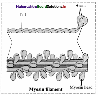
i. Myosin filament:
- Each myosin filament is a polymerized protein. Many meromyosins (monomeric proteins) constitute one thick filament.
- Myosin molecule consists of two heavy chains (heavy meromyosin / HMM) coiled around each other forming a double helix. One end of each of these chains is projected outwardly is known as cross bridge. This end folds to form a globular protein mass called myosin head.
- Two light chains are associated with each head forming 4 light chains/light meromyosin / LMM.
- Myosin head has a special ATPase activity. It can split ATP to produce energy.
- Myosin contributes 55% of muscle proteins.
- In sarcomere, myosin tails are arranged to point towards the centre of the sarcomere and the heads point to the sides of the myofilament band.
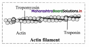
ii. Actin filament: It is a complex type of contractile protein. It is made up of three components:
- F actin: It forms the backbone of actin filament. F actin is made up of two helical strands. Each strand is composed of polymerized G actin molecules. One ADP molecule is attached to G actin molecule.
- Tropomyosin: The actin filament contains two additional protein strands that are polymers of tropomyosin molecules. Each strand is loosely attached to an F actin. In the resting stage, tropomyosin physically covers the active myosin-binding site of the actin strand.
- Troponin: It is a complex of three globular proteins, is attached approx. 2/3rd distance along each tropomyosin molecule. It has affinity for actin, tropomyosin and calcium ions. The troponin complex is believed to attach the tropomyosin to the actin. The strong affinity of troponin for calcium ions is believed to initiate the contraction process.
Question 14.
A muscle can only pull and not push the bone. Why?
Answer:
The fundamental characteristic of the muscle is contraction. Therefore, muscle can only pull and not push the bone.
Question 15.
Explain the physiology of muscle contraction.
Answer:
When the muscles are relaxed, the active sites remain covered with tropomyosin and troponin complex. Due to this, myosin cannot interact with active site of actin and thus contraction cannot occur.
- When an impulse (action potential) comes to muscle through motor end plate, it spreads throughout the sarcolemma of the myofibril.
- The transverse tubules of sarcoplasmic reticulum release large number of Ca++ ions into sarcoplasm. These calcium ions interact with the troponin molecules and the interaction inactivates troponin-tropomyosin complex. This causes change in the structure of tropomyosin.
- As a result, tropomyosin gets detached from the active site of actin (F actin) filament, exposing the active site for actin.
- The myosin head cleaves the ATP to derive energy and gets attached to the uncovered active site of actin. This results into the formation of acto-myosin complex.
- The myosin heads are now tilted backwards and pull the attached actin filament inwardly. This results in contraction of the muscle fibres.
Question 16.
Why do we shiver during winter?
Answer:
- Humans are homeotherms as they can regulate their body temperature with respect to the surrounding temperature. During winter, when temperature falls, the thermoreceptors detect the change in temperature and send signals to the brain.
- Shivering reflex i.e. rapid contraction of muscles is triggered by the brain to generate heat and raise the body temperature.
![]()
Question 17.
Write a short note on role of calcium ions in contraction and relaxation of muscles.
Answer:
Calcium ions play a major role in contraction and relaxation of muscles.
- Calcium ions are released from the sarcoplasm during muscle contraction and stored in sarcoplasmic reticulum during muscle relaxation.
- When a skeletal muscle is excited and an action potential travels along the T tubule, the concentration of calcium ions increases.
- These calcium ions bind to troponin which in turn undergoes a conformational change that causes tropomyosin to move away from the myosin-binding sites on actin. Once these binding sites are free, myosin heads bind to them to form cross-bridges and the muscle fiber contracts.
- The decrease in calcium ion concentration in the sarcoplasmic reticulum causes tropomyosin to slide back and block the myosin binding sites on actin. This causes the muscle to relax.
[Note: Students can scan the adjacent Q.R code to get conceptual clarity with the aid of a relevant video.]
Question 18.
Muscle contraction and relaxation are active processes. Give reason.
Answer:
Muscle contraction and relaxation are active processes as during both the processes energy is utilized by hydrolysis of ATP into ADP and inorganic phosphate by the enzyme ATPase.
Question 19.
Describe the properties of muscles observed on electrical stimulation.
Answer:
Properties of muscles observed on electrical excitation:
- Single muscle twitch: It is a muscle contraction initiated by a single brief-stimulation. It occurs in 3 stages: a latent period of no contraction, a contraction period and a relaxation period.
- Summation: If the muscle is stimulated before the end of the twitch, it generates greater tension i.e., summation or addition of effect takes place. Repeated stimuli will produce increasing strength of contraction (stair case phenomenon).
- Tetanus: If stimulation is very rapid and frequent the muscle does not have time to relax. It remains in a
state of contraction called tetanus. - Rcfraètory period: Immediately aller one stimulus, the muscle fibre cannot respond to another stimulus. This resting or refractory period is about 0.02 seconds.
- Threshold stimulus: For a muscle fibre to contract, a certain minimum strength or intensity of stimulus is required. This is called threshold stimulus.
- All or none principie: A stimulus below threshold will not result in contraction. A threshold stimulus will result in contraction. This contraction leads to maximum force. Higher stimulus will not increase force of contraction i.e. a muscle libre contracts either ftilly or not at all. This is ‘all or none’ principle. All types of muscle and nerve fibres obey this law.
- Oxygen debt: During strenuous exercise, the oxygen supply of muscle rapidly becomes insufficient to maintain oxidative phosphorylation of respiratory substrate. At this stage, muscles contract anaerobically and accumulate lactic acid produced by anaerobic glycolysis. Lactic acid produces less AlP and is toxic. It causes tiredness, pain and muscle cramps. During recovery, oxygen consumption of the muscle far exceeds than that in the resting state. This extra oxygen consumed during recovery is called oxygen debt of the muscle.
Note: The duration of refractory period varies with the muscle involved. Cardiac muscles have a longer refractory period of about 250 msec whereas skeletal muscles have a short refractory period of about I msec]
Question 20.
What is difference between endoskeleton and exoskeleton?
Answer:
The supportive structures present inside the body form the endoskeleton and when the supportive structures are present on the outer surface of the body they form exoskeleton.
Question 21.
Name the tissues that form the structural framework of the body.
Answer:
Cartilage and bone
Question 22.
What type of bones are present in our body?
Answer:
Long bones, short bones, flat bones, irregular bones and sesamoid bones.
Question 23.
How do bones help in various ways?
Answer:
- Bones form the framework of our body and thus provide shape to the body.
- They protect vital organs thus help in the smooth functioning of body.
- The joints between the bones help in movement and locomotion.
- They provide firm surface for attachment of muscles.
- They are reservoirs of calcium and form important site for hemopoiesis.
Question 24.
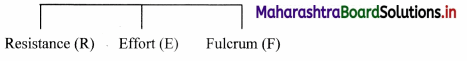
Explain the three types of lever found in human body.
Answer:
The three types of lever are as follows:
- Class I lever: The joint between the first vertebra and occipital condyle of skull is an example of Class I lever. The force is directed towards the joints (fulcrum); contraction of back muscle provides force while the part of head that is raised acts as resistance.

- Class II lever: Human body raised on toes is an example of Class II lever. Toe acts as fulcrum, contracting calf muscles provide the force while raised body acts as resistance.
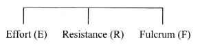
- Class III lever: Flexion of forearm at elbow exhibit lever of class III. Elbow joint acts as fulcrum and radius, ulna provides resistance. Contracting bicep muscles provides force for the movement.
Question 25.
Give an account of bones of human skull.
Answer:
Skull is made up of 22 bones. It is located at the superior end of vertebral column. The bones of skull are
joined by fixed or immovable joints except for jaw.
Skull consists of cranium or brain box and facial bones.
i. Cranium: It is made up of four median bones and two paired bones.
- Frontal bone: It is median bone (unpaired) forming forehead, roof of orbit (eye socket) and the most anterior part of cranium. It is connected to two parietals, sphenoid and ethmoid bone.
- Parietal bones: These paired bones form the roof of cranium and greater portion of sides of the cranium.
- Temporal bones: These paired bones are situated laterally just above the ear on either side. Each temporal bone gives out zygomatic process that joins zygomatic bone to form zygomatic arch. Just at the base of zygomatic process is mandibular fossa, a depression for mandibles (lower jaw bone) that forms the only movable joint of the skull. This bone harbors the ear canal that directs sound waves into the ear. The processes of temporal bones provide points for attachment for various muscles of neck and tongue.
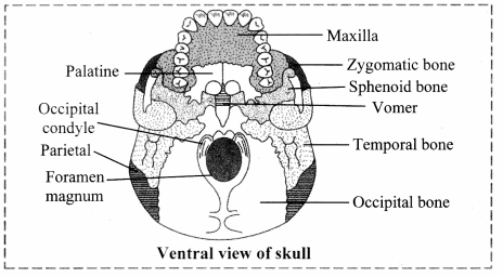
- Occipital bone: It is a single bone present at the back of the head. It forms the posterior part and most of the base of cranium. The inferior part of this bone shows foramen magnum, the opening through which medulla oblongata connects with spinal cord. On the either sides of foramen magnum are two prominent protuberances called occipital condyles. These fit into the corresponding depressions present in 1st vertebra.
- Sphenoid bone: Median bone present at the base of the skull that articulates with all other cranial bones and holds them together. This butterfly shaped bone has a saddle shaped region called sella turcica. In this hypophyseal fossa, the pituitary gland is lodged.
- Ethmoid bone: This median bone is spongy in appearance. It is located anterior to sphenoid and posterior to nasal bones. It contributes to formation of nasal septum and is major supporting structure of nasal cavity.
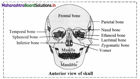
ii. Facial Bones: Fourteen facial bones give a characteristic shape to the face. The growth of face stops of the age of 16.
Following bones comprise the facial bones:
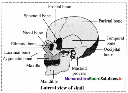
- Nasals: These are paired bones that form the bridge of nose.
- Maxillae: These form the upper jaw bones. They are paired bones that join with all facial bones except mandible. Upper row of teeth are lodged maxillae.
- Palatines: These are paired bones forming the roof of buccal cavity or floor of the nasal cavity.
- Zygomatic bones: They are commonly called as cheek bones.
- Lacrimal bones: These are the smallest amongst the facial bones.
These bones form the medial wall of each orbit. They have lacrimal fossa that houses lacrimal sacs. These sacs gather tears and send them to nasal cavity. - Inferior nasal conchae: They form the part of lateral wall of nasal cavity. They help to swirl and filter air before it passes to lungs.
- Vomer: The median, roughly triangular bone that forms the inferior portion of nasal septum.
- Mandible: This median bone forms the lower jaw. It is the largest and strongest facial bone. It is the only movable bone of skull. It has curved horizontal body and two perpendicular branches i.e. rami. These help in attachment of muscles. It has lower row of teeth lodged in it.
![]()
Question 26.
Give an account of hyoid bone.
Answer:
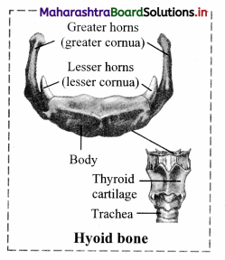
- It is a ‘U’ shaped bone that does not articulate with any other bone.
- It is suspended from temporal bone by ligaments and muscles.
- It is located between mandible and larynx.
- It has horizontal body and paired projections called horns.
- It provides site for attachment of some tongue muscles and muscles of neck and pharynx.
Question 27.
Sketch and label the anterior and ‘entraI view of skull.
Answer:
Question 28.
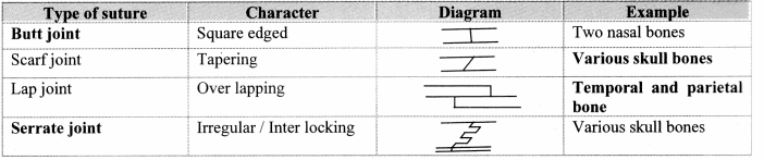
Mention the sutures in skull along with their location.
Answer:
Skull has many sutures (type of immovable joints) present, out of which four prominent ones are:
- Coronal suture; Joins frontal bone with parietals.
- Sagittal suture: Joins two parietal bones.
- Lambdiodal suture: Joins two parietal bones with occipital bone.
- Lateral/squamous sutures: Joins parietal and temporal bones on lateral side.
Question 29.
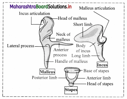
What are ear ossicles?
Answer:
The three tiny bones present in each middle ear namely malleus, incus and stapes, together are called ‘ear ossicles’
Question 30.
Draw a neat and labelled diagram of ear ossicles.
Answer:
Question 31.
Write a note on human vertebral column. Sketch and label the structure of the vertebral column.
Answer:
- Human vertebral column or backbone is a part of axial skeleton.
- It is made up of a chain of irregular bones called vertebrae.
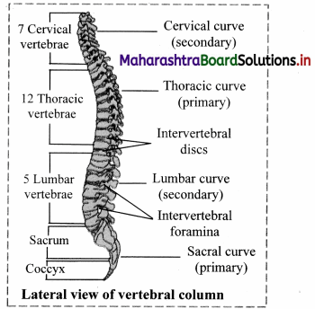
- Humans have 33 vertebrae in early years of growth, whereas adults have 26 vertebrae.
- In adults, five sacral vertebrae lìjse to tòrm one sacrum and four coccygeal vertebrae fuse to form single coccyx.
Question 32.
Explain the cervical vertebrae in detail.
Answer:
- Atlas vertebrae:
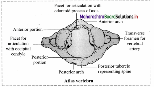
- It is the 1st cervical vertebra.
- It is a ring like bone and consists of anterior and posterior arches.
- It does not have centrum and spinous process.
- The transverse processes and transverse foramina of atlas are large.
- The vertebral foramen is large and divided into two parts by transverse ligament.
- Spinal cord passes through anterior compartment.
- Anterior zygopophyses are replaced by facets for attachment with occipital condyle of skull that forms ‘Yes joint’.
ii. Axis vertebrae:
- It is the 2nd cervical vertebrae.
- The centrum of this vertebra gives out tooth-like odontoid process.
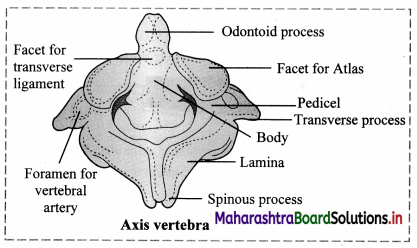
- Odontoid process fits into the anterior portion of vertebral foramen of atlas vertebra thereby forming pivot joint, also known as ‘No joint’.
iii. Typical cervical vertebrae:
- Vertebrae number 3 to 6 are called as typical cervical vertebrae.
- They show short centrum and bifid spinous process.
- The transverse processes of these vertebrae are reduced; each having large vertebrarterial canal at its base for the passage of vertebral artery.
iv. 7th cervical vertebra (Vertebra prominens): It is the largest cervical vertebra where the neural spine is straight.
Question 33.
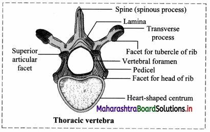
Write a note on thoracic vertebrae. Sketch and label thoracic vertebrae.
Answer:
- There are 12 thoracic vertebrae found in the chest region.
- The centrurn of thoracic vertebrae is heart shaped and all the processes are well developed.
- Except for 11th and 12th vertebrae; transverse process of other thoracic vertebrae show facets for attachment with ribs.
Question 34.
Elaborate on lumbar vertebrae with help of a neat and labelled diagram.
Answer:
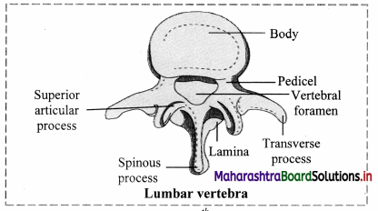
- There are 5 lumbar vertebrae.
- They are well developed vertebrae and exhibit all the characters of a typical vertebrae.
- The centrum of the lumbar vertebrae is kidney shaped.
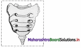
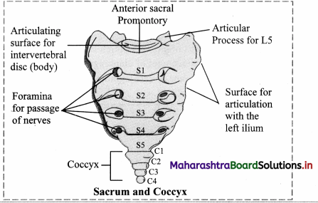
Question 35.
Give an account on:
i. Sacrum
ii. Coccyx
Answer:
i. Sacrum:
- It is a triangular bone formed by the fusion of five sacral vertebrae.
- It is located in pelvic cavity between two hip bones.
- The anterior end of sacrum is broad and posterior end is narrow.
- It consists of vertebral foramina formed by the fusion of vertebrae.
- The reduced neural spines can be observed projecting from dorsal aspect of sacrum.
- Function: It gives strength to pelvic girdle,
ii. Coccyx:
- It is a triangular bone which is formed by fusion of four coccygeal vertebrae.
- It is reduced and does not show vertebral foramina and spinous processes.
- The transverse processes of coccygeal vertebrae are reduced.
![]()
Question 36.
Identify the given vertebrae and label it.
Answer:
Question 37.
Describe in brief the structure of thoracic cage.
Answer:
Thoracic cage consists of 12 pairs of ribs, breast bone or sternum.
i. Ribs:
- A rib is a ‘C’ shaped bone. It is attached to respective thoracic vertebrae on dorsal side.
- Twelve pairs of ribs are attached to twelve thoracic vertebrae. For attachment to the vertebrae the posterior ends of ribs have two protuberances namely the head and tubercle.
- The head of rib attaches to facet formed by demifacets of adjacent thoracic vertebrae at the base of transverse processes.
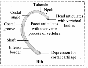
- The tip of transverse processes of these vertebrae also have facets for attachment of ribs where tubercles of ribs are attached.
- Ribs provide space for attachment of intercostal muscles.
- On ventral side, the ribs may or may not attach the sternum. Depending on their attachment, the ribs are classified into three types:
- True ribs: First seven pairs of ribs are attached directly to the sternum by means of their costal cartilages.
- False ribs: Costal cartilages of rib numbers 8, 9 and 10 are attached to rib number 7 on either side and not directly to the sternum. Hence, these are called false ribs.
- Floating ribs: Last two pairs of ribs have no ventral connection. Hence, they are called floating ribs.
ii. Sternum:
- It is a flat, narrow bone, around 15 cms in length.
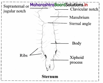
- It is placed medially in anterior thoracic ] jugular notch wall (chest region).
- It consists of three distinct parts – manubrium, body and xiphoid processes.
- Manubrium shows two notches on anterio-lateral side for attachment with clavicle of each side. It also shows two notches on each of the lateral side for attachment of first two pairs of ribs.
- Body of sternum is a flat bone that shows five notches on lateral aspect which are meant for direct or indirect attachment of ribs.
- Xiphoid process is lowermost part of sternum which is initially cartilaginous and gets ossified in adults. It provides space for the attachment of diaphragm and abdominal muscles.
- Ribs are attached to sternum by means of cartilaginous extensions called costal cartilages.
Question 38.
What is intercostal space?
Answer:
The space between the ribs is called as intercostal space.
Question 39.
Describe the bones of pectoral girdle.
Answer:
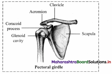
Pectoral girdle is called as shoulder girdle. It attaches forelimb skeleton with axial skeleton. There are two pectoral girdles, each consists of a shoulder blade or scapula and collar bone or clavicle.
i. Clavicle:
- It is a ‘S’ shaped slender bone.
- One end of clavicle is attached to acromion process of scapula. The other rounded end called sternal end attaches to manubrium of sternum.
- This connects upper arm skeleton to axial skeleton.
ii. Scapula:
- It is a large, flat, triangular bone that occupies posterior chest wall extending from 2nd to 7th ribs.
- It is attached to axial skeleton by muscles and tendons.
- Scapula bears a concave socket called glenoid cavity at its lateral angle.
- Head of humerus (the upper arm bone) fits into the glenoid cavity.
- A beak like coracoid process projects from lateral angle of scapula and acromion process arise from scapula. They can be easily felt as high point of shoulder. Both the processes are meant for attachment of muscles.
Question 40.
Describe the bones of forelimb.
Answer:
Forelimb consists of humerus, radius and ulna (together forming forearm bones), carpals (bones of wrist), metacarpals (bones of palm) and phalanges (bones of digits). It consists of 30 bones.
i. Humerus:
- This is the bone of upper arm.
- It has a hemispherical head at its proximal end. On either side of head of humerus are present a pair of projections termed greater and lesser tubercles.
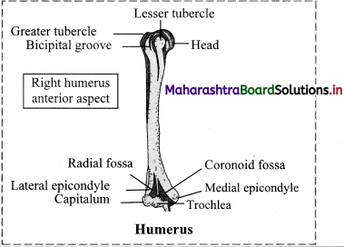
- Bicipital groove is a deep groove present between the tubercles where a tendon of biceps muscle is attached.
- The shaft of humerus shows deltoid tuberosity. Distal end of humerus shows pulley like part called trochlea that articulates with ulna.
ii. Radius and Ulna:
- Radius is located laterally on thumb side of the forearm.
- The proximal end of radius has disc like head that articulates with humerus bone.
- The shaft of radius widens distally to form styloid process.
- Ulna is located medially on little finger side of forearm.
- At the proximal end of ulna there is a prominent process called Olecranon process that forms elbow joint with humerus bone. On the lateral side, near the upper end of ulna is present the radial notch into which the side of head of radius is fixed.
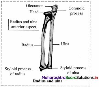
f. Radius and ulna articulate with each other at upper and lower extremities by superior and inferior radio-ulnar joints. In between the shaft of two bones, interosseous membrane is present.
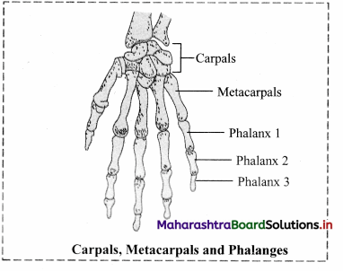
iii. Carpals: These are bones of wrist, arranged in two rows of four each.
iv. Metacarpals: Five elongated metacarpals form the bones of palm. Their proximal ends join with carpals and distal ends form knuckles.
v. Phalanges: Phalanges form the bones of fingers and thumb. There are 14 phalanges in each hand (Four fingers have three phalanges each and thumb has two).
Question 41.
Explain briefly the pelvic girdle with a neat and labelled diagram.
Answer:
- Pelvic girdle also known as hip girdle connects hind limb skeleton with axial skeleton.
- It is made up of two hip bones called coxal bones. These coxal bones unite posteriorly with sacrum.
- Coxal bone is a large irregularly shaped bone is made up of three parts – ilium, ischium and pubis.
- At the point of fusion of ilium, ischium and pubis, a cavity called acetabulum is present that forms ball and socket joint with the thigh bone.
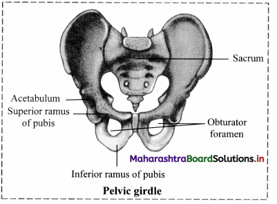
- The two pubis bones are joined medially by cartilaginous joint called pubic symphysis. Pubis and ischium together form a ring of bone that encloses a space called obturator foramen.
Question 42.
Describe the bones of lower limb.
Answer:
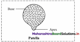
The bones of lower limb are femur, patella, tibia and fibula, tarsals, metatarsals and phalanges.
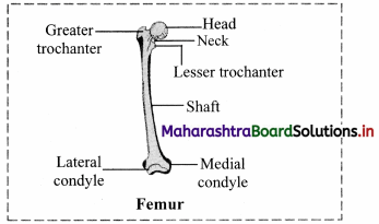
i. Femur: The thigh bone is the longest bone in the body. The head is joined to shaft at an angle by a short neck. It forms ball and socket joint with acetabulum cavity of coxal bone. The lower one third region of shaft is triangular flattened area called popliteal surface. Distal end has two condyles that articulate with tibia and fibula.
ii. Patella: It is also called as knee cap. ft is a sesamoid bone (bone embedded in tendon). It is a flat rounded bone with a pointed lower end.
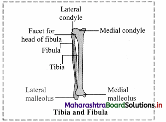
iii. Tibia and fibula: These are the two long bones of shank or lower leg. The two bones are connected to each other at the extremities. In between the two bones interosseous membrane is present.
- Tibia: It is much thicker and stronger than fibula. Its broad and expanded upper end articulates with femur and the lower end articulates with talus (tarsal bone).
- Fibula: It is a long slender bone on lateral side of tibia.
iv. Tarsals: These are the bones of ankle. Seven tarsals are arranged in three rows, two proximal, one intermediate and four distal.
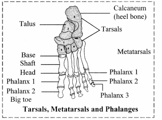
v. Metatarsals: Five metatarsal bones support the foot. Proximally they attach with distal row of tarsals and distally the metatarsals articulate with phalanges.
vi. Phalanges: These are the bones of the toes. Except the big toe which has two phalanges, the other four toes have three phalanges each.
Question 43.
What is arthrology?
Answer:
The study of joints is called arthrology.
Question 44.
What makes synovial joint freely movable?
Answer:
Synovial joint is characterised by synovial cavity between the articulating bones which allows free movement at the joint. This makes the joint freely movable.
Question 45.
Complete the given table.
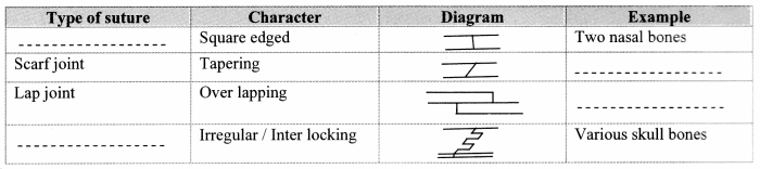
Answer:
Question 46.
State and explain the disorders related to muscles.
Answer:
i. Muscular dystrophy:
- It is a gradual wasting disease affecting various groups of muscles.
- It is a genetic disorder.
- Usually the voluntary skeletal muscles are weakened whereas internal muscles such as diaphragm are not affected in the patient suffering from this disorder.
- The Duchenne type of muscular dystrophy usually occurs in boys, affecting their lower limbs.
- Limb girdle muscular dystrophy affects the muscles of shoulders or hips and it usually starts in adults between 20 – 30 years.
- There is no cure for this disease.
ii. Myasthenia gravis:
- It is a weakness of skeletal muscles.
- It is caused by an abnormality at the neuromuscular junction that partially blocks muscle contraction.
- It is an autoimmune disorder caused by excessive production of certain antibodies in the blood stream. These antibodies bind to acetylcholine receptors of neuromuscular junction. Thus, the transmission of nerve impulses to the muscle fibres is blocked. This causes progressive and extensive muscle weakness.
- It may affect the eye and eyelid movements, facial expression and swallowing.
- The degree of muscle weakness varies from local to general.
- Symptoms: Ptosis (drooping or falling of upper eye lid), diplopia or double vision, difficulty in swallowing, chewing and speech.
![]()
Question 47.
Give an account on disorders related to bones.
Answer:
- Arthritis: It is an inflammation of joints. It is a painful disorder of bones, ligaments, tendons, etc. In this disorder, joints become swollen, stiff and painful. It can lead to disability.
- Arthritis is of three types:
- Osteoarthritis: In this disorder, the joint cartilage is degenerated. It is caused by various factors like aging, obesity, muscle weakness, etc. This is the most common type of arthritis that affects hands, knees and spine.
- Gouty arthritis (Gout): In this disorder, joint pain occurs due to the deposition of uric acid in joints. If uric acid is produced in excess or is not excreted, it accumulates in joints as sodium urate. The accumulated sodium urate degenerates the cartilage causing inflammation and pain. It generally affects the joints of feet.
- Rheumotoid arthritis: It is an autoimmune disorder where body’s immune system attacks its own tissues. In rheumatoid arthritis, synovial membrane swells up and starts secreting extra synovial fluid. This fluid exerts pressure on joint and makes it painful. Membrane may develop abnormal granulation tissue called pannus. Pannus may erode cartilage. Fibrous tissue gets ossified and may lead to stiffness in joints.
- Osteoporosis:
- In this disorder, bones become porous and hence brittle. It is primarily age related disease and is more common in women than men.
- As age advances, bone resorption outpaces bone formation. Hence, the bones lose mass and become brittle. More calcium is lost in urine, sweat, etc., than it is gained through diet. Thus, prevention of disease is better than treatment by consuming adequate amount of calcium and exercise at young age.
- Osteoporosis may be caused due to decreasing estrogen secretion after menopause, deficiency of vitamin D, low calcium diet, decreased secretion of sex hormones and thyrocalcitonin.
Question 48.
Name the following.
Question 1.
The striated muscles that are known as prime movers
Answer:
Agonists
Question 2.
The antagonistic muscles that lower the body part
Answer:
Depressor
Question 3.
The contractile proteins of sarcomere
Answer:
Actin and myosin
Question 4.
Sliding filament theory is also known as
Answer:
Walk along theory or Rachet theory
Question 5.
The smallest facial bone
Answer:
Lacrimal bones
Question 6.
Number of bones in thoracic cage
Answer:
25
Question 7.
The first cervical vertebrae
Answer:
Atlas
Question 8.
The three bones of pelvic girdle
Answer:
Ilium, ischium and pubis
Question 9.
The bone which is known as the knee cap
Answer:
Patella
Question 10.
Age related disorder more common in woman than man.
Answer:
Osteoporosis
Question 49.
Fill in the blanks.
Question 1.
The actin filament contains two additional proteins strands that are polymers of ___ molecules.
Answer:
tropomyosin
Question 2.
___ of sarcoplasmic reticulum release large number of Ca++ into sarcoplasm.
Answer:
Transverse (T) tubules
Question 3.
The joint between the first vertebra and occipital condyle of skull is an example of ____ class of lever.
Answer:
Class I
Question 4.
Endoskeleton of an adult human consists of ___ bones.
Answer:
206
![]()
Question 5.
___ are the bones that connect limbs to the axial skeleton.
Answer:
Girdles
Question 6.
___ bones form the roof of cranium.
Answer:
Parietal
Question 7.
Zveomatic bone is commonly known as ___ bone.
Answer:
Cheek
Question 8.
The largest and strongest facial bone is ___
Answer:
Mandible
Question 9.
There are ___ types of vertebrae.
Answer:
5
Question 10.
___ is also known as shoulder girdle.
Answer:
Pectoral girdle
Question 11.
___ are the bones of wrist.
Answer:
Carpals
Question 12.
___ is known as immovable or fixed joint.
Answer:
Synarthroses
![]()
Question 13.
Tibia and fibula are two long bones of ___.
Answer:
Shank or lower leg
Question 14.
___ is caused due to deposition of uric acid in joints.
Answer:
Gouty arthritis
Question 50.
State whether the following statements are True or False:
Question 1.
Saggital suture joins frontal bone with parietal bone.
Answer:
False. Saggital suture joins two parietal bones.
Question 2.
In human beings, there are 10 pairs of ribs.
Answer:
False. In human beings, there are 12 pairs of ribs.
Question 3.
Hyoid bone does not articulate with any other bone.
Answer:
True
Question 4.
Pivot joint is present between Atlas and Axis vertebrae.
Answer:
True
Question 5.
The two pubis bones are joined by cartilaginous joint called pubic symphysis.
Answer:
True
Question 6.
Rheumatoid arthritis is an autoimmune disorder.
Answer:
True
Question 50.
Identify the INCORRECTLY labelled part of pectoral girdle.
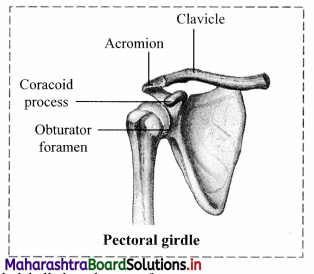
Answer:
Glenoid cavity is incorrectly labelled as obturator foramen.
Multiple Choice Questions
Question 1.
Locomotion in sperms takes place with the help of
(A) flagella
(B) cilia
(C) pseudopodia
(D) muscles
Answer:
(A) flagella
Question 2.
Levator muscles result into the action of
(A) lowering a body part
(B) tensing a body part
(C) relaxing a body part
(D) raising a body part
Answer:
(D) raising a body part
![]()
Question 3.
Which of these is not a skull bone?
(A) Frontal
(B) Scapula
(C) Occipital
(D) Temporal
Answer:
(B) Scapula
Question 4.
Immovable joint is in between the bones of
(A) frontal and parietal
(B) metacarpal and phalangeal
(C) femur and tibia
(D) radius and ulna
Answer:
(A) frontal and parietal
Question 5.
Total number of vertebrae in human beings is
(A) 22
(B) 33
(C) 24
(D) 12
Answer:
(B) 33
Question 6.
Which of the following is NOT a part of appendicular skeleton?
(A) Girdles
(B) Forelimb
(C) Hindlimb
(D) Vertebral column
Answer:
(D) Vertebral column
Question 7.
___ connects the upper arm to the axial skeleton.
(A) Clavicle
(B) Patella
(C) Tibia
(D) Femur
Answer:
(A) Clavicle
Question 8.
__ is a part of hind limb.
(A) Radius
(B) Ulna
(C) Humers
(D) Fibula
Answer:
(D) Fibula
Question 9.
Which of the following are toe bones?
(A) Tarsals
(B) Metatarsals
(C) Carpals
(D) Phalanges
Answer:
(D) Phalanges
Question 10.
Sutures on the skull are ___ joints.
(A) freely movable
(B) slightly movable
(C) fixed
(D) synovial
Answer:
(C) fixed
Question 11.
___ is an example of syndesmoses.
(A) Distal tibiofibular joint
(B) Rib-sternum junction
(C) Tooth and jaw bones
(D) Intervertebral disc
Answer:
(A) Distal tibiofibular joint
Question 12.
Elbow joint is
(A) ball and socket joint
(B) hinge joint
(C) suture joint
(D) gliding joint
Answer:
(B) hinge joint
Question 13.
Complete the analogy.
Hinge joint: Monoaxial movement:: ___ Biaxial movement
(A) Ball and socket joint
(B) Gliding joint
(C) Condyloid joint
(D) Both (A) and (B)
Answer:
(C) Condyloid joint
Question 14.
An autoimmune disorder in which an antibody reduces the efficiency of transmission between the motor neuron is called
(A) Myasthenia gravis
(B) Tetany
(C) Osteoarthritis
(D) Osteoporosis
Answer:
(A) Myasthenia gravis